Cyclin B1 (also CCNB1 and G2/mitotic-specific cyclin-B1) is a member of the cyclin AB subfamily, cyclin family of proteins. Although its predicted MW is 50 kDa, it runs anomalously at 62 kDa in SDS-PAGE. Cyclin B1 associates with both CDK1 and 2 providing substrate specificity to a phosphorylating complex. A phosphor‑CDK1:Cyclin B1 complex is inactive and cytosolic during interphase. At the beginning of mitosis, CDK1 is dephosphorylated and activated, and the CDK1:Cyclin B1 complex initiates formation of the mitotic scaffold. Human Cyclin B1 is 433 amino acids (aa) in length. It contains two cyclin box folds (aa 201‑290 and 298‑383) and two substrate binding sites (aa 298‑342 and 343‑380). Phosphorylation occurs at Ser9, Ser35, Ser69, and Thr321. There is one potential alternative start site at Met252 and deletions of aa 363‑399 and 365‑433. Over aa 1‑91, human Cyclin B1 shares 63% aa identity with mouse Cyclin B1.
Human Cyclin B1 Antibody (2061D)
R&D Systems | Catalog # MAB60001
Recombinant Monoclonal Antibody.

Key Product Details
Species Reactivity
Human
Applications
Immunohistochemistry, Western Blot, Intracellular Staining by Flow Cytometry, Simple Western
Label
Unconjugated
Antibody Source
Recombinant Monoclonal Rabbit IgG Clone # 2061D
Loading...
Product Specifications
Immunogen
E. coli-derived recombinant human Cyclin B1
Met1-Pro91
Accession # P14635
Met1-Pro91
Accession # P14635
Specificity
Detects human Cyclin B1 in direct ELISAs and Western blots.
Clonality
Monoclonal
Host
Rabbit
Isotype
IgG
Scientific Data Images for Human Cyclin B1 Antibody (2061D)
Detection of Human Cyclin B1 by Western Blot.
Western blot shows lysates of Jurkat human acute T cell leukemia cell line, K562 human chronic myelogenous leukemia cell line, and U2OS human osteosarcoma cell line. PVDF membrane was probed with 0.05 µg/mL of Rabbit Anti-Human Cyclin B1 Monoclonal Antibody (Catalog # MAB60001) followed by HRP-conjugated Anti-Rabbit IgG Secondary Antibody (Catalog # HAF008). A specific band was detected for Cyclin B1 at approximately 52 kDa (as indicated). This experiment was conducted under reducing conditions and using Immunoblot Buffer Group 1.
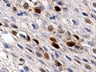
Cyclin B1 in Human Squamous Cell Carcinoma.
Cyclin B1 was detected in immersion fixed paraffin-embedded sections of human squamous cell carcinoma using Rabbit Anti-Human Cyclin B1 Monoclonal Antibody (Catalog # MAB60001) at 3 µg/mL for 1 hour at room temperature followed by incubation with the Anti-Rabbit IgG VisUCyte™ HRP Polymer Antibody (Catalog # VC003). Tissue was stained using DAB (brown) and counterstained with hematoxylin (blue). Specific staining was localized to nuclei. View our protocol for IHC Staining with VisUCyte HRP Polymer Detection Reagents.
Detection of Cyclin B1 in K562 Human Cell Line by Flow Cytometry.
K562 human chronic myelogenous leukemia cell line was stained with Rabbit Anti-Human Cyclin B1 Monoclonal Antibody (Catalog # MAB60001, filled histogram) or isotype control antibody (Catalog # AB-105-C, open histogram), followed by Phycoerythrin-conjugated Anti-Rabbit IgG Secondary Antibody (Catalog # F0110). To facilitate intracellular staining, cells were fixed with Flow Cytometry Fixation Buffer (Catalog # FC004) and permeabilized with Flow Cytometry Permeabilization/Wash Buffer I (Catalog # FC005). View our protocol for Staining Intracellular Molecules.
Detection of Human Cyclin B1 by Simple WesternTM.
Simple Western lane view shows lysates of Jurkat human acute T cell leukemia cell line, loaded at 0.2 mg/mL. A specific band was detected for Cyclin B1 at approximately 64 kDa (as indicated) using 0.5 µg/mL of Rabbit Anti-Human Cyclin B1 Monoclonal Antibody (Catalog # MAB60001). This experiment was conducted under reducing conditions and using the 12-230 kDa separation system.Applications for Human Cyclin B1 Antibody (2061D)
Application
Recommended Usage
Immunohistochemistry
3-25 µg/mL
Sample: Immersion fixed paraffin-embedded sections of human squamous cell carcinoma
Sample: Immersion fixed paraffin-embedded sections of human squamous cell carcinoma
Intracellular Staining by Flow Cytometry
0.25 µg/106 cells
Sample: K562 human chronic myelogenous leukemia cell line
Sample: K562 human chronic myelogenous leukemia cell line
Simple Western
0.5 µg/mL
Sample: Jurkat human acute T cell leukemia cell line
Sample: Jurkat human acute T cell leukemia cell line
Western Blot
0.05 µg/mL
Sample: Jurkat human acute T cell leukemia cell line, K562 human chronic myelogenous leukemia cell line, and U2OS human osteosarcoma cell line
Sample: Jurkat human acute T cell leukemia cell line, K562 human chronic myelogenous leukemia cell line, and U2OS human osteosarcoma cell line
Reviewed Applications
Read 2 reviews rated 4.5 using MAB60001 in the following applications:
Flow Cytometry Panel Builder
Bio-Techne Knows Flow Cytometry
Save time and reduce costly mistakes by quickly finding compatible reagents using the Panel Builder Tool.
Advanced Features
- Spectra Viewer - Custom analysis of spectra from multiple fluorochromes
- Spillover Popups - Visualize the spectra of individual fluorochromes
- Antigen Density Selector - Match fluorochrome brightness with antigen density

Formulation, Preparation, and Storage
Purification
Protein A or G purified from cell culture supernatant
Reconstitution
Reconstitute at 0.5 mg/mL in sterile PBS. For liquid material, refer to CoA for concentration.
Loading...
Formulation
Lyophilized from a 0.2 μm filtered solution in PBS with Trehalose. *Small pack size (SP) is supplied either lyophilized or as a 0.2 µm filtered solution in PBS.
Shipping
Lyophilized product is shipped at ambient temperature. Liquid small pack size (-SP) is shipped with polar packs. Upon receipt, store immediately at the temperature recommended below.
Stability & Storage
Use a manual defrost freezer and avoid repeated freeze-thaw cycles.
- 12 months from date of receipt, -20 to -70 °C as supplied.
- 1 month, 2 to 8 °C under sterile conditions after reconstitution.
- 6 months, -20 to -70 °C under sterile conditions after reconstitution.
Calculators
Background: Cyclin B1
Alternate Names
CCNB1
Gene Symbol
CCNB1
UniProt
Additional Cyclin B1 Products
Product Documents for Human Cyclin B1 Antibody (2061D)
Certificate of Analysis
To download a Certificate of Analysis, please enter a lot or batch number in the search box below.
Note: Certificate of Analysis not available for kit components.
Product Specific Notices for Human Cyclin B1 Antibody (2061D)
For research use only
Related Research Areas
Customer Reviews for Human Cyclin B1 Antibody (2061D) (2)
4.5 out of 5
2 Customer Ratings
Have you used Human Cyclin B1 Antibody (2061D)?
Submit a review and receive an Amazon gift card!
$25/€18/£15/$25CAN/¥2500 Yen for a review with an image
$10/€7/£6/$10CAN/¥1110 Yen for a review without an image
Submit a review
Customer Images
Showing
1
-
2 of
2 reviews
Showing All
Filter By:
-
Application: ImmunohistochemistrySample Tested: Oral mucosa tissueSpecies: HumanVerified Customer | Posted 01/28/2022

-
Application: Western BlotSample Tested: T47D human breast cancer cell line and MCF-7 human breast cancer cell lineSpecies: HumanVerified Customer | Posted 08/16/2017
There are no reviews that match your criteria.
Protocols
Find general support by application which include: protocols, troubleshooting, illustrated assays, videos and webinars.
- 7-Amino Actinomycin D (7-AAD) Cell Viability Flow Cytometry Protocol
- Antigen Retrieval Protocol (PIER)
- Antigen Retrieval for Frozen Sections Protocol
- Appropriate Fixation of IHC/ICC Samples
- Cellular Response to Hypoxia Protocols
- Chromogenic IHC Staining of Formalin-Fixed Paraffin-Embedded (FFPE) Tissue Protocol
- Chromogenic Immunohistochemistry Staining of Frozen Tissue
- ClariTSA™ Fluorophore Kits
- Detection & Visualization of Antibody Binding
- Extracellular Membrane Flow Cytometry Protocol
- Flow Cytometry Protocol for Cell Surface Markers
- Flow Cytometry Protocol for Staining Membrane Associated Proteins
- Flow Cytometry Staining Protocols
- Flow Cytometry Troubleshooting Guide
- Fluorescent IHC Staining of Frozen Tissue Protocol
- Graphic Protocol for Heat-induced Epitope Retrieval
- Graphic Protocol for the Preparation and Fluorescent IHC Staining of Frozen Tissue Sections
- Graphic Protocol for the Preparation and Fluorescent IHC Staining of Paraffin-embedded Tissue Sections
- Graphic Protocol for the Preparation of Gelatin-coated Slides for Histological Tissue Sections
- IHC Sample Preparation (Frozen sections vs Paraffin)
- Immunofluorescent IHC Staining of Formalin-Fixed Paraffin-Embedded (FFPE) Tissue Protocol
- Immunohistochemistry (IHC) and Immunocytochemistry (ICC) Protocols
- Immunohistochemistry Frozen Troubleshooting
- Immunohistochemistry Paraffin Troubleshooting
- Intracellular Flow Cytometry Protocol Using Alcohol (Methanol)
- Intracellular Flow Cytometry Protocol Using Detergents
- Intracellular Nuclear Staining Flow Cytometry Protocol Using Detergents
- Intracellular Staining Flow Cytometry Protocol Using Alcohol Permeabilization
- Intracellular Staining Flow Cytometry Protocol Using Detergents to Permeabilize Cells
- Preparing Samples for IHC/ICC Experiments
- Preventing Non-Specific Staining (Non-Specific Binding)
- Primary Antibody Selection & Optimization
- Propidium Iodide Cell Viability Flow Cytometry Protocol
- Protocol for Heat-Induced Epitope Retrieval (HIER)
- Protocol for Liperfluo
- Protocol for Making a 4% Formaldehyde Solution in PBS
- Protocol for VisUCyte™ HRP Polymer Detection Reagent
- Protocol for the Characterization of Human Th22 Cells
- Protocol for the Characterization of Human Th9 Cells
- Protocol for the Preparation & Fixation of Cells on Coverslips
- Protocol for the Preparation and Chromogenic IHC Staining of Frozen Tissue Sections
- Protocol for the Preparation and Chromogenic IHC Staining of Frozen Tissue Sections - Graphic
- Protocol for the Preparation and Chromogenic IHC Staining of Paraffin-embedded Tissue Sections
- Protocol for the Preparation and Chromogenic IHC Staining of Paraffin-embedded Tissue Sections - Graphic
- Protocol for the Preparation and Fluorescent IHC Staining of Frozen Tissue Sections
- Protocol for the Preparation and Fluorescent IHC Staining of Paraffin-embedded Tissue Sections
- Protocol for the Preparation of Gelatin-coated Slides for Histological Tissue Sections
- Protocol: Annexin V and PI Staining by Flow Cytometry
- Protocol: Annexin V and PI Staining for Apoptosis by Flow Cytometry
- R&D Systems Quality Control Western Blot Protocol
- TUNEL and Active Caspase-3 Detection by IHC/ICC Protocol
- The Importance of IHC/ICC Controls
- Troubleshooting Guide: Fluorokine Flow Cytometry Kits
- Troubleshooting Guide: Immunohistochemistry
- Troubleshooting Guide: Western Blot Figures
- Western Blot Conditions
- Western Blot Protocol
- Western Blot Protocol for Cell Lysates
- Western Blot Troubleshooting
- Western Blot Troubleshooting Guide
- View all Protocols, Troubleshooting, Illustrated assays and Webinars
Loading...