ErbB3, also called Her3 (human epidermal growth factor receptor 3), is a type I membrane glycoprotein that is a member of the ErbB family of tyrosine kinase receptors. ErbB family members serve as receptors for the epidermal growth factor (EGF) family of growth factors. Among ErbB family members, ErbB3 is unique in that it contains a defective kinase domain. ErbB3 is expressed in keratinocytes, melanocytes, skeletal muscle cells, embryonic myoblasts and Schwann cells. Monomeric ErbB3 serves as a low affinity receptor for the heregulins (HRG). ErbB3 heterodimerizes with ErbB2 to form a high affinity receptor complex. In contrast, ErbB3 homodimerization or heterodimerization with ErbB4 forms a low affinity heregulin-binding complex. Because ErbB3 contains a defective kinase domain, the kinase domain of ErbB2 is responsible for initiating the tyrosine phosphorylation signal through the heterodimeric receptor. It has been found that a discrete three amino acid signal in the ErbB3 cytoplasmic domain is critical for transactivation of ErbB2. The cytoplasmic domain of ErbB3 also contains six consensus binding motifs for the SH2 domain of the regulatory p85 subunit of phosphoinositide 3-kinase (PI 3-kinase, PI3K) as well as one proline-rich consensus binding motif for the SH3 domain of p85. Human ErbB3 consists of 1342 amino acids (aa) with a 19 aa signal sequence, a 624 aa extracellular domain, a 21 aa transmembrane region, and a 678 aa cytoplasmic domain. ErbB3 appears to play roles in development, cancer, communication at the neuromuscular junction, and regulation of cell growth and differentiation.
Human ErbB3/Her3 Antibody (66223)
R&D Systems | Catalog # MAB3481

Key Product Details
Species Reactivity
Validated:
Human
Cited:
Human
Applications
Validated:
ELISA Capture (Matched Antibody Pair), Neutralization, Flow Cytometry, CyTOF-ready
Cited:
Western Blot, Neutralization, Flow Cytometry, Immunoprecipitation, Dynamic Light Scattering, ELISA Capture, ELISA Development, ELISA Development (Capture), Luminex Development
Label
Unconjugated
Antibody Source
Monoclonal Mouse IgG1 Clone # 66223
Loading...
Product Specifications
Immunogen
Mouse myeloma cell line NS0-derived recombinant human ErbB3/Her3
Ser20-Thr643
Accession # P21860
Ser20-Thr643
Accession # P21860
Specificity
Detects human ErbB3/Her3 in ELISAs. Does not cross-react with recombinant EGF R.
Clonality
Monoclonal
Host
Mouse
Isotype
IgG1
Endotoxin Level
<0.10 EU per 1 μg of the antibody by the LAL method.
Scientific Data Images for Human ErbB3/Her3 Antibody (66223)
Cell Proliferation Induced by NRG1‑ beta 1/HRG1‑ beta 1 and Neutralization by Human ErbB3/Her3 Antibody.
Recombinant Human NRG1-1 beta 1/HRG1-1 beta 1 (Catalog # 377-HB) stimulates proliferation in the MCF-17 human breast cancer cell line in a dose-dependent manner (orange line). Proliferation elicited by Recombinant Human NRG1-1 beta 1/HRG1-1 beta 1 (10 ng/mL) is neutralized (green line) by increasing concentrations of Human ErbB3/Her3 Monoclonal Antibody (Catalog # MAB3481). The ND50 is typically 0.0075-0.03 µg/mL.Applications for Human ErbB3/Her3 Antibody (66223)
Application
Recommended Usage
CyTOF-ready
Ready to be labeled using established conjugation methods. No BSA or other carrier proteins that could interfere with conjugation.
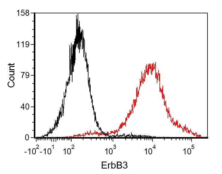
Flow Cytometry
2.5 µg/106 cells
Sample: MCF‑7 human breast cancer cell line
Sample: MCF‑7 human breast cancer cell line
Neutralization
Measured by its ability to neutralize NRG1‑ beta 1/HRG1‑ beta 1-induced proliferation in the MCF‑7 human breast cancer cell line. The Neutralization Dose (ND50) is typically 0.0075-0.03 µg/mL in the presence of 10 ng/mL Recombinant Human NRG1‑ beta 1/HRG1‑ beta 1 Extracellular Domain.
Human ErbB3/Her3 Sandwich Immunoassay
Please Note: Optimal dilutions of this antibody should be experimentally determined.
Reviewed Applications
Read 3 reviews rated 5 using MAB3481 in the following applications:
Flow Cytometry Panel Builder
Bio-Techne Knows Flow Cytometry
Save time and reduce costly mistakes by quickly finding compatible reagents using the Panel Builder Tool.
Advanced Features
- Spectra Viewer - Custom analysis of spectra from multiple fluorochromes
- Spillover Popups - Visualize the spectra of individual fluorochromes
- Antigen Density Selector - Match fluorochrome brightness with antigen density

Formulation, Preparation, and Storage
Purification
Protein A or G purified from hybridoma culture supernatant
Reconstitution
Reconstitute at 0.5 mg/mL in sterile PBS. For liquid material, refer to CoA for concentration.
Loading...
Formulation
Lyophilized from a 0.2 μm filtered solution in PBS with Trehalose. *Small pack size (SP) is supplied either lyophilized or as a 0.2 µm filtered solution in PBS.
Shipping
Lyophilized product is shipped at ambient temperature. Liquid small pack size (-SP) is shipped with polar packs. Upon receipt, store immediately at the temperature recommended below.
Stability & Storage
Use a manual defrost freezer and avoid repeated freeze-thaw cycles.
- 12 months from date of receipt, -20 to -70 °C as supplied.
- 1 month, 2 to 8 °C under sterile conditions after reconstitution.
- 6 months, -20 to -70 °C under sterile conditions after reconstitution.
Calculators
Background: ErbB3/Her3
References
- Kraus, M.H. et. al. (1989) Proc. Natl. Acad. Sci. USA 86:9193.
- Plowman, G.D. et. al. (1990) Proc. Natl. Acad. Sci. USA 87:4905.
- Carraway, K.L. 3rd et. al. (1994) J. Biol. Chem. 269:14303.
- Emkey, R. and C.R. Kahn (1997) J. Biol. Chem. 272:31172.
- Sundaresan, S. et. al. (1998) Endocrinology 139:4756.
- Hellyer, N.J. et. al. (1998) Biochem. J. 333:757.
- Schaefer, G. et. al. (1999) J. Biol. Chem. 274:859.
- Hellyer, N.J. et. al. (2001) J. Biol. Chem. 276:42153.
- Schlessinger, J. (2000) Cell 103:211.
- Daly, R.J. (1999) Growth Factors 16:255.
Long Name
Receptor Tyrosine Protein Kinase ErbB3
Alternate Names
HER3
Gene Symbol
ERBB3
UniProt
Additional ErbB3/Her3 Products
Product Documents for Human ErbB3/Her3 Antibody (66223)
Certificate of Analysis
To download a Certificate of Analysis, please enter a lot or batch number in the search box below.
Note: Certificate of Analysis not available for kit components.
Product Specific Notices for Human ErbB3/Her3 Antibody (66223)
For research use only
Related Research Areas
Citations for Human ErbB3/Her3 Antibody (66223)
 Powered by Bioz
Customer Reviews for Human ErbB3/Her3 Antibody (66223) (3)
5 out of 5
3 Customer Ratings
Have you used Human ErbB3/Her3 Antibody (66223)?
Submit a review and receive an Amazon gift card!
$25/€18/£15/$25CAN/¥2500 Yen for a review with an image
$10/€7/£6/$10CAN/¥1110 Yen for a review without an image
Submit a review
Customer Images
Showing
1
-
3 of
3 reviews
Showing All
Filter By:
-
Application: Flow CytometrySample Tested: SK-Mel-28 human malignant melanoma cell lineSpecies: HumanVerified Customer | Posted 10/13/2021

-
Application: Block/NeutralizeSample Tested: ?TC-6 mouse beta cell insulinoma cell lineSpecies: MouseVerified Customer | Posted 05/07/2019
-
Application: Affinity PurificationSample Tested: 3T3-L1 mouse embryonic fibroblast adipose-like cell lineSpecies: MouseVerified Customer | Posted 04/05/2019
There are no reviews that match your criteria.
Protocols
Find general support by application which include: protocols, troubleshooting, illustrated assays, videos and webinars.
- 7-Amino Actinomycin D (7-AAD) Cell Viability Flow Cytometry Protocol
- Extracellular Membrane Flow Cytometry Protocol
- Flow Cytometry Protocol for Cell Surface Markers
- Flow Cytometry Protocol for Staining Membrane Associated Proteins
- Flow Cytometry Staining Protocols
- Flow Cytometry Troubleshooting Guide
- Intracellular Flow Cytometry Protocol Using Alcohol (Methanol)
- Intracellular Flow Cytometry Protocol Using Detergents
- Intracellular Nuclear Staining Flow Cytometry Protocol Using Detergents
- Intracellular Staining Flow Cytometry Protocol Using Alcohol Permeabilization
- Intracellular Staining Flow Cytometry Protocol Using Detergents to Permeabilize Cells
- Propidium Iodide Cell Viability Flow Cytometry Protocol
- Protocol for Liperfluo
- Protocol for the Characterization of Human Th22 Cells
- Protocol for the Characterization of Human Th9 Cells
- Protocol: Annexin V and PI Staining by Flow Cytometry
- Protocol: Annexin V and PI Staining for Apoptosis by Flow Cytometry
- Troubleshooting Guide: Fluorokine Flow Cytometry Kits
- View all Protocols, Troubleshooting, Illustrated assays and Webinars
Loading...
Associated Pathways


