Human Fas Ligand/TNFSF6 Antibody
R&D Systems | Catalog # MAB126

Key Product Details
Species Reactivity
Validated:
Human
Cited:
Human
Applications
Validated:
ELISA Capture (Matched Antibody Pair), Neutralization, Flow Cytometry
Cited:
Neutralization, Functional Assay
Label
Unconjugated
Antibody Source
Monoclonal Mouse IgG2B Clone # 100419
Loading...
Product Specifications
Immunogen
Chinese hamster ovary cell line CHO-derived recombinant human Fas Ligand/TNFSF6
Pro134-Leu281
Accession # P48023
Pro134-Leu281
Accession # P48023
Specificity
Detects human Fas Ligand/TNFSF6 in ELISAs.
Clonality
Monoclonal
Host
Mouse
Isotype
IgG2B
Endotoxin Level
<0.10 EU per 1 μg of the antibody by the LAL method.
Scientific Data Images for Human Fas Ligand/TNFSF6 Antibody
Detection of Fas Ligand/TNFSF6 in HEK293 Human Cell Line Transfected with Human Fas Ligand/TNFSF6 and eGFP by Flow Cytometry.
HEK293 human embryonic kidney cell line transfected with (A) Fas Ligand/TNFSF6 or (B) irrelevant protein, and eGFP were stained with Mouse Anti-Human Fas Ligand/TNFSF6 Monoclonal Antibody (Catalog # MAB126) followed by Allophycocyanin-conjugated Anti-Mouse IgG Secondary Antibody (Catalog # F0101B). Quadrant markers were set based Mouse IgG2B Isotype Control Antibody staining (Catalog # MAB0041, data not shown). View our protocol for Staining Membrane-associated Proteins.
Cytotoxicity Induced by Fas Ligand/TNFSF6 and Neutralization by Human Fas Ligand/TNFSF6 Antibody.
In the presence of a cross-linking antibody, Mouse polyHistidine Monoclonal Antibody (Catalog # MAB050), Recombinant Human Fas Ligand/TNFSF6 (Catalog # 126-FL) induces cytotoxicity in the Jurkat human acute T cell leukemia cell line in a dose-dependent manner (orange line). Under these conditions, cytotoxicity elicited by Recombinant Human Fas Ligand/TNFSF6 (2 ng/mL) is neutralized (green line) by increasing concentrations of Human Fas Ligand/TNFSF6 Monoclonal Antibody (Catalog # MAB126). The ND50 is typically 1-5 ng/mL.Applications for Human Fas Ligand/TNFSF6 Antibody
Application
Recommended Usage
Flow Cytometry
0.25 µg/106 cells
Sample: HEK293 Human Cell Line Transfected with Human Fas Ligand/TNFSF6 and eGFP
Sample: HEK293 Human Cell Line Transfected with Human Fas Ligand/TNFSF6 and eGFP
Neutralization
Measured by its ability to neutralize Fas Ligand/TNFSF6-induced cytotoxicity in the Jurkat human acute T cell leukemia cell line. The Neutralization Dose (ND50) is typically 1-5 ng/mL in the presence of 2 ng/mL Recombinant Human Fas Ligand/TNFSF6 and 10 µg/mL of a cross-linking antibody, Mouse poly-Histidine Monoclonal Antibody.
Human Fas Ligand/TNFSF6 Sandwich Immunoassay
Please Note: Optimal dilutions of this antibody should be experimentally determined.
Reviewed Applications
Read 2 reviews rated 4.5 using MAB126 in the following applications:
Flow Cytometry Panel Builder
Bio-Techne Knows Flow Cytometry
Save time and reduce costly mistakes by quickly finding compatible reagents using the Panel Builder Tool.
Advanced Features
- Spectra Viewer - Custom analysis of spectra from multiple fluorochromes
- Spillover Popups - Visualize the spectra of individual fluorochromes
- Antigen Density Selector - Match fluorochrome brightness with antigen density

Formulation, Preparation, and Storage
Purification
Protein A or G purified from hybridoma culture supernatant
Reconstitution
Reconstitute at 0.5 mg/mL in sterile PBS. For liquid material, refer to CoA for concentration.
Loading...
Formulation
Lyophilized from a 0.2 μm filtered solution in PBS with Trehalose. *Small pack size (SP) is supplied either lyophilized or as a 0.2 µm filtered solution in PBS.
Shipping
Lyophilized product is shipped at ambient temperature. Liquid small pack size (-SP) is shipped with polar packs. Upon receipt, store immediately at the temperature recommended below.
Stability & Storage
Use a manual defrost freezer and avoid repeated freeze-thaw cycles.
- 12 months from date of receipt, -20 to -70 °C as supplied.
- 1 month, 2 to 8 °C under sterile conditions after reconstitution.
- 6 months, -20 to -70 °C under sterile conditions after reconstitution.
Calculators
Background: Fas Ligand/TNFSF6
References
- Lettau, M. et al. (2008) Curr. Med. Chem. 15:1684.
- Takahashi, T. et al. (1994) Int. Immunol. 6:1567.
- Seino, K-I. et al. (1998) J. Immunol. 161:4484.
- Suda, T. et al. (1993) Cell 75:1169.
- Pitti, R.M. et al. (1998) Nature 396:699.
- Schneider, P. et al. (1998) J. Exp. Med. 187:1205.
- Tanaka, M. et al. (1998) Nature Med. 4:31.
- Chen, J-J. et al. (1998) Science 282:1714.
- Suzuki, I. and P.J. Fink (2000) Proc. Natl. Acad. Sci. USA 97:1707.
- Ferguson, T.A. and T.S. Griffith (2006) Immunol. Rev. 213:228.
- Ryan, A.E. et al. (2005) Cancer Res. 65:9817.
- Takahashi, T. et al. (1994) Cell 76:969.
- Lynch, D.H. et al. (1994) Immunity 1:131.
Alternate Names
CD178, CD95L, FASLG, TNFSF6
Gene Symbol
FASLG
UniProt
Additional Fas Ligand/TNFSF6 Products
Product Documents for Human Fas Ligand/TNFSF6 Antibody
Certificate of Analysis
To download a Certificate of Analysis, please enter a lot or batch number in the search box below.
Note: Certificate of Analysis not available for kit components.
Product Specific Notices for Human Fas Ligand/TNFSF6 Antibody
For research use only
Citations for Human Fas Ligand/TNFSF6 Antibody
 Powered by Bioz
Customer Reviews for Human Fas Ligand/TNFSF6 Antibody (2)
4.5 out of 5
2 Customer Ratings
Have you used Human Fas Ligand/TNFSF6 Antibody?
Submit a review and receive an Amazon gift card!
$25/€18/£15/$25CAN/¥2500 Yen for a review with an image
$10/€7/£6/$10CAN/¥1110 Yen for a review without an image
Submit a review
Customer Images
Showing
1
-
2 of
2 reviews
Showing All
Filter By:
-
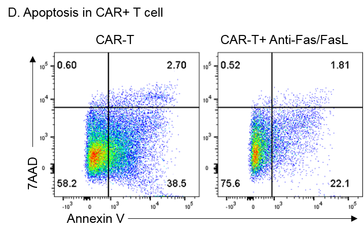
Application: Block/NeutralizeSample Tested: Human NKT cell, Human CAR-T cell and activated CAR-T cellSpecies: HumanVerified Customer | Posted 07/17/2018the Fas/FasL pathways in CAR-T cell apoptosis is significant rescued upon blockade by combined Anti-Fas and Anti-FasL Antibody.

-
Application: ImmunohistochemistrySample Tested: Adult lungSpecies: HumanVerified Customer | Posted 11/04/2016
There are no reviews that match your criteria.
Protocols
Find general support by application which include: protocols, troubleshooting, illustrated assays, videos and webinars.
- 7-Amino Actinomycin D (7-AAD) Cell Viability Flow Cytometry Protocol
- Extracellular Membrane Flow Cytometry Protocol
- Flow Cytometry Protocol for Cell Surface Markers
- Flow Cytometry Protocol for Staining Membrane Associated Proteins
- Flow Cytometry Staining Protocols
- Flow Cytometry Troubleshooting Guide
- Intracellular Flow Cytometry Protocol Using Alcohol (Methanol)
- Intracellular Flow Cytometry Protocol Using Detergents
- Intracellular Nuclear Staining Flow Cytometry Protocol Using Detergents
- Intracellular Staining Flow Cytometry Protocol Using Alcohol Permeabilization
- Intracellular Staining Flow Cytometry Protocol Using Detergents to Permeabilize Cells
- Propidium Iodide Cell Viability Flow Cytometry Protocol
- Protocol for Liperfluo
- Protocol for the Characterization of Human Th22 Cells
- Protocol for the Characterization of Human Th9 Cells
- Protocol: Annexin V and PI Staining by Flow Cytometry
- Protocol: Annexin V and PI Staining for Apoptosis by Flow Cytometry
- Troubleshooting Guide: Fluorokine Flow Cytometry Kits
- View all Protocols, Troubleshooting, Illustrated assays and Webinars
Loading...
Associated Pathways


