The galectins constitute a large family of carbohydrate-binding proteins with specificity for N-acetyl-lactosamine-containing glycoproteins. At least 14 mammalian galectins, which share structural similarities in their carbohydrate recognition domains (CRD), have been identified. The galectins have been classified into the prototype galectins (-1, -2, -5, -7, -10, -11, -13, -14), which contain one CRD and exist either as a monomer or a noncovalent homodimer; the chimera galectins (Galectin-3) containing one CRD linked to a nonlectin domain; and the tandem-repeat galectins (-4, -6, -8, -9, -12) consisting of two CRDs joined by a linker peptide. Galectins lack a classical signal peptide and can be localized to the cytosolic compartments where they have intracellular functions. However, via one or more as yet unidentified non-classical secretory pathways, galectins can also be secreted to function extracellularly. Individual members of the galectin family have different tissue distribution profiles and exhibit subtle differences in their carbohydrate-binding specificities. Each family member may preferentially bind to a unique subset of cell-surface glycoproteins (1-4). Human Galectin-7 is a prototype monomeric galectin. It is specifically expressed in stratified epithelia, notably in epidermis, but is barely detectable in epidermal tumors and significantly down regulated or absent from squamous carconima cell lines. The Galectin-7 gene is induced by tumor suppressor protein p53 transcriptional activity following genotoxic events. A pro-apoptotic protein, Galectin-7 functions intracellularly upstream of JNK activation and cytochrome-c release. This protein has been shown to increase the susceptibility of keratinocytes to UVB induced apoptosis, an essential processss in the maintenance of epidermal homeostasis. Cell lines transfected with the Galectin-7 gene localized the protein in the nucleus and intracellularly. Human and mouse Galectin-7 share 79% amino acid homology (4-6).

Key Product Details
Species Reactivity
Human
Applications
Immunohistochemistry, Western Blot, Flow Cytometry, Immunocytochemistry, CyTOF-ready
Label
Unconjugated
Antibody Source
Monoclonal Mouse IgG1 Clone # 950723
Loading...
Product Specifications
Immunogen
E. coli-derived recombinant human Galectin-7
Ser2-Phe136
Accession # NP_002298
Ser2-Phe136
Accession # NP_002298
Specificity
Detects human Galectin-7 in direct ELISAs and Western blots.
Clonality
Monoclonal
Host
Mouse
Isotype
IgG1
Scientific Data Images for Human Galectin-7 Antibody (950723)
Detection of Human Galectin‑7 by Western Blot.
Western blot shows lysates of human skin tissue and human skin cancer tissue. PVDF membrane was probed with 2 µg/mL of Mouse Anti-Human Galectin-7 Monoclonal Antibody (Catalog # MAB13392) followed by HRP-conjugated Anti-Mouse IgG Secondary Antibody (Catalog # HAF018). A specific band was detected for Galectin-7 at approximately 14 kDa (as indicated). This experiment was conducted under reducing conditions and using Immunoblot Buffer Group 1.
Detection of Galectin‑7 in A431 Human Cell Line by Flow Cytometry.
A431 human epithelial carcinoma cell line was stained with Mouse Anti-Human Galectin-7 Monoclonal Antibody (Catalog # MAB13392, filled histogram) or isotype control antibody (Catalog # MAB002, open histogram), followed by Allophycocyanin-conjugated Anti-Mouse IgG Secondary Antibody (Catalog # F0101B). To facilitate intracellular staining, cells were fixed with Flow Cytometry Fixation Buffer (Catalog # FC004) and permeabilized with Flow Cytometry Permeabilization/Wash Buffer I (Catalog # FC005). View our protocol for Staining Intracellular Molecules.
Galectin‑7 in HEK001 Human Cell Line.
Galectin-7 was detected in immersion fixed HEK001 human epidermal keratinocyte cell line using Mouse Anti-Human Galectin-7 Monoclonal Antibody (Catalog # MAB13392) at 5 µg/mL for 3 hours at room temperature. Cells were stained using the NorthernLights™ 557-conjugated Anti-Mouse IgG Secondary Antibody (red; Catalog # NL007) and counterstained with DAPI (blue). Specific staining was localized to cytoplasm and nuclei. View our protocol for Fluorescent ICC Staining of Cells on Coverslips.
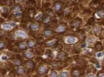
Galectin‑7 in Human Cervical Cancer Tissue.
Galectin-7 was detected in immersion fixed paraffin-embedded sections of human cervical cancer tissue using Mouse Anti-Human Galectin-7 Monoclonal Antibody (Catalog # MAB13392) at 5 µg/mL for 1 hour at room temperature followed by incubation with the Anti-Mouse IgG VisUCyte™ HRP Polymer Antibody (Catalog # VC001). Tissue was stained using DAB (brown) and counterstained with hematoxylin (blue). Specific staining was localized to cytoplasm and nuclei. View our protocol for IHC Staining with VisUCyte HRP Polymer Detection Reagents.Applications for Human Galectin-7 Antibody (950723)
Application
Recommended Usage
CyTOF-ready
Ready to be labeled using established conjugation methods. No BSA or other carrier proteins that could interfere with conjugation.
Flow Cytometry
0.25 µg/106 cells
Sample: A431 human epithelial carcinoma cell line fixed with Flow Cytometry Fixation Buffer (Catalog # FC004) and permeabilized with Flow Cytometry Permeabilization/Wash Buffer I (Catalog # FC005)
Sample: A431 human epithelial carcinoma cell line fixed with Flow Cytometry Fixation Buffer (Catalog # FC004) and permeabilized with Flow Cytometry Permeabilization/Wash Buffer I (Catalog # FC005)
Immunocytochemistry
5-25 µg/mL
Sample: Immersion fixed HEK001 human epidermal keratinocyte cell line
Sample: Immersion fixed HEK001 human epidermal keratinocyte cell line
Immunohistochemistry
5-25 µg/mL
Sample: Immersion fixed paraffin-embedded sections of human cervical cancer tissue
Sample: Immersion fixed paraffin-embedded sections of human cervical cancer tissue
Western Blot
2 µg/mL
Sample: Human skin tissue and human skin cancer tissue
Sample: Human skin tissue and human skin cancer tissue
Reviewed Applications
Read 2 reviews rated 4.5 using MAB13392 in the following applications:
Flow Cytometry Panel Builder
Bio-Techne Knows Flow Cytometry
Save time and reduce costly mistakes by quickly finding compatible reagents using the Panel Builder Tool.
Advanced Features
- Spectra Viewer - Custom analysis of spectra from multiple fluorochromes
- Spillover Popups - Visualize the spectra of individual fluorochromes
- Antigen Density Selector - Match fluorochrome brightness with antigen density

Formulation, Preparation, and Storage
Purification
Protein A or G purified from hybridoma culture supernatant
Reconstitution
Reconstitute at 0.5 mg/mL in sterile PBS. For liquid material, refer to CoA for concentration.
Loading...
Formulation
Lyophilized from a 0.2 μm filtered solution in PBS with Trehalose. *Small pack size (SP) is supplied either lyophilized or as a 0.2 µm filtered solution in PBS.
Shipping
Lyophilized product is shipped at ambient temperature. Liquid small pack size (-SP) is shipped with polar packs. Upon receipt, store immediately at the temperature recommended below.
Stability & Storage
Use a manual defrost freezer and avoid repeated freeze-thaw cycles.
- 12 months from date of receipt, -20 to -70 °C as supplied.
- 1 month, 2 to 8 °C under sterile conditions after reconstitution.
- 6 months, -20 to -70 °C under sterile conditions after reconstitution.
Calculators
Background: Galectin-7
References
- Rabinovich, A. et al. (2002) TRENDS in Immunol. 23:313.
- Rabinovich, A. et al. (2002) J. Leukocyte Biology 71:741.
- Hughes, R.C. (2002) Biochimie 83:667.
- R&D Systems Cytokine Bulletin; Summer 2002.
- Bernerd, F. et al. (1999) Proc. Natl. Acad. Sci. USA 96:11329.
- Kuwabara, I. et al. (2002) J. Biol. Chem. 277:3487.
Alternate Names
GAL7, Galectin7, LGALS7
Gene Symbol
LGALS7
UniProt
Additional Galectin-7 Products
Product Documents for Human Galectin-7 Antibody (950723)
Certificate of Analysis
To download a Certificate of Analysis, please enter a lot or batch number in the search box below.
Note: Certificate of Analysis not available for kit components.
Product Specific Notices for Human Galectin-7 Antibody (950723)
For research use only
Related Research Areas
Customer Reviews for Human Galectin-7 Antibody (950723) (2)
4.5 out of 5
2 Customer Ratings
Have you used Human Galectin-7 Antibody (950723)?
Submit a review and receive an Amazon gift card!
$25/€18/£15/$25CAN/¥2500 Yen for a review with an image
$10/€7/£6/$10CAN/¥1110 Yen for a review without an image
Submit a review
Customer Images
Showing
1
-
2 of
2 reviews
Showing All
Filter By:
-
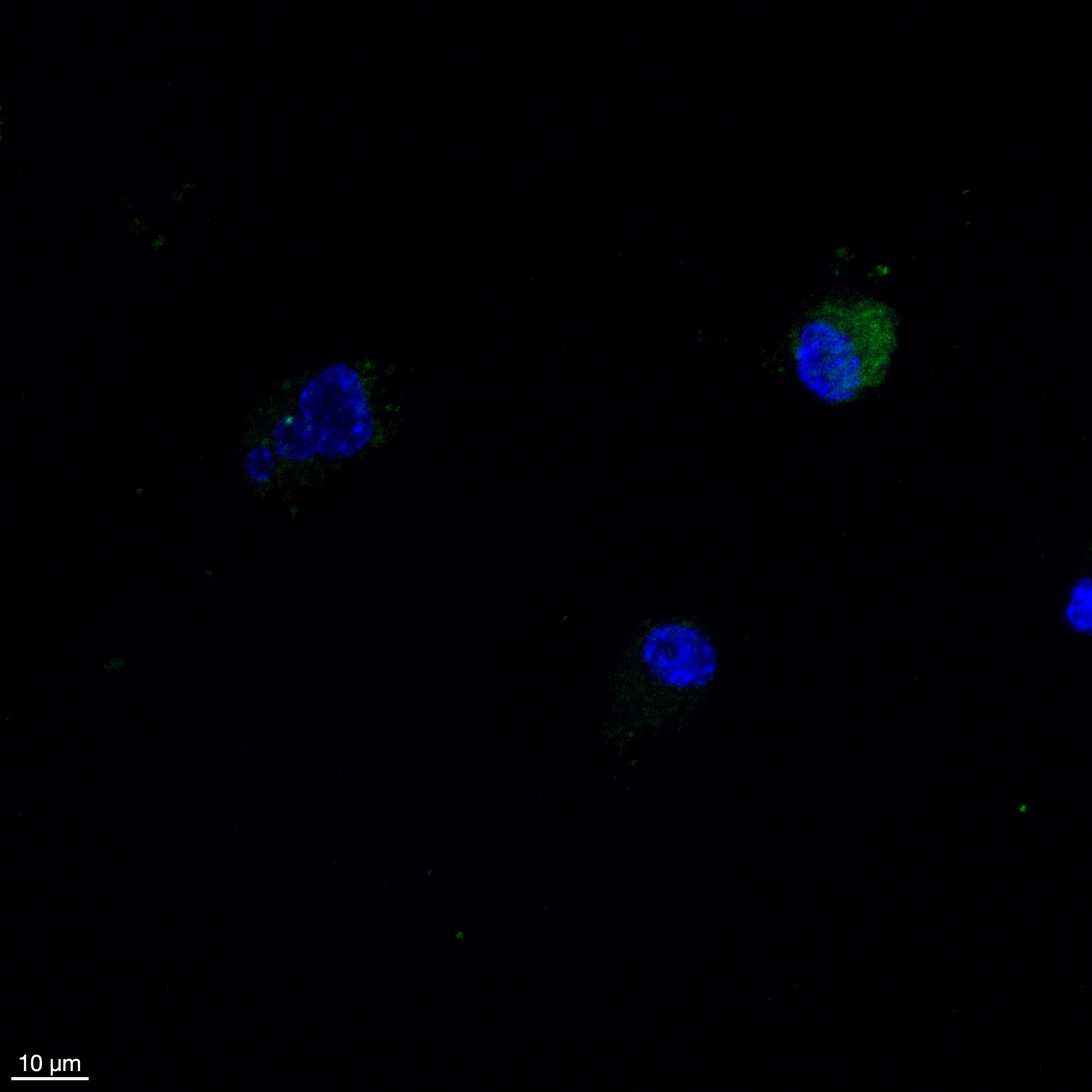
Application: ImmunocytochemistrySample Tested: ccd-841-conSpecies: HumanVerified Customer | Posted 04/18/2026Dapi (blue) and Galectin-7 (green)In vitro immunostaining of galectin-7 (1:1000)

-
Application: ImmunohistochemistrySample Tested: Cervical cancer tissueSpecies: HumanVerified Customer | Posted 03/11/2022
There are no reviews that match your criteria.
Protocols
Find general support by application which include: protocols, troubleshooting, illustrated assays, videos and webinars.
- 7-Amino Actinomycin D (7-AAD) Cell Viability Flow Cytometry Protocol
- Antigen Retrieval Protocol (PIER)
- Antigen Retrieval for Frozen Sections Protocol
- Appropriate Fixation of IHC/ICC Samples
- Cellular Response to Hypoxia Protocols
- Chromogenic IHC Staining of Formalin-Fixed Paraffin-Embedded (FFPE) Tissue Protocol
- Chromogenic Immunohistochemistry Staining of Frozen Tissue
- ClariTSA™ Fluorophore Kits
- Detection & Visualization of Antibody Binding
- Extracellular Membrane Flow Cytometry Protocol
- Flow Cytometry Protocol for Cell Surface Markers
- Flow Cytometry Protocol for Staining Membrane Associated Proteins
- Flow Cytometry Staining Protocols
- Flow Cytometry Troubleshooting Guide
- Fluorescent IHC Staining of Frozen Tissue Protocol
- Graphic Protocol for Heat-induced Epitope Retrieval
- Graphic Protocol for the Preparation and Fluorescent IHC Staining of Frozen Tissue Sections
- Graphic Protocol for the Preparation and Fluorescent IHC Staining of Paraffin-embedded Tissue Sections
- Graphic Protocol for the Preparation of Gelatin-coated Slides for Histological Tissue Sections
- ICC Cell Smear Protocol for Suspension Cells
- ICC Immunocytochemistry Protocol Videos
- ICC for Adherent Cells
- IHC Sample Preparation (Frozen sections vs Paraffin)
- Immunocytochemistry (ICC) Protocol
- Immunocytochemistry Troubleshooting
- Immunofluorescence of Organoids Embedded in Cultrex Basement Membrane Extract
- Immunofluorescent IHC Staining of Formalin-Fixed Paraffin-Embedded (FFPE) Tissue Protocol
- Immunohistochemistry (IHC) and Immunocytochemistry (ICC) Protocols
- Immunohistochemistry Frozen Troubleshooting
- Immunohistochemistry Paraffin Troubleshooting
- Intracellular Flow Cytometry Protocol Using Alcohol (Methanol)
- Intracellular Flow Cytometry Protocol Using Detergents
- Intracellular Nuclear Staining Flow Cytometry Protocol Using Detergents
- Intracellular Staining Flow Cytometry Protocol Using Alcohol Permeabilization
- Intracellular Staining Flow Cytometry Protocol Using Detergents to Permeabilize Cells
- Preparing Samples for IHC/ICC Experiments
- Preventing Non-Specific Staining (Non-Specific Binding)
- Primary Antibody Selection & Optimization
- Propidium Iodide Cell Viability Flow Cytometry Protocol
- Protocol for Heat-Induced Epitope Retrieval (HIER)
- Protocol for Liperfluo
- Protocol for Making a 4% Formaldehyde Solution in PBS
- Protocol for VisUCyte™ HRP Polymer Detection Reagent
- Protocol for the Characterization of Human Th22 Cells
- Protocol for the Characterization of Human Th9 Cells
- Protocol for the Fluorescent ICC Staining of Cell Smears - Graphic
- Protocol for the Fluorescent ICC Staining of Cultured Cells on Coverslips - Graphic
- Protocol for the Preparation & Fixation of Cells on Coverslips
- Protocol for the Preparation and Chromogenic IHC Staining of Frozen Tissue Sections
- Protocol for the Preparation and Chromogenic IHC Staining of Frozen Tissue Sections - Graphic
- Protocol for the Preparation and Chromogenic IHC Staining of Paraffin-embedded Tissue Sections
- Protocol for the Preparation and Chromogenic IHC Staining of Paraffin-embedded Tissue Sections - Graphic
- Protocol for the Preparation and Fluorescent ICC Staining of Cells on Coverslips
- Protocol for the Preparation and Fluorescent ICC Staining of Non-adherent Cells
- Protocol for the Preparation and Fluorescent ICC Staining of Stem Cells on Coverslips
- Protocol for the Preparation and Fluorescent IHC Staining of Frozen Tissue Sections
- Protocol for the Preparation and Fluorescent IHC Staining of Paraffin-embedded Tissue Sections
- Protocol for the Preparation of Gelatin-coated Slides for Histological Tissue Sections
- Protocol for the Preparation of a Cell Smear for Non-adherent Cell ICC - Graphic
- Protocol: Annexin V and PI Staining by Flow Cytometry
- Protocol: Annexin V and PI Staining for Apoptosis by Flow Cytometry
- R&D Systems Quality Control Western Blot Protocol
- TUNEL and Active Caspase-3 Detection by IHC/ICC Protocol
- The Importance of IHC/ICC Controls
- Troubleshooting Guide: Fluorokine Flow Cytometry Kits
- Troubleshooting Guide: Immunohistochemistry
- Troubleshooting Guide: Western Blot Figures
- Western Blot Conditions
- Western Blot Protocol
- Western Blot Protocol for Cell Lysates
- Western Blot Troubleshooting
- Western Blot Troubleshooting Guide
- View all Protocols, Troubleshooting, Illustrated assays and Webinars
Loading...