Key Product Details

Species Reactivity

Validated:

Human

Cited:

Human, Mouse, Complex Species Category

Applications

Validated:

Immunohistochemistry, Western Blot, Simple Western

Cited:

Immunohistochemistry-Paraffin, Western Blot, Flow Cytometry, Immunocytochemistry, Immunoprecipitation, ELISA Detection, ELISA Development (Detection)

Label

Unconjugated

Antibody Source

Polyclonal Goat IgG
Loading...

Product Specifications

Immunogen

E. coli-derived recombinant human Galectin-8
Met1-Trp317
Accession # O00214

Specificity

Detects human Galectin-8 in direct ELISAs and Western blots. In direct ELISAs, less than 5% cross‑reactivity with recombinant human (rh) rhGalectin-1 and rhGalectin-3 is observed, and less than 1% cross-reactivity with rhGalectin-2, rhGalectin-4 and rhGalectin-7 is observed.

Clonality

Polyclonal

Host

Goat

Isotype

IgG

Scientific Data Images for Human Galectin‑8 Antibody

Detection of Human Galectin‑8 antibody by Western Blot.

Detection of Human Galectin‑8 by Western Blot.

Western blot shows lysates of LNCaP human prostate cancer cell line. PVDF membrane was probed with 1 µg/mL of Goat Anti-Human Galectin-8 Antigen Affinity-purified Polyclonal Antibody (Catalog # AF1305) followed by HRP-conjugated Anti-Goat IgG Secondary Antibody (Catalog # HAF019). A specific band was detected for Galectin-8 at approximately 35 kDa (as indicated). This experiment was conducted under reducing conditions and using Immunoblot Buffer Group 1.

Galectin-8 antibody in Human Lung by Immunohistochemistry (IHC-P).

Galectin‑8 in Human Lung.

Galectin-8 was detected in immersion fixed paraffin-embedded sections of human lung using Goat Anti-Human Galectin-8 Antigen Affinity-purified Polyclonal Antibody (Catalog # AF1305) at 15 µg/mL overnight at 4 °C. Before incubation with the primary antibody, tissue was subjected to heat-induced epitope retrieval using Antigen Retrieval Reagent-Basic (Catalog # CTS013). Tissue was stained using the Anti-Goat HRP-DAB Cell & Tissue Staining Kit (brown; Catalog # CTS008) and counterstained with hematoxylin (blue). Specific staining was localized to cell surfaces and cytoplasm. View our protocol for Chromogenic IHC Staining of Paraffin-embedded Tissue Sections.

Detection of Human Galectin-8 antibody by Simple WesternTM.

Detection of Human Galectin‑8 by Simple WesternTM.

Simple Western lane view shows lysates of LNCaP human prostate cancer cell line, loaded at 0.2 mg/mL. A specific band was detected for Galectin-8 at approximately 40 kDa (as indicated) using 10 µg/mL of Goat Anti-Human Galectin-8 Antigen Affinity-purified Polyclonal Antibody (Catalog # AF1305) followed by 1:50 dilution of HRP-conjugated Anti-Goat IgG Secondary Antibody (Catalog # HAF109). This experiment was conducted under reducing conditions and using the 12-230 kDa separation system.

Applications for Human Galectin‑8 Antibody

Application
Recommended Usage

Immunohistochemistry

5-15 µg/mL
Sample: Immersion fixed paraffin-embedded sections of human lung subjected to Antigen Retrieval Reagent-Basic (Catalog # CTS013)

Simple Western

10 µg/mL
Sample: LNCaP human prostate cancer cell line

Western Blot

1 µg/mL
Sample: LNCaP human prostate cancer cell line

Reviewed Applications

Read 6 reviews rated 4.5 using AF1305 in the following applications:

Formulation, Preparation, and Storage

Purification

Antigen Affinity-purified

Reconstitution

Reconstitute at 0.2 mg/mL in sterile PBS. For liquid material, refer to CoA for concentration.


Loading...

Formulation

Lyophilized from a 0.2 μm filtered solution in PBS with Trehalose. *Small pack size (SP) is supplied either lyophilized or as a 0.2 µm filtered solution in PBS.

Shipping

Lyophilized product is shipped at ambient temperature. Liquid small pack size (-SP) is shipped with polar packs. Upon receipt, store immediately at the temperature recommended below.

Stability & Storage

Use a manual defrost freezer and avoid repeated freeze-thaw cycles.
  • 12 months from date of receipt, -20 to -70 °C as supplied.
  • 1 month, 2 to 8 °C under sterile conditions after reconstitution.
  • 6 months, -20 to -70 °C under sterile conditions after reconstitution.

Calculators

The reconstitution calculator allows you to quickly calculate the volume of a reagent to reconstitute your vial. Simply enter the mass of reagent and the target concentration and the calculator will determine the rest.

=
÷

Background: Galectin-8

The galectins constitute a large family of carbohydrate-binding proteins with specificity for N-acetyl-lactosamine-containing glycoproteins. At least 14 mammalian galectins, which share structural similarities in their carbohydrate recognition domains (CRD), have been identified to date. The galectins have been classified into the prototype galectins (-1, -2, -5, -7, -10, -11, -13, -14), which contain one CRD and exist either as a monomer or a noncovalent homodimer. The chimera galectins (Galectin-3) containing one CRD linked to a nonlectin domain, and the tandem-repeat Galectins (-4, -6, -8, -9, -12) consisting of two CRDs joined by a linker peptide. Galectins lack a classical signal peptide and can be localized to the cytosolic compartments where they have intracellular functions. However, via one or more as yet unidentified non-classical secretory pathways, galectins can also be secreted to function extracellularly. Individual members of the galectin family have different tissue distribution profiles and exhibit subtle differences in their carbohydrate-binding specificities. Each family member may preferentially bind to a unique subset of cell-surface glycoproteins (1‑4).

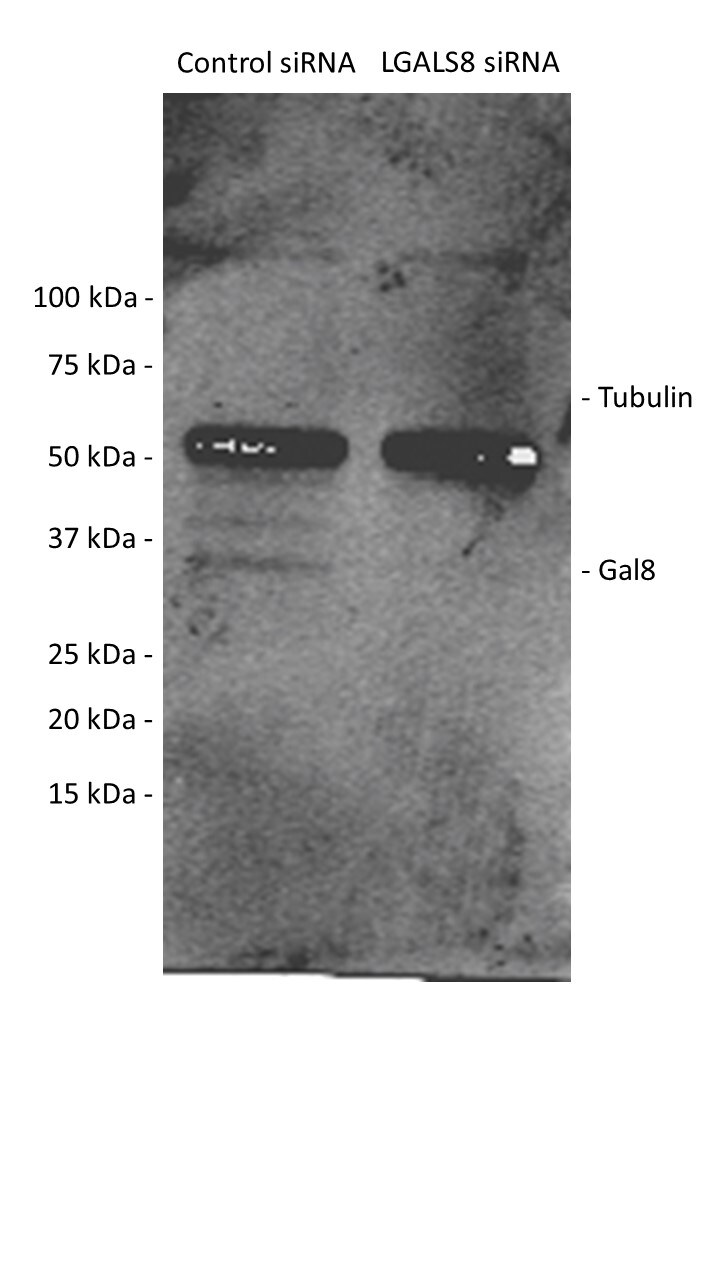
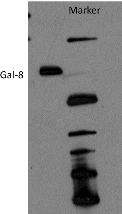
Galectin-8, also known as prostate carcinoma tumor antigen 1 (PCTA1) in human, is a tandem repeat-type galectin. Prototype (single CRD) isoforms arising through alternate gene splicing have also been identified (5). Galectin-8 is highly expressed in lung carcinomas, certain forms of prostate carcinomas, as well as other tumor cells. It binds to a subset of cell surface integrins to modulate ECM-integrin interactions. As a soluble ligand, Galectin-8 can inhibit cell adhesion (6). Immobilized Galectin-8, however, has also been shown to promote cell adhesion (7). Human and mouse Galectin-8 share approximately 80% amino acid homology (4).

References

  1. Rabinovich, A. et al. (2002) TRENDS in Immunol. 23:313.
  2. Rabinovich, A. et al. (2002) J. Leukocyte Biology 71:741.
  3. Hughes, R.C. (2002) Biochimie 83:667.
  4. R&D Systems’ Cytokine Bulletin, Summer, 2002.
  5. Bidon, N. et al. (2001) Gene 274:253.
  6. Hadari, Y. et al. (1995) J. Biol. Chem. 270:3447.
  7. Levy, Y. et al. (2001) J. Biol. Chem. 276:31285.

Alternate Names

GAL8, Galectin8, LGALS8, PCTA1

Entrez Gene IDs

3964 (Human); 56048 (Mouse)

Gene Symbol

LGALS8

UniProt

Additional Galectin-8 Products

Product Documents for Human Galectin‑8 Antibody

Certificate of Analysis

To download a Certificate of Analysis, please enter a lot or batch number in the search box below.

Note: Certificate of Analysis not available for kit components.

Product Specific Notices for Human Galectin‑8 Antibody

For research use only

Related Research Areas

Citations for Human Galectin‑8 Antibody

Customer Reviews for Human Galectin‑8 Antibody (6)

4.5 out of 5
6 Customer Ratings
5 Stars
50%
4 Stars
50%
3 Stars
0%
2 Stars
0%
1 Stars
0%

Have you used Human Galectin‑8 Antibody?

Submit a review and receive an Amazon gift card!

$25/€18/£15/$25CAN/¥2500 Yen for a review with an image

$10/€7/£6/$10CAN/¥1110 Yen for a review without an image

Submit a review
Amazon Gift Card

Customer Images


Showing  1 - 5 of 6 reviews Showing All
Filter By:
  • Human Galectin-8 Antibody
    Name: Anonymous
    Application: Western Blot
    Sample Tested: Huh-7 human hepatoma cell line
    Species: Human
    Verified Customer | Posted 08/20/2018
    Human Galectin‑8 Antibody AF1305
  • Human Galectin‑8 Antibody
    Name: Anonymous
    Application: Immunocytochemistry/Immunofluorescence
    Sample Tested: Cancer Cells
    Species: Human
    Verified Customer | Posted 10/26/2015
    The antibody is good for IFC also. <br />Specificity: Reasonably specific<br />Sensitivity: Reasonably sensitive<br />Buffer: PBS<br />Dilution: 1:1000
    Human Galectin‑8 Antibody AF1305
  • Human Galectin‑8 Antibody
    Name: Anonymous
    Application: Western Blot
    Sample Tested: Human recombinant galectin-8
    Species: Human
    Verified Customer | Posted 10/26/2015
    The antibody can detect the higher oligomeric form of galectin-8 here. The 36 kDa version was not detectable due to increasing oligomerization of the protein. <br />Specificity: Specific<br />Sensitivity: Sensitive<br />Buffer: Loading buffer<br />Dilution: 1:1000
    Human Galectin‑8 Antibody AF1305
  • Human Galectin‑8 Antibody
    Name: Anonymous
    Application: Immunohistochemistry
    Sample Tested: Colon tissue
    Species: Human
    Verified Customer | Posted 10/26/2015
    At 1:2000 dilution there was very specific binding and the detection was limited to crypts as observed here. <br />Specificity: Specific<br />Sensitivity: Sensitive<br />Buffer: PBS<br />Dilution: 1:1500 to 1:2000
    Human Galectin‑8 Antibody AF1305
  • Human Galectin‑8 Antibody
    Name: Anonymous
    Application: Immunocytochemistry/Immunofluorescence
    Sample Tested: Human cells
    Species: Human
    Verified Customer | Posted 10/26/2015
    This experiment, the cells were stained for galectin-8 antibody and then red color secondary was used to detect it. <br />Specificity: Specific<br />Sensitivity: Sensitive<br />Buffer: PBS<br />Dilution: 1:1000
    Human Galectin‑8 Antibody AF1305
  • Name: Anonymous
    Application: Western Blot
    Sample Tested: See PMID 20876211
    Species: Human
    Verified Customer | Posted 01/05/2015

There are no reviews that match your criteria.

Showing  1 - 5 of 6 reviews Showing All

Protocols

Find general support by application which include: protocols, troubleshooting, illustrated assays, videos and webinars.

FAQs

No product specific FAQs exist for this product.

View all FAQs for Antibodies