Herpesvirus entry mediator (HVEM), also referred to as TR2 (TNF receptor-like molecule) and ATAR (another TRAF-associated receptor), is a type I membrane protein belonging to the TNF/NGF receptor superfamily. In the TNF superfamily nomenclature, HVEM is referred to as TNFRSF14. Human HVEM cDNA encodes a 283 amino acid (aa) protein with a probable 36 aa signal peptide, a 166 aa extracellular domain, a 23 aa transmembrane region and a 58 aa cytoplasmic region. The extracellular domain of HVEM contains several cysteine-rich repeats characteristic of TNF receptor superfamily members. The short cytoplasmic region lacks a death domain present in some TNF receptor family members, but contains a TRAF (TNF receptor-associated factor) interaction domain. HVEM expression has been detected in peripheral blood T cells, B cells, monocytes and in various tissues enriched in lymphoid cells. The extracellular domain of HVEM has been shown to interact directly with the herpes simplex virus envelope glycoprotein D. Two TNF superfamily ligands, including the secreted TNF‑ beta (lymphotoxin alpha ) and the membrane protein LIGHT (lymphotoxins, exhibits inducible expression, and competes with HSV glycoprotein D for HVEM, a receptor expressed by T lymphocytes), have been shown to be the cellular ligands for HVEM. Besides HVEM, LIGHT can also interact with LT beta R, the receptor for lymphotoxin alpha beta heterotrimer.
Human HVEM/TNFRSF14 Antibody (94801)
R&D Systems | Catalog # MAB356

Key Product Details
Species Reactivity
Validated:
Human
Cited:
Human
Applications
Validated:
Western Blot, ELISA Capture (Matched Antibody Pair), Flow Cytometry, CyTOF-ready
Cited:
Immunohistochemistry-Frozen, Neutralization, Flow Cytometry
Label
Unconjugated
Antibody Source
Monoclonal Mouse IgG1 Clone # 94801
Loading...
Product Specifications
Immunogen
Mouse myeloma cell line NS0-derived recombinant human HVEM/TNFRSF14
Pro37-Val202 (Ser108Thr and Ala140Arg)
Accession # AAB58354
Pro37-Val202 (Ser108Thr and Ala140Arg)
Accession # AAB58354
Specificity
Detects human HVEM/TNFRSF14 in direct ELISAs and Western blots. In direct ELISAs, approximately 5% cross-reactivity with recombinant human (rh) CD30 and no cross-reactivity with rh4-1BB, rhCD27, rhCD40, rhDR3, rhDR6, rhFas, rhGITR, rhNGF-R, rhOPG, rhRANK, recombinant mouse (rm) RANK, rhTAJ, rhTNF RI, rhTNF RII, or rmTNF RI is observed.
Clonality
Monoclonal
Host
Mouse
Isotype
IgG1
Scientific Data Images for Human HVEM/TNFRSF14 Antibody (94801)
Detection of HVEM/TNFRSF14 in Human Blood Lymphocytes by Flow Cytometry.
Human peripheral blood lymphocytes were stained with Mouse Anti-Human HVEM/TNFRSF14 Monoclonal Antibody (Catalog # MAB356, filled histogram) or isotype control antibody (Catalog # MAB002, open histogram), followed by Allophycocyanin-conjugated Anti-Mouse IgG Secondary Antibody (Catalog # F0101B). View our protocol for Staining Membrane-associated Proteins.
Detection of Human HVEM/TNFRSF14 by Block/Neutralize
Differential inhibition of HVEM/CD160 binding with benchmark tool antibodies. TRF assay measuring the potency of different antibodies to inhibit the binding of recombinant human HVEM-Fc chimera to CD160+ CHO-K1 cells. A) CD160 monoclonal antibodies inhibit binding of HVEM-Fc to both CD160-GPI and CD160-TM isoforms. B) Polyclonal HVEM (left panel) and monoclonal HVEM (right panel) antibodies both enhance binding of HVEM-Fc to CD160-TM isoform. The polyclonal anti-HVEM inhibits HVEM-Fc binding to CD160-GPI (left panel). Antibody concentrations are plotted on the X axis whereas, the calculated percentage of inhibition of binding is plotted on the Y axis. Matched isotype control antibody for each individual antibody candidate was also used in the assay (empty circles and squares). CTL = control, mAb = monoclonal antibody, pAb = polyclonal antibody. Image collected and cropped by CiteAb from the following publication (https://pubmed.ncbi.nlm.nih.gov/25179432), licensed under a CC-BY license. Not internally tested by R&D Systems.
Human HVEM / TNFRSF14 ELISA Standard Curve
Human HVEM/TNFRSF14 was serially diluted and captured by Mouse Anti-Human HVEM/TNFRSF14 Monoclonal Antibody (Catalog # MAB356) coated on a Clear Polystyrene Microplate (Catalog # DY990). Goat Anti-Human HVEM/TNFRSF14 Antigen Affinity-purified Polyclonal Antibody (Catalog # AF356) was biotinylated and incubated with the protein captured on the plate. Detection of the standard curve was achieved by incubating Streptavidin-HRP (Catalog # DY998)Applications for Human HVEM/TNFRSF14 Antibody (94801)
Application
Recommended Usage
CyTOF-ready
Ready to be labeled using established conjugation methods. No BSA or other carrier proteins that could interfere with conjugation.
Flow Cytometry
0.25 µg/106 cells
Sample: Human peripheral blood lymphocytes
Sample: Human peripheral blood lymphocytes
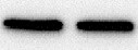
Western Blot
1 µg/mL
Sample: Recombinant Human HVEM/TNFRSF14 Fc Chimera (Catalog # 356-HV)
Sample: Recombinant Human HVEM/TNFRSF14 Fc Chimera (Catalog # 356-HV)
Human HVEM/TNFRSF14 Sandwich Immunoassay
Please Note: Optimal dilutions of this antibody should be experimentally determined.
Reviewed Applications
Read 1 review rated 5 using MAB356 in the following applications:
Flow Cytometry Panel Builder
Bio-Techne Knows Flow Cytometry
Save time and reduce costly mistakes by quickly finding compatible reagents using the Panel Builder Tool.
Advanced Features
- Spectra Viewer - Custom analysis of spectra from multiple fluorochromes
- Spillover Popups - Visualize the spectra of individual fluorochromes
- Antigen Density Selector - Match fluorochrome brightness with antigen density

Formulation, Preparation, and Storage
Purification
Protein A or G purified from ascites
Reconstitution
Reconstitute at 0.5 mg/mL in sterile PBS. For liquid material, refer to CoA for concentration.
Loading...
Formulation
Lyophilized from a 0.2 μm filtered solution in PBS with Trehalose. *Small pack size (SP) is supplied either lyophilized or as a 0.2 µm filtered solution in PBS.
Shipping
Lyophilized product is shipped at ambient temperature. Liquid small pack size (-SP) is shipped with polar packs. Upon receipt, store immediately at the temperature recommended below.
Stability & Storage
Use a manual defrost freezer and avoid repeated freeze-thaw cycles.
- 12 months from date of receipt, -20 to -70 °C as supplied.
- 1 month, 2 to 8 °C under sterile conditions after reconstitution.
- 6 months, -20 to -70 °C under sterile conditions after reconstitution.
Calculators
Background: HVEM/TNFRSF14
References
- Hsu, H. et al. (1997) J. Biol. Chem. 272:13471.
- Mauri, D.N. et al. (1998) Immunity 8:21.
- Montgomery, R.I. et al. (1996) Cell 87:427.
Long Name
Herpesvirus Entry Mediator
Alternate Names
ATAR, CD270, LIGHTR, TNFRSF14
Gene Symbol
TNFRSF14
UniProt
Additional HVEM/TNFRSF14 Products
Product Documents for Human HVEM/TNFRSF14 Antibody (94801)
Certificate of Analysis
To download a Certificate of Analysis, please enter a lot or batch number in the search box below.
Note: Certificate of Analysis not available for kit components.
Product Specific Notices for Human HVEM/TNFRSF14 Antibody (94801)
For research use only
Citations for Human HVEM/TNFRSF14 Antibody (94801)
 Powered by Bioz
Customer Reviews for Human HVEM/TNFRSF14 Antibody (94801) (1)
5 out of 5
1 Customer Rating
Have you used Human HVEM/TNFRSF14 Antibody (94801)?
Submit a review and receive an Amazon gift card!
$25/€18/£15/$25CAN/¥2500 Yen for a review with an image
$10/€7/£6/$10CAN/¥1110 Yen for a review without an image
Submit a review
Customer Images
Showing
1
-
1 of
1 review
Showing All
Filter By:
-
Application: Western BlotSample Tested: B lymphocytesSpecies: HumanVerified Customer | Posted 11/09/2021B lymphocytes, Human HVEM/TNFRSF14 Antibody

There are no reviews that match your criteria.
Protocols
Find general support by application which include: protocols, troubleshooting, illustrated assays, videos and webinars.
- 7-Amino Actinomycin D (7-AAD) Cell Viability Flow Cytometry Protocol
- Cellular Response to Hypoxia Protocols
- Extracellular Membrane Flow Cytometry Protocol
- Flow Cytometry Protocol for Cell Surface Markers
- Flow Cytometry Protocol for Staining Membrane Associated Proteins
- Flow Cytometry Staining Protocols
- Flow Cytometry Troubleshooting Guide
- Intracellular Flow Cytometry Protocol Using Alcohol (Methanol)
- Intracellular Flow Cytometry Protocol Using Detergents
- Intracellular Nuclear Staining Flow Cytometry Protocol Using Detergents
- Intracellular Staining Flow Cytometry Protocol Using Alcohol Permeabilization
- Intracellular Staining Flow Cytometry Protocol Using Detergents to Permeabilize Cells
- Propidium Iodide Cell Viability Flow Cytometry Protocol
- Protocol for Liperfluo
- Protocol for the Characterization of Human Th22 Cells
- Protocol for the Characterization of Human Th9 Cells
- Protocol: Annexin V and PI Staining by Flow Cytometry
- Protocol: Annexin V and PI Staining for Apoptosis by Flow Cytometry
- R&D Systems Quality Control Western Blot Protocol
- Troubleshooting Guide: Fluorokine Flow Cytometry Kits
- Troubleshooting Guide: Western Blot Figures
- Western Blot Conditions
- Western Blot Protocol
- Western Blot Protocol for Cell Lysates
- Western Blot Troubleshooting
- Western Blot Troubleshooting Guide
- View all Protocols, Troubleshooting, Illustrated assays and Webinars
Loading...
Associated Pathways
