IL-12 is produced by macrophages and B lymphocytes and has been shown to have multiple effects on T cells and natural killer (NK) cells. These effects include inducing production of IFN-gamma and TNF by resting and activated T and NK cells, synergizing with other IFN-gamma inducers at both the transcriptional and post-transcriptional levels. This interaction induces IFN-gamma gene expression, enhancing the cytotoxic activity of resting NK and T cells, inducing and synergizing with IL-2 in the generation of lymphokine-activated killer (LAK) cells, acting as a co-mitogen to stimulate proliferation of resting T cells, and inducing proliferation of activated T and NK cells. Current evidence indicates that IL-12, produced by macrophages in response to infectious agents, is a central mediator of the cell-mediated immune response by its actions on the development, proliferation, and activities of TH1 cells. In its role as the initiator of cell-mediated immunity, it has been suggested that IL-12 has therapeutic potential as a stimulator of cell-mediated immune responses to microbial pathogens, metastatic cancers, and viral infections such as AIDS.

Key Product Details
Species Reactivity
Validated:
Human
Cited:
Human
Applications
Validated:
Western Blot, Neutralization, Immunocytochemistry
Cited:
Immunohistochemistry, Western Blot, Neutralization, Immunocytochemistry, Affinity Chromatography, Bioassay
Label
Unconjugated
Antibody Source
Polyclonal Goat IgG
Loading...
Product Specifications
Immunogen
S. frugiperda insect ovarian cell line Sf 21-derived recombinant human IL‑12 heterodimer
Arg23-Ser219 of p35; Ile23-Ser328 of p40
Accession # P29459 (p35) & P29460 (p40)
Arg23-Ser219 of p35; Ile23-Ser328 of p40
Accession # P29459 (p35) & P29460 (p40)
Specificity
Detects human IL-12 in direct ELISAs and Western blots.
Clonality
Polyclonal
Host
Goat
Isotype
IgG
Endotoxin Level
<0.10 EU per 1 μg of the antibody by the LAL method.
Scientific Data Images for Human IL-12 Antibody
Cell Proliferation Induced by IL‑12 and Neutralization by Human IL‑12 Antibody.
Recombinant Human IL-12 (219-IL) stimulates proliferation in PHA-activated human peripheral blood mononuclear cells (PBMC) in a dose-dependent manner (orange line). Proliferation elicited by Recombinant Human IL-12 (1 ng/mL) is neutralized (green line) by increasing concentrations of Goat Anti-Human IL-12 Antigen Affinity-purified Polyclonal Antibody (Catalog # AF-219-NA). The ND50 is typically 5-50 ng/mL.
Detection of IL‑12 in Human PBMCs.
IL‑12 was detected in immersion fixed Human PBMCs using Goat Anti-Human IL‑12 Antigen Affinity-purified Polyclonal Antibody (Catalog # AF-219-NA) at 15 µg/mL for 3 hours at room temperature. Cells were stained using the NorthernLights™ 557-conjugated Anti-Goat IgG Secondary Antibody (red; Catalog # NL001) and counterstained with DAPI (blue). Specific staining was localized to cytoplasm. View our protocol for Fluorescent ICC Staining of Cells on Coverslips.
Human IL-12 / IL-23 p40 ELISA Standard Curve
Recombinant Human IL‑12/IL‑23 p40 Monomer (Catalog # 309-IL) was serially diluted and captured by Mouse Anti-Human IL‑12/IL‑23 p40 Monoclonal Antibody (Catalog # MAB609) coated on a Clear Polystyrene Microplate (Catalog # DY990). Goat Anti-Human IL‑12 Antigen Affinity-purified Polyclonal Antibody (Catalog # AF-219-NA) was biotinylated and incubated with the protein captured on the plate. Detection of the standard curve was achieved by incubating Streptavidin-HRP (Catalog # DY998)
Human IL-12 p70 ELISA Standard Curve
Recombinant Human IL‑12 (Catalog # 219-IL) was serially diluted and captured by Mouse Anti-Human IL‑12 p70 Monoclonal Antibody (Catalog # MAB611R) coated on a Clear Polystyrene Microplate (Catalog # DY990). Goat Anti-Human IL‑12 Antigen Affinity-purified Polyclonal Antibody (Catalog # AF-219-NA) was biotinylated and incubated with the protein captured on the plate. Detection of the standard curve was achieved by incubating Streptavidin-HRP (Catalog # DY998)Applications for Human IL-12 Antibody
Application
Recommended Usage
Immunocytochemistry
5-15 µg/mL
Sample: Immersion fixed human PBMCs
Sample: Immersion fixed human PBMCs
Western Blot
0.1 µg/mL
Sample: Recombinant Human IL‑12 (Catalog # 219-IL)
Sample: Recombinant Human IL‑12 (Catalog # 219-IL)
Neutralization
Measured by its ability to neutralize IL‑12-induced proliferation in PHA-activated human peripheral blood mononuclear cells (PBMC). Yokota, T. et al. (1986) Proc. Natl. Acad. Sci. USA 83:5894. The Neutralization Dose (ND50) is typically 5-50 ng/mL in the presence of 1 ng/mL Recombinant Human IL‑12.
Reviewed Applications
Read 1 review rated 5 using AF-219-NA in the following applications:
Formulation, Preparation, and Storage
Purification
Antigen Affinity-purified
Reconstitution
Reconstitute at 0.2 mg/mL in sterile PBS. For liquid material, refer to CoA for concentration.
Loading...
Formulation
Lyophilized from a 0.2 μm filtered solution in PBS with Trehalose. *Small pack size (SP) is supplied either lyophilized or as a 0.2 µm filtered solution in PBS.
Shipping
Lyophilized product is shipped at ambient temperature. Liquid small pack size (-SP) is shipped with polar packs. Upon receipt, store immediately at the temperature recommended below.
Stability & Storage
Use a manual defrost freezer and avoid repeated freeze-thaw cycles.
- 12 months from date of receipt, -20 to -70 °C as supplied.
- 1 month, 2 to 8 °C under sterile conditions after reconstitution.
- 6 months, -20 to -70 °C under sterile conditions after reconstitution.
Calculators
Background: IL-12
Long Name
Interleukin 12
Alternate Names
CLMF1, IL12, IL12A, NKSF1
Entrez Gene IDs
Gene Symbol
IL12A
UniProt
Additional IL-12 Products
Product Documents for Human IL-12 Antibody
Certificate of Analysis
To download a Certificate of Analysis, please enter a lot or batch number in the search box below.
Note: Certificate of Analysis not available for kit components.
Product Specific Notices for Human IL-12 Antibody
For research use only
Related Research Areas
Citations for Human IL-12 Antibody
 Powered by Bioz
Customer Reviews for Human IL-12 Antibody (1)
5 out of 5
1 Customer Rating
Have you used Human IL-12 Antibody?
Submit a review and receive an Amazon gift card!
$25/€18/£15/$25CAN/¥2500 Yen for a review with an image
$10/€7/£6/$10CAN/¥1110 Yen for a review without an image
Submit a review
Customer Images
Showing
1
-
1 of
1 review
Showing All
Filter By:
-
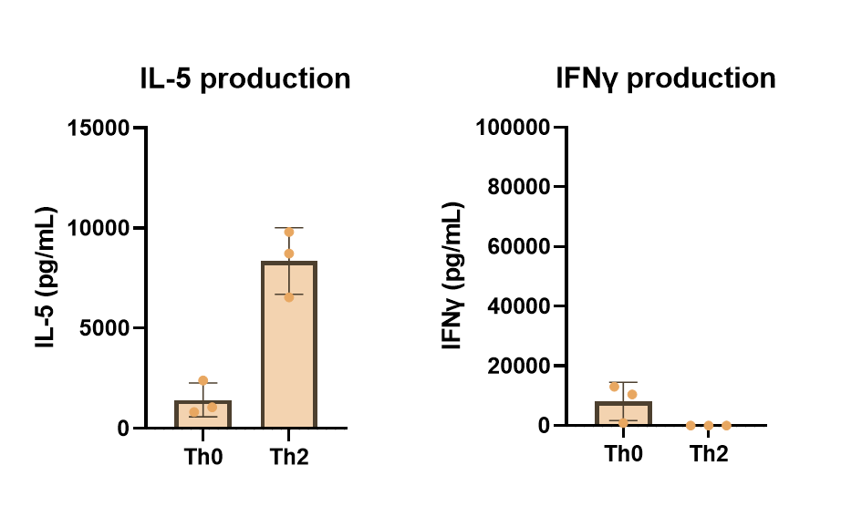
Application: Block/NeutralizeSample Tested: T cellsSpecies: HumanVerified Customer | Posted 08/10/2023Utilized during in vitro Th2 polarization cultures to prevent unwanted differentiation of IFNg-producing Th1 cells. Naive CD4 T cells were stimulated with anti-CD3/anti-CD28 antibodies and polarized with culture media that included 20 ng/mL IL-4, 5 ug/mL anti-IL-12 antibody, and 5 ug/mL anti-IFNg antibody. After 5 days of culture, supernatants were analyzed AlphaLISA to assess IL-5 and IFNg production. As expected, the Th2 cultures did not produce unwanted IFNg indicating Th1 polarization was inhibited.

There are no reviews that match your criteria.
Protocols
Find general support by application which include: protocols, troubleshooting, illustrated assays, videos and webinars.
- Appropriate Fixation of IHC/ICC Samples
- Cellular Response to Hypoxia Protocols
- ClariTSA™ Fluorophore Kits
- Detection & Visualization of Antibody Binding
- ICC Cell Smear Protocol for Suspension Cells
- ICC Immunocytochemistry Protocol Videos
- ICC for Adherent Cells
- Immunocytochemistry (ICC) Protocol
- Immunocytochemistry Troubleshooting
- Immunofluorescence of Organoids Embedded in Cultrex Basement Membrane Extract
- Immunohistochemistry (IHC) and Immunocytochemistry (ICC) Protocols
- Preparing Samples for IHC/ICC Experiments
- Preventing Non-Specific Staining (Non-Specific Binding)
- Primary Antibody Selection & Optimization
- Protocol for VisUCyte™ HRP Polymer Detection Reagent
- Protocol for the Fluorescent ICC Staining of Cell Smears - Graphic
- Protocol for the Fluorescent ICC Staining of Cultured Cells on Coverslips - Graphic
- Protocol for the Preparation and Fluorescent ICC Staining of Cells on Coverslips
- Protocol for the Preparation and Fluorescent ICC Staining of Non-adherent Cells
- Protocol for the Preparation and Fluorescent ICC Staining of Stem Cells on Coverslips
- Protocol for the Preparation of a Cell Smear for Non-adherent Cell ICC - Graphic
- R&D Systems Quality Control Western Blot Protocol
- TUNEL and Active Caspase-3 Detection by IHC/ICC Protocol
- The Importance of IHC/ICC Controls
- Troubleshooting Guide: Western Blot Figures
- Western Blot Conditions
- Western Blot Protocol
- Western Blot Protocol for Cell Lysates
- Western Blot Troubleshooting
- Western Blot Troubleshooting Guide
- View all Protocols, Troubleshooting, Illustrated assays and Webinars


