
LAG-3 is an activation-induced molecule, expressed on activated T cells and NK cells, but not on resting T cells. Studies using LAG-3 -/- mice have shown significant delay of T cell apoptosis following antigen stimulation and increased size of memory T cells pool following infection (3, 4). It also has been reported that anti-LAG-3 antibodies up-regulate T cell activation by blocking interaction of LAG-3 and MHC class II. The study has demonstrated that LAG-3 is selectively expressed on activated CD4+CD25+ TReg cells and plays a role in their suppressive activity (5). This evidence indicated, unlike the interaction of CD4 with MHC class II that plays a positive role in T cell activation, LAG-3 binds to MHC class II and negatively regulates T cell activation through LAG-3 signaling. On the other hand, studies have shown that binding of LAG-3 to MHC class II molecules on antigen presenting cells induce maturation of dendritic cells and cytokine secretion by monocytes through MHC class II signal transduction (6). Taken together, LAG-3 may have two major functions, it negatively regulates T cells activation through LAG-3 signaling and stimulates antigen presenting cells which express MHC class II.

Scientific images may be resized, cropped, or adjusted for brightness or contrast for presentation purposes. These updates are limited to presentation and do not affect the underlying scientific interpretation of the data. Where available, additional source data may be provided upon request.
Key Product Details
Validated by
Species Reactivity
Applications
Label
Antibody Source
Product Specifications
Immunogen
Met1-Leu450
Accession # P18627
Specificity
Clonality
Host
Isotype
Scientific Data Images for Human LAG-3 Antibody (1009614)
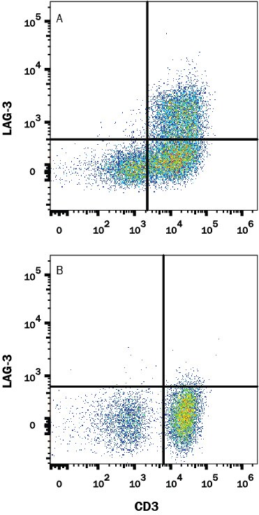
Detection of LAG‑3 in Human PBMCs by Flow Cytometry.
Human peripheral blood mononuclear cells (PBMCs) were either untreated (bottom panel) or treated with with 5 µg/mL PHA (top panel) for 5 days. PBMCs were stained with Mouse Anti-Human LAG-3 Monoclonal Antibody (Catalog # MAB23194) followed by Allophycocyanin-conjugated Anti-Mouse IgG Secondary Antibody (Catalog # F0101B) and Mouse Anti-Human CD3 epsilon PE-conjugated Monoclonal Antibody (Catalog # FAB100P). Quadrant markers were set based on isotype control antibody staining (Catalog # MAB003) (data not shown). View our protocol for Staining Membrane-associated Proteins.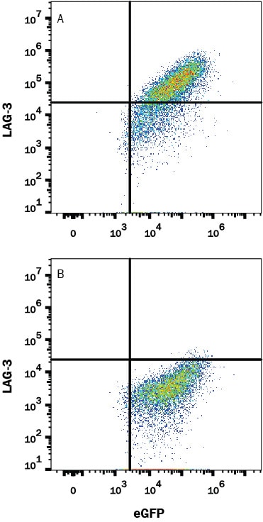
Detection of LAG-3 in HEK293 Human Cell Line Transfected with Human LAG-3 and eGFP by Flow Cytometry.
HEK293 human embryonic kidney cell line transfected with either (A) human LAG-3 or (B) irrelevant transfectants and eGFP was stained with Mouse Anti-Human LAG-3 Monoclonal Antibody (Catalog # MAB23194) followed by APC-conjugated Anti-Mouse IgG Secondary Antibody (Catalog # F0101B). Quadrant markers were set based on control antibody staining (Catalog # MAB003, data not shown). View our protocol for Staining Membrane-associated Proteins.Applications for Human LAG-3 Antibody (1009614)
CyTOF-ready
Flow Cytometry
Sample: Human PBMC treated with PHA and HEK293 Human Cell Line Transfected with Human LAG-3 and eGFP
Flow Cytometry Panel Builder
Bio-Techne Knows Flow Cytometry
Save time and reduce costly mistakes by quickly finding compatible reagents using the Panel Builder Tool.
Advanced Features
- Spectra Viewer - Custom analysis of spectra from multiple fluorochromes
- Spillover Popups - Visualize the spectra of individual fluorochromes
- Antigen Density Selector - Match fluorochrome brightness with antigen density

Formulation, Preparation, and Storage
Purification
Reconstitution
Reconstitute at 0.5 mg/mL in sterile PBS. For liquid material, refer to CoA for concentration.
Formulation
Shipping
Stability & Storage
- 12 months from date of receipt, -20 to -70 °C as supplied.
- 1 month, 2 to 8 °C under sterile conditions after reconstitution.
- 6 months, -20 to -70 °C under sterile conditions after reconstitution.
Calculators
Background: LAG-3
References
- Triebel, F. et al. (1990) J. Exp. Med. 171:1393.
- Baixeras, E. et al. (1992) J. Exp. Med 176:327.
- Workman, C.J. and D.A. Vignali (2003) Eur. J. Immunol. 33:970.
- Workman, C.J. et al. (2004) J. Immunol. 172:5450.
- Huang, C.T. et al. (2004) Immunity 21:503.
- Andreae, S. et al. (2003) Blood 102:2130.
Long Name
Alternate Names
Gene Symbol
UniProt
Additional LAG-3 Products
Product Documents for Human LAG-3 Antibody (1009614)
Certificate of Analysis
To download a Certificate of Analysis, please enter a lot or batch number in the search box below.
Note: Certificate of Analysis not available for kit components.
Product Specific Notices for Human LAG-3 Antibody (1009614)
For research use only
Related Research Areas
Customer Reviews for Human LAG-3 Antibody (1009614)
There are currently no reviews for this product. Be the first to review Human LAG-3 Antibody (1009614) and earn rewards!
Have you used Human LAG-3 Antibody (1009614)?
Submit a review and receive an Amazon gift card!
$25/€18/£15/$25CAN/¥2500 for a review with an image
$10/€7/£6/$10CAN/¥1110 for a review without an image
Submit a review
Protocols
Find general support by application which include: protocols, troubleshooting, illustrated assays, videos and webinars.
- 7-Amino Actinomycin D (7-AAD) Cell Viability Flow Cytometry Protocol
- Extracellular Membrane Flow Cytometry Protocol
- Flow Cytometry Protocol for Cell Surface Markers
- Flow Cytometry Protocol for Staining Membrane Associated Proteins
- Flow Cytometry Staining Protocols
- Flow Cytometry Troubleshooting Guide
- Intracellular Flow Cytometry Protocol Using Alcohol (Methanol)
- Intracellular Flow Cytometry Protocol Using Detergents
- Intracellular Nuclear Staining Flow Cytometry Protocol Using Detergents
- Intracellular Staining Flow Cytometry Protocol Using Alcohol Permeabilization
- Intracellular Staining Flow Cytometry Protocol Using Detergents to Permeabilize Cells
- Propidium Iodide Cell Viability Flow Cytometry Protocol
- Protocol for Liperfluo
- Protocol for the Characterization of Human Th22 Cells
- Protocol for the Characterization of Human Th9 Cells
- Protocol: Annexin V and PI Staining by Flow Cytometry
- Protocol: Annexin V and PI Staining for Apoptosis by Flow Cytometry
- Troubleshooting Guide: Fluorokine Flow Cytometry Kits
- View all Protocols, Troubleshooting, Illustrated assays and Webinars
FAQs for Human LAG-3 Antibody (1009614)
-
Q: Can this LAG-3 antibody be used in ELISA?
A: We have not validated this particular LAG-3 antibody for ELISA in mouse; however, it has been cited as being used as a detection antibody in a publicatoin (PMID 15946792).
-
Q: I'm using your LAG-3 antibody in western blot but the observed band is a different size than the predicted band size, do you know what the bands are?
A: There are several alternative splice varients of LAG-3 that could explain the different sized proteins stained by the LAG-3 antibody. It is also believed that there are two secreted forms of LAG-3, one that is about 36 kDa and another at 52 kDa.
-
Q: Can this LAG-3 antibody be used in ELISA?
A: We have not validated this particular LAG-3 antibody for ELISA in mouse; however, it has been cited as being used as a detection antibody in a publicatoin (PMID 15946792).
-
Q: I'm using your LAG-3 antibody in western blot but the observed band is a different size than the predicted band size, do you know what the bands are?
A: There are several alternative splice varients of LAG-3 that could explain the different sized proteins stained by the LAG-3 antibody. It is also believed that there are two secreted forms of LAG-3, one that is about 36 kDa and another at 52 kDa.