The Low Density Lipoprotein Receptor (LDLR) is the founding member of the LDLR family of scavenger receptors (1, 2). This family contains transmembrane molecules that are characterized by the presence of EGF repeats, complement-like repeats, and YWTD motifs that form beta -propellers. Although members of the family were originally thought to be endocytic receptors, it is now clear that some members interact with adjacent cell-surface molecules, expanding their range of activities (2). Human LDLR is synthesized as an 860 amino acid (aa) precursor that contains a 21 aa signal sequence, a 767 aa extracellular region, a 22 aa transmembrane segment and a 50 aa cytoplasmic tail (3). The extracellular region is complex. It consists of seven N-terminal complement-like cysteine-rich repeats that bind ligand. Cysteine residues in this region participate in intrachain disulfide bonds. This region is followed by three EGF-like repeats with a beta -propeller YWTD containing motif. The EGF-like repeats are responsible for ligand bonding and dissociation. Finally, there is a 50 aa membrane proximal Ser/Thr-rich region that serves as a carbohydrate attachment point (1, 3, 4). There is extensive O-linked and modest N-linked glycosylation. Thus the receptor’s predicted molecular weight of 93 kDa is increased to a native molecular weight of 120-160 kDa (3, 4). Within the 50 aa cytoplasmic tail, there is an NPXY motif that links the receptor to clathrin pits (1). The extracellular region of human LDLR is 51% aa identical to the extracellular region of human VLDLR, and 79% aa identical to the extracellular region of mouse LDLR. LDLR is constitutively expressed and binds ApoB of LDL and ApoE of VLDL (5). It is responsible for clearing 70% of plasma LDL in liver (5). Mutations in the LDLR gene cause the autosomal dominant disorder, familial hypercholesterolemia (6).

Key Product Details
Species Reactivity
Validated:
Human
Cited:
Human
Applications
Validated:
Western Blot, ELISA Capture (Matched Antibody Pair), Flow Cytometry, Immunoprecipitation, CyTOF-ready
Cited:
Immunohistochemistry, Western Blot, Flow Cytometry, ELISA Capture, ELISA Development, Functional Assay
Label
Unconjugated
Antibody Source
Monoclonal Mouse IgG1 Clone # 472413
Loading...
Product Specifications
Immunogen
Chinese hamster ovary cell line CHO-derived recombinant human LDLR
Ala22-Arg788
Accession # P01130
Ala22-Arg788
Accession # P01130
Specificity
Detects human LDLR in ELISAs and Western blots. In direct ELISAs and Western blots, no cross-reactivity with recombinant mouse (rm) LDLR, recombinant human LRP-5, or rmLRP-6 is observed.
Clonality
Monoclonal
Host
Mouse
Isotype
IgG1
Scientific Data Images for Human LDLR Antibody (472413)
Detection of LDLR in HepG2 Human Cell Line by Flow Cytometry.
HepG2 human hepatocellular carcinoma cell line was stained with Mouse Anti-Human LDLR Monoclonal Antibody (Catalog # MAB2148, filled histogram) or isotype control antibody (MAB002, open histogram), followed by PE-conjugated Anti-Mouse IgG F(ab')2Secondary Antibody (F0102B).
Detection of LDLR in A172 cells by Flow Cytometry.
A172 cells were stained with Mouse Anti-Human LDLR Monoclonal Antibody (Catalog # MAB2148, filled histogram) or isotype control antibody (Catalog # MAB002, open histogram), followed by Fluorescein-conjugated Anti-Mouse IgG Secondary Antibody (Catalog # F0103B). View our protocol for Staining Membrane-associated Proteins.
Detection of LDLR in U-118-MG cells by Flow Cytometry
U-118-MG cells were stained with Mouse Anti-Human LDLR Monoclonal Antibody (Catalog # MAB2148, filled histogram) or isotype control antibody (Catalog # MAB002, open histogram) followed by Allophycocyanin-conjugated Anti-Mouse IgG Secondary Antibody (Catalog # F0101B). View our protocol for Staining Membrane-associated Proteins.Applications for Human LDLR Antibody (472413)
Application
Recommended Usage
CyTOF-ready
Ready to be labeled using established conjugation methods. No BSA or other carrier proteins that could interfere with conjugation.
Flow Cytometry
0.25 µg/106 cells
Sample: HepG2 human hepatocellular carcinoma cell line, A172 human glioblastoma cell line, and U-118-MG human glioblastoma/astrocytoma cell line
Sample: HepG2 human hepatocellular carcinoma cell line, A172 human glioblastoma cell line, and U-118-MG human glioblastoma/astrocytoma cell line
Immunoprecipitation
25 µg/mL
Sample: Conditioned cell culture medium spiked with Recombinant Human LDLR (Catalog # 2148‑LD), see our available Western blot detection antibodies
Sample: Conditioned cell culture medium spiked with Recombinant Human LDLR (Catalog # 2148‑LD), see our available Western blot detection antibodies
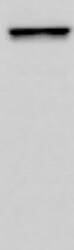
Western Blot
1 µg/mL
Sample: Recombinant Human LDLR (Catalog # 2148-LD) under non-reducing conditions only
Sample: Recombinant Human LDLR (Catalog # 2148-LD) under non-reducing conditions only
Human LDLR Sandwich Immunoassay
Please Note: Optimal dilutions of this antibody should be experimentally determined.
Reviewed Applications
Read 1 review rated 5 using MAB2148 in the following applications:
Flow Cytometry Panel Builder
Bio-Techne Knows Flow Cytometry
Save time and reduce costly mistakes by quickly finding compatible reagents using the Panel Builder Tool.
Advanced Features
- Spectra Viewer - Custom analysis of spectra from multiple fluorochromes
- Spillover Popups - Visualize the spectra of individual fluorochromes
- Antigen Density Selector - Match fluorochrome brightness with antigen density

Formulation, Preparation, and Storage
Purification
Protein A or G purified from hybridoma culture supernatant
Reconstitution
Reconstitute at 0.5 mg/mL in sterile PBS. For liquid material, refer to CoA for concentration.
Loading...
Formulation
Lyophilized from a 0.2 μm filtered solution in PBS with Trehalose. *Small pack size (SP) is supplied either lyophilized or as a 0.2 µm filtered solution in PBS.
Shipping
Lyophilized product is shipped at ambient temperature. Liquid small pack size (-SP) is shipped with polar packs. Upon receipt, store immediately at the temperature recommended below.
Stability & Storage
Use a manual defrost freezer and avoid repeated freeze-thaw cycles.
- 12 months from date of receipt, -20 to -70 °C as supplied.
- 1 month, 2 to 8 °C under sterile conditions after reconstitution.
- 6 months, -20 to -70 °C under sterile conditions after reconstitution.
Calculators
Background: LDLR
References
- Strickland, D.K. et al. (2002) Trends Endocrinol. Metab. 13:66.
- Nykjaer, A. and T.E. Willnow (2002) Trends Cell Biol. 12:273.
- Yamamoto, T. et al. (1984) Cell 39:27.
- Davis, C.G. et al. (1986) J. Biol. Chem. 261:2828.
- Defesche, J.C. (2004) Semin. Vasc. Med. 4:5.
- Varret, M. et al. (2008) Clin Genet. 73:1.
Long Name
Low Density Lipoprotein Receptor
Alternate Names
LDL R
Entrez Gene IDs
Gene Symbol
LDLR
UniProt
Additional LDLR Products
Product Documents for Human LDLR Antibody (472413)
Certificate of Analysis
To download a Certificate of Analysis, please enter a lot or batch number in the search box below.
Note: Certificate of Analysis not available for kit components.
Product Specific Notices for Human LDLR Antibody (472413)
For research use only
Related Research Areas
Citations for Human LDLR Antibody (472413)
 Powered by Bioz
Customer Reviews for Human LDLR Antibody (472413) (1)
5 out of 5
1 Customer Rating
Have you used Human LDLR Antibody (472413)?
Submit a review and receive an Amazon gift card!
$25/€18/£15/$25CAN/¥2500 Yen for a review with an image
$10/€7/£6/$10CAN/¥1110 Yen for a review without an image
Submit a review
Customer Images
Showing
1
-
1 of
1 review
Showing All
Filter By:
-
Application: Western BlotSample Tested: MacrophagesSpecies: HumanVerified Customer | Posted 10/27/2021

There are no reviews that match your criteria.
Protocols
Find general support by application which include: protocols, troubleshooting, illustrated assays, videos and webinars.
- 7-Amino Actinomycin D (7-AAD) Cell Viability Flow Cytometry Protocol
- Cellular Response to Hypoxia Protocols
- Extracellular Membrane Flow Cytometry Protocol
- Flow Cytometry Protocol for Cell Surface Markers
- Flow Cytometry Protocol for Staining Membrane Associated Proteins
- Flow Cytometry Staining Protocols
- Flow Cytometry Troubleshooting Guide
- Immunoprecipitation Protocol
- Intracellular Flow Cytometry Protocol Using Alcohol (Methanol)
- Intracellular Flow Cytometry Protocol Using Detergents
- Intracellular Nuclear Staining Flow Cytometry Protocol Using Detergents
- Intracellular Staining Flow Cytometry Protocol Using Alcohol Permeabilization
- Intracellular Staining Flow Cytometry Protocol Using Detergents to Permeabilize Cells
- Propidium Iodide Cell Viability Flow Cytometry Protocol
- Protocol for Liperfluo
- Protocol for the Characterization of Human Th22 Cells
- Protocol for the Characterization of Human Th9 Cells
- Protocol: Annexin V and PI Staining by Flow Cytometry
- Protocol: Annexin V and PI Staining for Apoptosis by Flow Cytometry
- R&D Systems Quality Control Western Blot Protocol
- Troubleshooting Guide: Fluorokine Flow Cytometry Kits
- Troubleshooting Guide: Western Blot Figures
- Western Blot Conditions
- Western Blot Protocol
- Western Blot Protocol for Cell Lysates
- Western Blot Troubleshooting
- Western Blot Troubleshooting Guide
- View all Protocols, Troubleshooting, Illustrated assays and Webinars
Loading...
Associated Pathways
