Human LIGHT, also known as TNFSF14, is a type II membrane protein that is a member of the TNF superfamily. LIGHT is an acronym which stands for "is homologous to lymphotoxins, exhibits inducible expression, and competes with HSV glycoprotein D for HVEM, a receptor expressed by T lymphocytes". LIGHT has also been called HVEM-L and LT-gamma. LIGHT is a 240 amino acid (aa) protein that contains a 37 aa cytoplasmic domain, a 22 aa transmembrane region, and a 181 aa extracellular domain. Similar to other TNF ligand family members, LIGHT is predicted to assemble as a homotrimer. LIGHT is produced by activated T cells and was first identified by its ability to compete with HSV glycoprotein D for HVEM binding. LIGHT has also been shown to bind to the lymphotoxin beta receptor (LT beta R) and the decoy receptor (DcR3/TR6). LIGHT overexpression in tumor cells induces apoptosis, which can be enhanced by IFN-gamma.
Human LIGHT/TNFSF14 Antibody (115520)
R&D Systems | Catalog # MAB664
Clone 115520 was used by HLDA to establish CD designation

Key Product Details
Species Reactivity
Validated:
Human
Cited:
Human
Applications
Validated:
ELISA Capture (Matched Antibody Pair), Flow Cytometry, CyTOF-ready
Cited:
Immunohistochemistry-Paraffin, Flow Cytometry
Label
Unconjugated
Antibody Source
Monoclonal Mouse IgG1 Clone # 115520
Loading...
Product Specifications
Immunogen
Mouse myeloma cell line NS0-derived recombinant human LIGHT/TNFSF14
Asp74-Val240
Accession # O43557
Asp74-Val240
Accession # O43557
Specificity
Detects human LIGHT/TNFSF14 in ELISAs. In sandwich immunoassays, no significant cross-reactivity or interference with recombinant human (rh) Fas Ligand, rhAPRIL, recombinant mouse TRANCE, rhTNF-alpha or rhTRAIL is observed.
Clonality
Monoclonal
Host
Mouse
Isotype
IgG1
Scientific Data Images for Human LIGHT/TNFSF14 Antibody (115520)
Detection of LIGHT/TNFSF14 in PBMC treated with PMA and Calcium Ionomycin vs resting PBMC by Flow Cytometry
PBMC treated with 50ng/mL PMA and 500ng/mL Calcium Ionomycin for 48 hours (A) vs resting PBMC (B) were stained with Mouse Anti-Human LIGHT/TNFSF14 Monoclonal Antibody (Catalog # MAB664) and Mouse Anti-Human CD8 alpha PE‑conjugated Monoclonal Antibody (Catalog # FAB1509P) isotype control antibody (Catalog # MAB002) followed by Allophycocyanin-conjugated Anti-Mouse IgG Secondary Antibody (Catalog # F0101B). View our protocol for Staining Membrane-associated Proteins.
Human LIGHT / TNFSF14 ELISA Standard Curve
Recombinant Human LIGHT/TNFSF14 (Catalog # 664-LI) was serially diluted and captured by Mouse Anti-Human LIGHT/TNFSF14 Monoclonal Antibody (Catalog # MAB664) coated on a Clear Polystyrene Microplate (Catalog # DY990). Goat Anti-Human LIGHT/TNFSF14 Antigen Affinity-purified Polyclonal Antibody (Catalog # AF664) was biotinylated and incubated with the protein captured on the plate. Detection of the standard curve was achieved by incubating Streptavidin-HRP (Catalog # DY998)Applications for Human LIGHT/TNFSF14 Antibody (115520)
Application
Recommended Usage
CyTOF-ready
Ready to be labeled using established conjugation methods. No BSA or other carrier proteins that could interfere with conjugation.
Flow Cytometry
0.25 µg/106 cells
Sample: Human PBMC treated with PMA and Ca2+ ionomycin
Sample: Human PBMC treated with PMA and Ca2+ ionomycin
Human LIGHT/TNFSF14 Sandwich Immunoassay
Please Note: Optimal dilutions of this antibody should be experimentally determined.
Reviewed Applications
Read 1 review rated 5 using MAB664 in the following applications:
Flow Cytometry Panel Builder
Bio-Techne Knows Flow Cytometry
Save time and reduce costly mistakes by quickly finding compatible reagents using the Panel Builder Tool.
Advanced Features
- Spectra Viewer - Custom analysis of spectra from multiple fluorochromes
- Spillover Popups - Visualize the spectra of individual fluorochromes
- Antigen Density Selector - Match fluorochrome brightness with antigen density

Formulation, Preparation, and Storage
Purification
Protein A or G purified from ascites
Reconstitution
Reconstitute at 0.5 mg/mL in sterile PBS. For liquid material, refer to CoA for concentration.
Loading...
Formulation
Lyophilized from a 0.2 μm filtered solution in PBS with Trehalose. See Certificate of Analysis for details.
*Small pack size (-SP) is supplied either lyophilized or as a 0.2 µm filtered solution in PBS.
*Small pack size (-SP) is supplied either lyophilized or as a 0.2 µm filtered solution in PBS.
Shipping
Lyophilized product is shipped at ambient temperature. Liquid small pack size (-SP) is shipped with polar packs. Upon receipt, store immediately at the temperature recommended below.
Stability & Storage
Use a manual defrost freezer and avoid repeated freeze-thaw cycles.
- 12 months from date of receipt, -20 to -70 °C as supplied.
- 1 month, 2 to 8 °C under sterile conditions after reconstitution.
- 6 months, -20 to -70 °C under sterile conditions after reconstitution.
Calculators
Background: LIGHT/TNFSF14
References
- Mauri, D.N. et al. (1998) Immunity 8:21.
- Zhai, Y. et al. (1998) J. Clin. Invest. 102:1142.
- Harrop, J.A. et al. (1998) J. Biol. Chem. 273:27548.
- Yu, K-Y. et al. (1999) J. Biol. Chem. 274:13733.
Long Name
TNF Ligand Superfamily Member 14
Alternate Names
CD258, TNFSF14
Gene Symbol
TNFSF14
UniProt
Additional LIGHT/TNFSF14 Products
Product Documents for Human LIGHT/TNFSF14 Antibody (115520)
Certificate of Analysis
To download a Certificate of Analysis, please enter a lot or batch number in the search box below.
Note: Certificate of Analysis not available for kit components.
Product Specific Notices for Human LIGHT/TNFSF14 Antibody (115520)
For research use only
Citations for Human LIGHT/TNFSF14 Antibody (115520)
 Powered by Bioz
Customer Reviews for Human LIGHT/TNFSF14 Antibody (115520) (1)
5 out of 5
1 Customer Rating
Have you used Human LIGHT/TNFSF14 Antibody (115520)?
Submit a review and receive an Amazon gift card!
$25/€18/£15/$25CAN/¥2500 Yen for a review with an image
$10/€7/£6/$10CAN/¥1110 Yen for a review without an image
Submit a review
Customer Images
Showing
1
-
1 of
1 review
Showing All
Filter By:
-
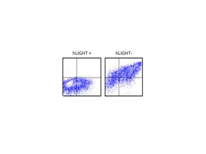
Application: Flow CytometrySample Tested: HEK293 human embryonic kidney cell lineSpecies: HumanVerified Customer | Posted 05/22/2018Cells were transfected with hLIGHT (IRES GFP) or a negative control. Cells were stained with MAB664 using a 1/500 dilution and binding was detected with an anti-mouse 594 secondary antibody.

There are no reviews that match your criteria.
Protocols
Find general support by application which include: protocols, troubleshooting, illustrated assays, videos and webinars.
- 7-Amino Actinomycin D (7-AAD) Cell Viability Flow Cytometry Protocol
- Extracellular Membrane Flow Cytometry Protocol
- Flow Cytometry Protocol for Cell Surface Markers
- Flow Cytometry Protocol for Staining Membrane Associated Proteins
- Flow Cytometry Staining Protocols
- Flow Cytometry Troubleshooting Guide
- Intracellular Flow Cytometry Protocol Using Alcohol (Methanol)
- Intracellular Flow Cytometry Protocol Using Detergents
- Intracellular Nuclear Staining Flow Cytometry Protocol Using Detergents
- Intracellular Staining Flow Cytometry Protocol Using Alcohol Permeabilization
- Intracellular Staining Flow Cytometry Protocol Using Detergents to Permeabilize Cells
- Propidium Iodide Cell Viability Flow Cytometry Protocol
- Protocol for Liperfluo
- Protocol for the Characterization of Human Th22 Cells
- Protocol for the Characterization of Human Th9 Cells
- Protocol: Annexin V and PI Staining by Flow Cytometry
- Protocol: Annexin V and PI Staining for Apoptosis by Flow Cytometry
- Troubleshooting Guide: Fluorokine Flow Cytometry Kits
- View all Protocols, Troubleshooting, Illustrated assays and Webinars
Loading...
Associated Pathways
