Mesothelin is a 40 kDa glycosylphosphatidylinositol-anchored glycoprotein that is expressed on mesothelial cells in the pleura, pericardium and peritoneum and overexpressed in mesotheliomas and ovarian or pancreatic adenocarcinoma. Mesothelin is a product of the CAK-1 gene, which also encodes megakaryocyte-potentiating factor (MPF). Mature human mesothelin shares 60% amino acid identity with either mouse or rat mesothelin. Two variant forms exist; variant 1 has an eight amino acid (1 kDa) insertion and is rarely expressed, while variant 2 is a truncated form secreted in the majority of ovarian cancers but rarely found in normal individuals.
Human Mesothelin Antibody (420411)
R&D Systems | Catalog # MAB32652

Key Product Details
Validated by
Knockout/Knockdown
Species Reactivity
Validated:
Human
Cited:
Human, Mouse, Transgenic Mouse
Applications
Validated:
Knockout Validated, ELISA, Flow Cytometry, CyTOF-ready
Cited:
Flow Cytometry, Immunocytochemistry, CAR-T (Flow Cytometry), Mass Cytometry
Label
Unconjugated
Antibody Source
Monoclonal Rat IgG2A Clone # 420411
Loading...
Product Specifications
Immunogen
Mouse myeloma cell line NS0-derived recombinant human Mesothelin
Glu296-Gly580
Accession # AAH09272
Glu296-Gly580
Accession # AAH09272
Specificity
Detects human Mesothelin in direct ELISAs.
Clonality
Monoclonal
Host
Rat
Isotype
IgG2A
Scientific Data Images for Human Mesothelin Antibody (420411)
Detection of Mesothelin in HeLa Human Cell Line by Flow Cytometry.
HeLa human cervical epithelial carcinoma cell line was stained with Rat Anti-Human Mesothelin Monoclonal Antibody (Catalog # MAB32652, filled histogram) or isotype control antibody (Catalog # MAB006, open histogram), followed by Phycoerythrin-conjugated Anti-Rat IgG F(ab')2Secondary Antibody (Catalog # F0105B).
Mesothelin Specificity is Shown by Flow Cytometry in Knockout Cell Line
Mesothelin knockout HeLa human cervical epithelial carcinoma cell line was stained with Rat Anti-Human Mesothelin Monoclonal Antibody (Catalog # MAB32652, filled histogram) or isotype control antibody (Catalog # MAB006, open histogram) followed by Phycoerythrin-conjugated Anti-Rat IgG F(ab')2 Secondary Antibody (Catalog # F0105B). No staining in the Mesothelin knockout HeLa cell line was observed. View our protocol for Staining Membrane-associated Proteins.
Human Mesothelin ELISA Standard Curve.
Recombinant Human Mesothelin protein was serially diluted 2-fold and captured by Mouse Anti-Human Mesothelin Monoclonal Antibody (Catalog # MAB32655) coated on a Clear Polystyrene Microplate (Catalog # DY990). Rat Anti-Human Mesothelin Monoclonal Antibody (Catalog # MAB32652) was biotinylated and incubated with the protein captured on the plate. Detection of the standard curve was achieved by incubating Streptavidin-HRP (Catalog # DY998) followed by Substrate Solution (Catalog # DY999) and stopping the enzymatic reaction with Stop Solution (Catalog # DY994).Applications for Human Mesothelin Antibody (420411)
Application
Recommended Usage
CyTOF-ready
Ready to be labeled using established conjugation methods. No BSA or other carrier proteins that could interfere with conjugation.
ELISA
This antibody functions as an ELISA detection antibody when paired with Mouse Anti-Human Mesothelin Monoclonal Antibody (Catalog # MAB32655).
This product is intended for assay development on various assay platforms requiring antibody pairs. We recommend the Human Mesothelin DuoSet ELISA Kit (Catalog # DY3265) for convenient development of a sandwich ELISA or the Human Mesothelin Quantikine ELISA Kit (Catalog # DMSLN0) for a complete optimized ELISA.
Flow Cytometry
0.25 µg/106 cells
Sample: HeLa human cervical epithelial carcinoma cell line
Sample: HeLa human cervical epithelial carcinoma cell line
Knockout Validated
Mesothelin is specifically detected in HeLa human cervical epithelial carcinoma parental cell line but is not detectable in Mesothelin knockout HeLa cell line.
Reviewed Applications
Read 4 reviews rated 4.3 using MAB32652 in the following applications:
Flow Cytometry Panel Builder
Bio-Techne Knows Flow Cytometry
Save time and reduce costly mistakes by quickly finding compatible reagents using the Panel Builder Tool.
Advanced Features
- Spectra Viewer - Custom analysis of spectra from multiple fluorochromes
- Spillover Popups - Visualize the spectra of individual fluorochromes
- Antigen Density Selector - Match fluorochrome brightness with antigen density

Formulation, Preparation, and Storage
Purification
Protein A or G purified from hybridoma culture supernatant
Reconstitution
Reconstitute at 0.5 mg/mL in sterile PBS. For liquid material, refer to CoA for concentration.
Loading...
Formulation
Lyophilized from a 0.2 μm filtered solution in PBS with Trehalose. *Small pack size (SP) is supplied either lyophilized or as a 0.2 µm filtered solution in PBS.
Shipping
Lyophilized product is shipped at ambient temperature. Liquid small pack size (-SP) is shipped with polar packs. Upon receipt, store immediately at the temperature recommended below.
Stability & Storage
Use a manual defrost freezer and avoid repeated freeze-thaw cycles.
- 12 months from date of receipt, -20 to -70 °C as supplied.
- 1 month, 2 to 8 °C under sterile conditions after reconstitution.
- 6 months, -20 to -70 °C under sterile conditions after reconstitution.
Calculators
Background: Mesothelin
Alternate Names
CAK1, MPF, MSLN, SMR
Gene Symbol
MSLN
UniProt
Additional Mesothelin Products
Product Documents for Human Mesothelin Antibody (420411)
Certificate of Analysis
To download a Certificate of Analysis, please enter a lot or batch number in the search box below.
Note: Certificate of Analysis not available for kit components.
Product Specific Notices for Human Mesothelin Antibody (420411)
For research use only
Related Research Areas
Citations for Human Mesothelin Antibody (420411)
 Powered by Bioz
Customer Reviews for Human Mesothelin Antibody (420411) (4)
4.3 out of 5
4 Customer Ratings
Have you used Human Mesothelin Antibody (420411)?
Submit a review and receive an Amazon gift card!
$25/€18/£15/$25CAN/¥2500 Yen for a review with an image
$10/€7/£6/$10CAN/¥1110 Yen for a review without an image
Submit a review
Customer Images

Showing
1
-
4 of
4 reviews
Showing All
Filter By:
-
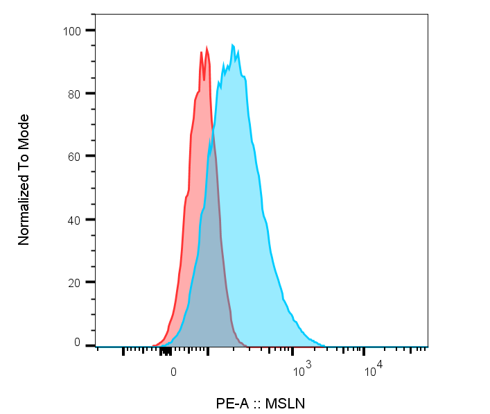
Application: Flow CytometrySample Tested: HeLa cellsSpecies: HumanVerified Customer | Posted 12/08/2022Used the antibody at 1:100 dilution to stain HeLa cells. The secondary antibody was Rat F(ab)2 IgG (H+L) PE-conjugated antibody. Pink is the isotype control stained sample, and blue is the MSLN antibody stained sample. Iso control is Rat IgG2A.

-
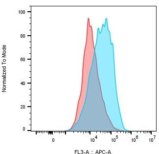
Application: Flow CytometrySample Tested: NCI-H226 lung squamous cell carcinoma cellsSpecies: HumanVerified Customer | Posted 12/23/2020Detection of Mesothelin on NCI-H226 lung squamous cell carcinoma cells. NCI-H226 cells were treated with 67 nM of the Human Mesothelin Antibody (catalog # MAB32652) or rat IgG2a isotype control, followed by a secondary APC AffiniPure F(ab')₂ Fragment Goat Anti-Rat IgG, F(ab')₂ fragment specific. Isotype control (RED), human TROP2 antibody (Blue).
-
Application: Affinity PurificationSample Tested: Ovarian cancer tissueSpecies: MouseVerified Customer | Posted 07/27/2018
-
Application: Flow CytometrySample Tested: AsPC1 human pancreatic cancer cell lineSpecies: HumanVerified Customer | Posted 02/22/2016
There are no reviews that match your criteria.
Protocols
Find general support by application which include: protocols, troubleshooting, illustrated assays, videos and webinars.
- 7-Amino Actinomycin D (7-AAD) Cell Viability Flow Cytometry Protocol
- ELISA Sample Preparation & Collection Guide
- ELISA Troubleshooting Guide
- Extracellular Membrane Flow Cytometry Protocol
- Flow Cytometry Protocol for Cell Surface Markers
- Flow Cytometry Protocol for Staining Membrane Associated Proteins
- Flow Cytometry Staining Protocols
- Flow Cytometry Troubleshooting Guide
- How to Run an R&D Systems DuoSet ELISA
- How to Run an R&D Systems Quantikine ELISA
- How to Run an R&D Systems Quantikine™ QuicKit™ ELISA
- Intracellular Flow Cytometry Protocol Using Alcohol (Methanol)
- Intracellular Flow Cytometry Protocol Using Detergents
- Intracellular Nuclear Staining Flow Cytometry Protocol Using Detergents
- Intracellular Staining Flow Cytometry Protocol Using Alcohol Permeabilization
- Intracellular Staining Flow Cytometry Protocol Using Detergents to Permeabilize Cells
- Propidium Iodide Cell Viability Flow Cytometry Protocol
- Protocol for Liperfluo
- Protocol for the Characterization of Human Th22 Cells
- Protocol for the Characterization of Human Th9 Cells
- Protocol: Annexin V and PI Staining by Flow Cytometry
- Protocol: Annexin V and PI Staining for Apoptosis by Flow Cytometry
- Quantikine HS ELISA Kit Assay Principle, Alkaline Phosphatase
- Quantikine HS ELISA Kit Principle, Streptavidin-HRP Polymer
- Sandwich ELISA (Colorimetric) – Biotin/Streptavidin Detection Protocol
- Sandwich ELISA (Colorimetric) – Direct Detection Protocol
- Troubleshooting Guide: ELISA
- Troubleshooting Guide: Fluorokine Flow Cytometry Kits
- View all Protocols, Troubleshooting, Illustrated assays and Webinars
Loading...