
Key Product Details
Species Reactivity
Validated:
Human
Cited:
Human, Mouse
Applications
Validated:
Western Blot, Intracellular Staining by Flow Cytometry, CyTOF-ready
Cited:
Immunohistochemistry, Immunohistochemistry-Paraffin, Western Blot, Flow Cytometry, Immunocytochemistry, Bioassay, Complex Application
Label
Unconjugated
Antibody Source
Monoclonal Mouse IgG2A Clone # 278918
Loading...
Product Specifications
Immunogen
Mouse myeloma cell line NS0-derived recombinant human MFG-E8
Leu24-Cys387
Accession # Q08431
Leu24-Cys387
Accession # Q08431
Specificity
Detects human MFG-E8 in direct ELISAs and Western blots.
Clonality
Monoclonal
Host
Mouse
Isotype
IgG2A
Scientific Data Images for Human MFG‑E8 Antibody
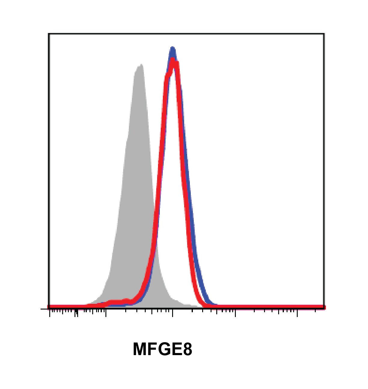
Detection of MFG‑E8 in Human Immature Dendritic Cells by Flow Cytometry.
Human immature dendritic cells were stained with Mouse Anti-Human MFG-E8 Monoclonal Antibody (Catalog # MAB27671, filled histogram) or isotype control antibody (MAB003, open histogram), followed by Phycoerythrin-conjugated Anti-Mouse IgG Secondary Antibody (F0102B). To facilitate intracellular staining, cells were fixed with Flow Cytometry Fixation Buffer (FC004) and permeabilized with Flow Cytometry Permeabilization/Wash Buffer I (FC005). View our protocol for Staining Intracellular Molecules.
Detection of MFG‑E8 in TF-1 cells by Flow Cytometry.
TF-1 cells were stained with Mouse Anti-Human MFG‑E8 Monoclonal Antibody (Catalog # MAB27671, filled histogram) or isotype control antibody (Catalog # MAB003, open histogram), followed by Fluorescein-conjugated Anti-Mouse IgG Secondary Antibody (Catalog # F0103B). To facilitate intracellular staining, cells were fixed with FC012 and permeabilized with FoxP3 Perm. View our protocol for Staining Intracellular Molecules.Applications for Human MFG‑E8 Antibody
Application
Recommended Usage
CyTOF-ready
Ready to be labeled using established conjugation methods. No BSA or other carrier proteins that could interfere with conjugation.
Intracellular Staining by Flow Cytometry
0.25 µg/106 cells
Sample: Human immature dendritic cells fixed with Flow Cytometry Fixation Buffer (Catalog # FC004) and permeabilized with Flow Cytometry Permeabilization/Wash Buffer I (Catalog # FC005) and TF-1 cells
Sample: Human immature dendritic cells fixed with Flow Cytometry Fixation Buffer (Catalog # FC004) and permeabilized with Flow Cytometry Permeabilization/Wash Buffer I (Catalog # FC005) and TF-1 cells
Western Blot
1 µg/mL
Sample: Recombinant Human MFG-E8 (Catalog # 2767-MF) under non-reducing conditions only
Sample: Recombinant Human MFG-E8 (Catalog # 2767-MF) under non-reducing conditions only
Reviewed Applications
Read 1 review rated 4 using MAB27671 in the following applications:
Flow Cytometry Panel Builder
Bio-Techne Knows Flow Cytometry
Save time and reduce costly mistakes by quickly finding compatible reagents using the Panel Builder Tool.
Advanced Features
- Spectra Viewer - Custom analysis of spectra from multiple fluorochromes
- Spillover Popups - Visualize the spectra of individual fluorochromes
- Antigen Density Selector - Match fluorochrome brightness with antigen density

Formulation, Preparation, and Storage
Purification
Protein A or G purified from hybridoma culture supernatant
Reconstitution
Reconstitute at 0.5 mg/mL in sterile PBS. For liquid material, refer to CoA for concentration.
Loading...
Formulation
Lyophilized from a 0.2 μm filtered solution in PBS with Trehalose. *Small pack size (SP) is supplied either lyophilized or as a 0.2 µm filtered solution in PBS.
Shipping
Lyophilized product is shipped at ambient temperature. Liquid small pack size (-SP) is shipped with polar packs. Upon receipt, store immediately at the temperature recommended below.
Stability & Storage
Use a manual defrost freezer and avoid repeated freeze-thaw cycles.
- 12 months from date of receipt, -20 to -70 °C as supplied.
- 1 month, 2 to 8 °C under sterile conditions after reconstitution.
- 6 months, -20 to -70 °C under sterile conditions after reconstitution.
Calculators
Background: MFG-E8
References
- Raymond, A. et al. (2009) J. Cell. Biochem. 106:957.
- Couto, J.R. et al. (1996) DNA Cell Biol. 15:281.
- Yamaguchi, H. et al. (2010) Eur. J. Immunol. 40:1778.
- Haggqvist, B. et al. (1999) Proc. Natl. Acad. Sci. USA 96:8669.
- Silvestre, J.-S. et al. (2005) Nat. Med. 11:499.
- Borges, E. et al. (2000) J. Biol. Chem. 275:39867.
- Hanayama, R. et al. (2002) Nature 417:182.
- Ait-Oufella, H. et al. (2007) Circulation 115:2168.
- Kranich, J. et al. (2010) J. Exp. Med. 207:2271.
- Hanayama, R. et al. (2004) Science 304:1147.
- Kranich, J. et al. (2010) J. Exp. Med. 205:1293.
- Atabai, K. et al. (2009) J. Clin. Invest. 119:3713.
- Bu, H.-F. et al. (2007) J. Clin. Invest. 117:3673.
- Jinushi, M. et al. (2009) J. Exp. Med. 206:1317.
- Kvistgaard, A.S. et al. (2004) J. Dairy Sci. 87:4088.
Long Name
Milk Fat Globule EGF Factor 8
Alternate Names
BA46, Breast epithelial antigen BA46, Lactahedrin, Medin, MFGE8, SED1
Gene Symbol
MFGE8
UniProt
Additional MFG-E8 Products
Product Documents for Human MFG‑E8 Antibody
Certificate of Analysis
To download a Certificate of Analysis, please enter a lot or batch number in the search box below.
Note: Certificate of Analysis not available for kit components.
Product Specific Notices for Human MFG‑E8 Antibody
For research use only
Citations for Human MFG‑E8 Antibody
 Powered by Bioz
Customer Reviews for Human MFG‑E8 Antibody (1)
4 out of 5
1 Customer Rating
Have you used Human MFG‑E8 Antibody?
Submit a review and receive an Amazon gift card!
$25/€18/£15/$25CAN/¥2500 Yen for a review with an image
$10/€7/£6/$10CAN/¥1110 Yen for a review without an image
Submit a review
Customer Images
Showing
1
-
1 of
1 review
Showing All
Filter By:
-
Application: Flow CytometrySample Tested: Bone marrow cellsSpecies: HumanVerified Customer | Posted 08/01/2016

There are no reviews that match your criteria.
Protocols
Find general support by application which include: protocols, troubleshooting, illustrated assays, videos and webinars.
- 7-Amino Actinomycin D (7-AAD) Cell Viability Flow Cytometry Protocol
- Cellular Response to Hypoxia Protocols
- Extracellular Membrane Flow Cytometry Protocol
- Flow Cytometry Protocol for Cell Surface Markers
- Flow Cytometry Protocol for Staining Membrane Associated Proteins
- Flow Cytometry Staining Protocols
- Flow Cytometry Troubleshooting Guide
- Intracellular Flow Cytometry Protocol Using Alcohol (Methanol)
- Intracellular Flow Cytometry Protocol Using Detergents
- Intracellular Nuclear Staining Flow Cytometry Protocol Using Detergents
- Intracellular Staining Flow Cytometry Protocol Using Alcohol Permeabilization
- Intracellular Staining Flow Cytometry Protocol Using Detergents to Permeabilize Cells
- Propidium Iodide Cell Viability Flow Cytometry Protocol
- Protocol for Liperfluo
- Protocol for the Characterization of Human Th22 Cells
- Protocol for the Characterization of Human Th9 Cells
- Protocol: Annexin V and PI Staining by Flow Cytometry
- Protocol: Annexin V and PI Staining for Apoptosis by Flow Cytometry
- R&D Systems Quality Control Western Blot Protocol
- Troubleshooting Guide: Fluorokine Flow Cytometry Kits
- Troubleshooting Guide: Western Blot Figures
- Western Blot Conditions
- Western Blot Protocol
- Western Blot Protocol for Cell Lysates
- Western Blot Troubleshooting
- Western Blot Troubleshooting Guide
- View all Protocols, Troubleshooting, Illustrated assays and Webinars
Loading...