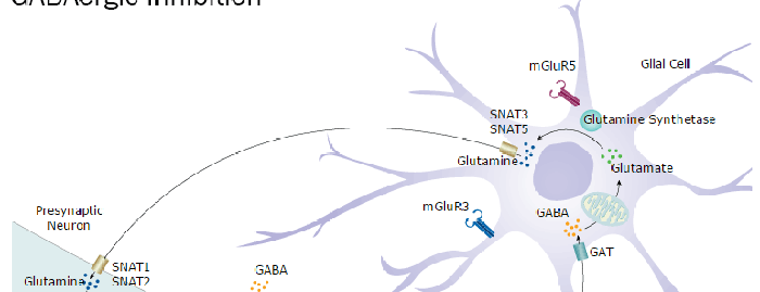
Metabotropic glutamate receptor 3 (mGluR3) is a 90-100 kDa, 7-transmembrane glycoprotein that belongs to group II of the C-family of G-protein coupled receptors. It is a presynaptic receptor expressed on both neurons and glia, whose activation reduces adenylate cyclase activity. Mature human mGluR3 is 857 amino acids in length and contains a 554 amino acid (aa) N-terminal extracellular domain (ECD) (aa 23-576). The ECD binds glutamate and forms homodimers. There is one alternative splice form that is soluble, 515 aa in length and shows a 96 aa substitution for aa 442-879. Over aa 25-507, human mGluR3 shares 97% aa sequence identity with mouse and rat mGluR3 and 67% aa sequence identity with hGluR2.

Key Product Details
Species Reactivity
Human
Applications
Flow Cytometry, CyTOF-ready
Label
Unconjugated
Antibody Source
Monoclonal Mouse IgG2B Clone # 530510
Loading...
Product Specifications
Immunogen
Chinese hamster ovary cell line CHO-derived recombinant human mGluR3
Asp25-Ser507
Accession # Q14832
Asp25-Ser507
Accession # Q14832
Specificity
Detects human mGluR3 in direct ELISAs.
Clonality
Monoclonal
Host
Mouse
Isotype
IgG2B
Scientific Data Images for Human mGluR3 Antibody (530510)
Detection of mGluR3 in HEK293 Human Cell Line Transfected with Human mGluR3 and eGFP by Flow Cytometry.
HEK293 human embryonic kidney cell line transfected with human mGluR3 and eGFP was stained with (A) Mouse Anti-Human mGluR3 Monoclonal Antibody (Catalog # MAB46681) or (B) Mouse IgG2B isotype control antibody (Catalog # MAB0041) followed by APC-conjugated Anti-Mouse IgG Secondary Antibody (Catalog # F0101B). Staining was performed using our Staining Membrane-associated Proteins protocol.Applications for Human mGluR3 Antibody (530510)
Application
Recommended Usage
CyTOF-ready
Ready to be labeled using established conjugation methods. No BSA or other carrier proteins that could interfere with conjugation.
Flow Cytometry
0.25 µg/106 cells
Sample: HEK293 Human Cell Line Transfected with Human MGLUR3 and eGFP
Sample: HEK293 Human Cell Line Transfected with Human MGLUR3 and eGFP
Flow Cytometry Panel Builder
Bio-Techne Knows Flow Cytometry
Save time and reduce costly mistakes by quickly finding compatible reagents using the Panel Builder Tool.
Advanced Features
- Spectra Viewer - Custom analysis of spectra from multiple fluorochromes
- Spillover Popups - Visualize the spectra of individual fluorochromes
- Antigen Density Selector - Match fluorochrome brightness with antigen density

Formulation, Preparation, and Storage
Purification
Protein A or G purified from hybridoma culture supernatant
Reconstitution
Reconstitute at 0.5 mg/mL in sterile PBS. For liquid material, refer to CoA for concentration.
Loading...
Formulation
Lyophilized from a 0.2 μm filtered solution in PBS with Trehalose. *Small pack size (SP) is supplied either lyophilized or as a 0.2 µm filtered solution in PBS.
Shipping
Lyophilized product is shipped at ambient temperature. Liquid small pack size (-SP) is shipped with polar packs. Upon receipt, store immediately at the temperature recommended below.
Stability & Storage
Use a manual defrost freezer and avoid repeated freeze-thaw cycles.
- 12 months from date of receipt, -20 to -70 °C as supplied.
- 1 month, 2 to 8 °C under sterile conditions after reconstitution.
- 6 months, -20 to -70 °C under sterile conditions after reconstitution.
Calculators
Background: mGluR3
Long Name
Metabotropic Glutamate Receptor 3
Alternate Names
GPRC1C, GRM3
Gene Symbol
GRM3
UniProt
Additional mGluR3 Products
Product Documents for Human mGluR3 Antibody (530510)
Certificate of Analysis
To download a Certificate of Analysis, please enter a lot or batch number in the search box below.
Note: Certificate of Analysis not available for kit components.
Product Specific Notices for Human mGluR3 Antibody (530510)
For research use only
Related Research Areas
Customer Reviews for Human mGluR3 Antibody (530510)
There are currently no reviews for this product. Be the first to review Human mGluR3 Antibody (530510) and earn rewards!
Have you used Human mGluR3 Antibody (530510)?
Submit a review and receive an Amazon gift card!
$25/€18/£15/$25CAN/¥2500 Yen for a review with an image
$10/€7/£6/$10CAN/¥1110 Yen for a review without an image
Submit a review
Protocols
Find general support by application which include: protocols, troubleshooting, illustrated assays, videos and webinars.
- 7-Amino Actinomycin D (7-AAD) Cell Viability Flow Cytometry Protocol
- Extracellular Membrane Flow Cytometry Protocol
- Flow Cytometry Protocol for Cell Surface Markers
- Flow Cytometry Protocol for Staining Membrane Associated Proteins
- Flow Cytometry Staining Protocols
- Flow Cytometry Troubleshooting Guide
- Intracellular Flow Cytometry Protocol Using Alcohol (Methanol)
- Intracellular Flow Cytometry Protocol Using Detergents
- Intracellular Nuclear Staining Flow Cytometry Protocol Using Detergents
- Intracellular Staining Flow Cytometry Protocol Using Alcohol Permeabilization
- Intracellular Staining Flow Cytometry Protocol Using Detergents to Permeabilize Cells
- Propidium Iodide Cell Viability Flow Cytometry Protocol
- Protocol for Liperfluo
- Protocol for the Characterization of Human Th22 Cells
- Protocol for the Characterization of Human Th9 Cells
- Protocol: Annexin V and PI Staining by Flow Cytometry
- Protocol: Annexin V and PI Staining for Apoptosis by Flow Cytometry
- Troubleshooting Guide: Fluorokine Flow Cytometry Kits
- View all Protocols, Troubleshooting, Illustrated assays and Webinars
Loading...
Associated Pathways