Matrix metalloproteinases are a family of zinc and calcium dependent endopeptidases with the combined ability to degrade all the components of the extracellular matrix. MMP-13 (Collagenase-3) has been demonstrated to degrade a range of extracellular matrix proteins, including collagen types I, II, III, IV, IX, X and XIV, gelatin, aggrecan, perlecan and fibronectin. MMP-13 is distinguished from the other human collagenases by its effecient degradation of type II collagen. MMP-13 is expressed by fibroblasts, chrondrocytes and squamous epithelial cells. Structurally, MMP-13 may be divided into several distinct domains; a pro-domain which is cleaved upon activation; a catalytic domain containing the zinc binding site; a short hinge region and a carboxyl terminal (hemopexin-like) domain.

Key Product Details
Validated by
Biological Validation
Species Reactivity
Validated:
Human
Cited:
Human, Mouse, Rat, Bovine, Canine, Rabbit
Applications
Validated:
Immunohistochemistry, Western Blot, Immunocytochemistry, Immunoprecipitation, Immunoaffinity Purification
Cited:
Immunohistochemistry, Immunohistochemistry-Paraffin, Immunohistochemistry-Frozen, Western Blot, Neutralization, Immunocytochemistry, Immunocytochemistry/ Immunofluorescence, ELISA Detection, ELISA Development
Label
Unconjugated
Antibody Source
Monoclonal Mouse IgG1 Clone # 87512
Loading...
Product Specifications
Immunogen
Chinese hamster ovary cell line CHO-derived recombinant human MMP-13
Leu20-Cys471
Accession # P45452
Leu20-Cys471
Accession # P45452
Specificity
Detects human MMP-13 in direct ELISAs and Western blots. Detects both pro and active forms of human MMP-13 in Western blots. In direct ELISAs and Western blots, no cross-reactivity with recombinant human MMP-1, -2, -3, -7, -8, -9, -10, or -12 is observed.
Clonality
Monoclonal
Host
Mouse
Isotype
IgG1
Scientific Data Images for Human MMP-13 Antibody (87512)
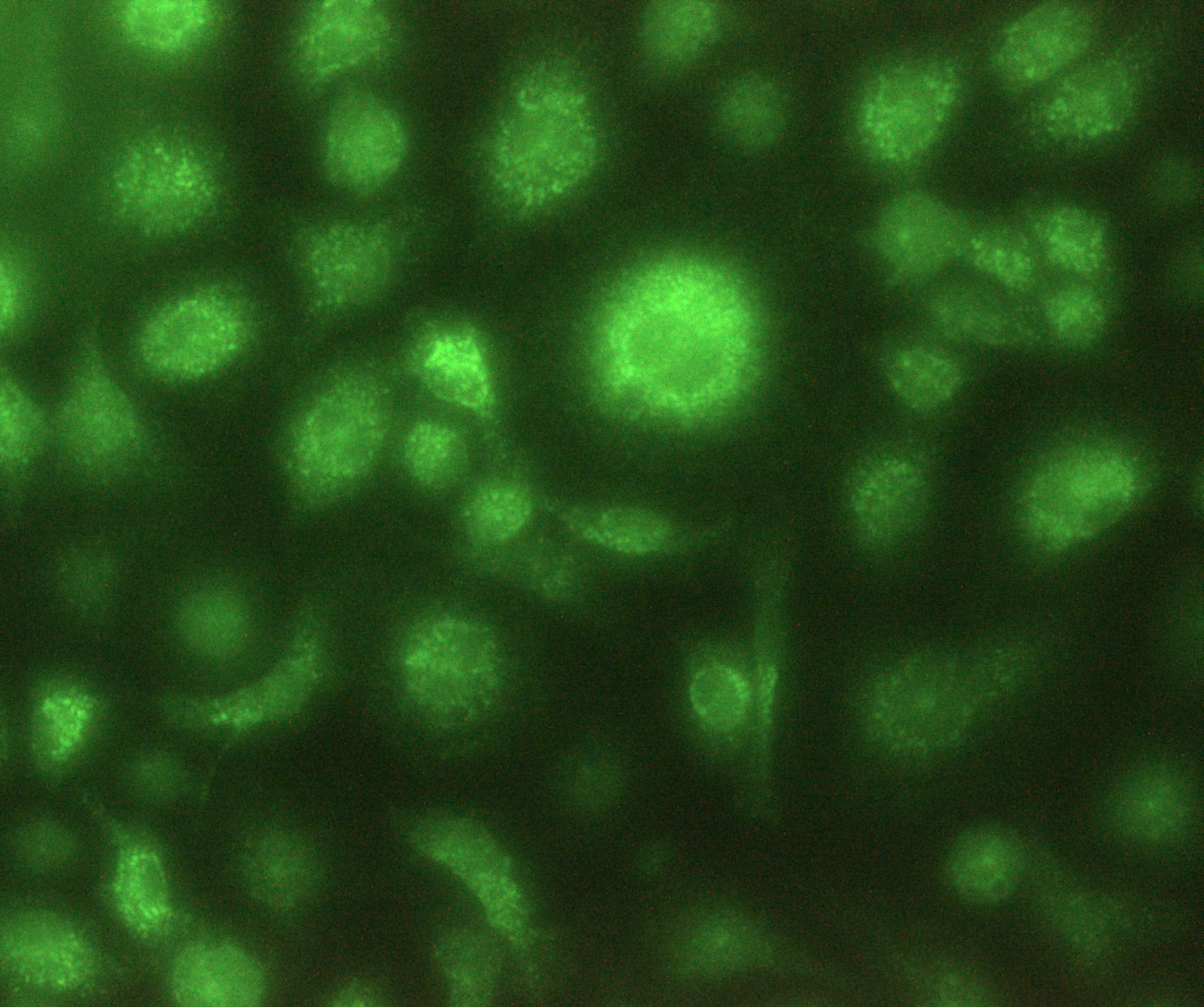
MMP‑13 in MDA‑MB‑231 Human Cell Line.
MMP-13 was detected in immersion fixed MDA-MB-231 human breast cancer cell line using Human MMP-13 Monoclonal Antibody (Catalog # MAB511) at 5 µg/mL for 3 hours at room temperature. Cells were stained using the NorthernLights™ 493-conjugated Anti-Mouse IgG Secondary Antibody (green; Catalog # NL009) and counterstained with DAPI (blue). View our protocol for Fluorescent ICC Staining of Cells on Coverslips.
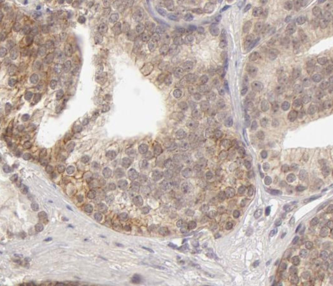
MMP‑13 in Human Ovarian Cancer Tissue.
MMP‑13 was detected in immersion fixed paraffin-embedded sections of human ovarian cancer tissue using 8 µg/mL Mouse Anti-Human MMP‑13 Monoclonal Antibody (Catalog # MAB511) overnight at 4 °C. Tissue was stained with the Anti-Mouse HRP-DAB Cell & Tissue Staining Kit (brown; Catalog # CTS002) and counterstained with hematoxylin (blue). View our protocol for Chromogenic IHC Staining of Paraffin-embedded Tissue Sections.
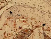
Detection of Canine MMP-13 by Western Blot
Effects of IGF-1 or/and PDGF-bb on IL-1 beta -induced NF-kappa B-dependent pro-inflammatory, pro-apoptotic and matrix degrading gene products in chondrocytes.To determine whether IGF-1 or/and PDGF-bbexert effects on IL-1 beta -induced NF-kappa B-dependent expression of pro-inflammatory, pro-apoptotic and matrix degrading gene products, primary chondrocytes were either stimulated with 10 ng/ml IL-1 beta, 10 ng/ml PDGF-bb, 10 ng/ml IGF-1 or combination of both growth factors (5 ng/ml each) or pre-stimulated for 12 h with 10 ng/ml PDGF-bb, 10 ng/ml IGF-1 or combination of both growth factors (5 ng/ml each) followed by 10 ng/ml IL-1 beta for 24. Equal amounts of total proteins were separated by SDS-PAGE and analyzed by immunoblotting using antibodies raised against COX-2, MMP-9 and MMP-13 and active caspase-3. Stimulation with IL-1 beta resulted in production of COX-2, MMP-9, MMP-13 and caspase-3 cleavage. Pre-treatment with a combination of both IGF-1 or/and PDGF-bb downregulated COX-2, MMP-9, MMP-13 and cleaved caspase-3. Image collected and cropped by CiteAb from the following publication (https://dx.plos.org/10.1371/journal.pone.0028663), licensed under a CC-BY license. Not internally tested by R&D Systems.
Detection of Human MMP-13 by Immunohistochemistry
Enhanced MMP13 gene expression and production due to 3D osteogenic differentiation. (A) Multivariate analysis of the sixty-six tested genes (NanoString® kit) displayed as a volcano plot (created with nSolver® software) with the 2D or 3D condition set as covariate. The plot shows each gene’s −log10 (p-value) and log2 fold change associated to the 2D or 3D condition. Highly statistically significant genes fall at the top of the plot above the indicated p-value lines, and highly differentially expressed genes fall to either side (right = upregulated, left = downregulated). The only significantly (p-value < 0.01) affected gene that was upregulated was MMP13. (B) Relative gene expression of MMP13 is shown in the four various conditions: 2D undifferentiated, 2D differentiated, 3D undifferentiated, 3D differentiated, after 5 weeks of culture. The significance levels for relative gene expression difference are indicated as ** (p-value < 0.01), *** (p-value < 0.001). (C) Immunohistochemistry of 5-week undifferentiated vs. differentiated (osteogenic) 3D Col I gels against MMP13 and nuclear DAPI. Scale bar = 50 μm. Image collected and cropped by CiteAb from the following publication (https://pubmed.ncbi.nlm.nih.gov/34948393), licensed under a CC-BY license. Not internally tested by R&D Systems.
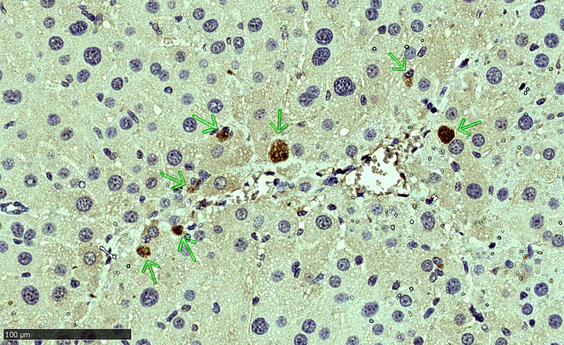
Detection of MMP-13 by Immunohistochemistry
Immunohistochemistry of metalloproteinase in meniscal samples. (a) number of patients with different expression of MMP-1 and -13; (b) one representative patient for MMP-1 (grade 3) and MMP-13 (grade 1), (magnification 10×, inset 40×, respectively). Image collected and cropped by CiteAb from the following open publication (https://www.mdpi.com/1422-0067/23/7/3903), licensed under a CC-BY license. Not internally tested by R&D Systems.Applications for Human MMP-13 Antibody (87512)
Application
Recommended Usage
Immunoaffinity Purification
Sepharose beads coupled with this antibody have been used to immunoprecipitate and immunopurify Recombinant Human MMP-13 (Catalog # 511-MM) from conditioned media.
Immunocytochemistry
8-25 µg/mL
Sample: Immersion fixed MDA-MB-231 human breast cancer cell line
Sample: Immersion fixed MDA-MB-231 human breast cancer cell line
Immunohistochemistry
8-25 µg/mL
Sample: Immersion fixed paraffin-embedded sections of human ovarian cancer tissue
Sample: Immersion fixed paraffin-embedded sections of human ovarian cancer tissue
Immunoprecipitation
25 µg/mL
Sample: Conditioned cell culture medium spiked with Recombinant Human MMP‑13 (Catalog # 511‑MM), see our available Western blot detection antibodies
Sample: Conditioned cell culture medium spiked with Recombinant Human MMP‑13 (Catalog # 511‑MM), see our available Western blot detection antibodies
Western Blot
1 µg/mL
Sample: Recombinant Human MMP‑13 Western Blot Standard (Catalog # WBC020)
Sample: Recombinant Human MMP‑13 Western Blot Standard (Catalog # WBC020)
Reviewed Applications
Read 6 reviews rated 4.7 using MAB511 in the following applications:
Formulation, Preparation, and Storage
Purification
Protein A or G purified from hybridoma culture supernatant
Reconstitution
Reconstitute at 0.5 mg/mL in sterile PBS. For liquid material, refer to CoA for concentration.
Loading...
Formulation
Lyophilized from a 0.2 μm filtered solution in PBS with Trehalose. *Small pack size (SP) is supplied either lyophilized or as a 0.2 µm filtered solution in PBS.
Shipping
Lyophilized product is shipped at ambient temperature. Liquid small pack size (-SP) is shipped with polar packs. Upon receipt, store immediately at the temperature recommended below.
Stability & Storage
Use a manual defrost freezer and avoid repeated freeze-thaw cycles.
- 12 months from date of receipt, -20 to -70 °C as supplied.
- 1 month, 2 to 8 °C under sterile conditions after reconstitution.
- 6 months, -20 to -70 °C under sterile conditions after reconstitution.
Calculators
Background: MMP-13
References
- Jeffery, J.J. (1998) in Collagenase 3. A.J. Barrett, et al. (eds): Handbook of Proteolytic Enzymes, San Diego: Academic Press, p. 1167.
Long Name
Matrix Metalloproteinase 13
Alternate Names
MMP13
Entrez Gene IDs
4322 (Human)
Gene Symbol
MMP13
UniProt
Additional MMP-13 Products
Product Documents for Human MMP-13 Antibody (87512)
Certificate of Analysis
To download a Certificate of Analysis, please enter a lot or batch number in the search box below.
Note: Certificate of Analysis not available for kit components.
Product Specific Notices for Human MMP-13 Antibody (87512)
For research use only
Related Research Areas
Citations for Human MMP-13 Antibody (87512)
 Powered by Bioz
Customer Reviews for Human MMP-13 Antibody (87512) (6)
4.7 out of 5
6 Customer Ratings
Have you used Human MMP-13 Antibody (87512)?
Submit a review and receive an Amazon gift card!
$25/€18/£15/$25CAN/¥2500 Yen for a review with an image
$10/€7/£6/$10CAN/¥1110 Yen for a review without an image
Submit a review
Customer Images
Showing
1
-
5 of
6 reviews
Showing All
Filter By:
-
Application: ImmunocytochemistrySample Tested: Bone Marrrow Derived MacrophagesSpecies: MouseVerified Customer | Posted 02/07/2022Macrophages in vitro produce MMP131:200 Dilution Overnight incubation at RT
 Bio-Techne ResponseThis review was submitted through the legacy Novus Innovators Program, reflecting a new species or application tested on a primary antibody.
 -
Application: Immunocytochemistry/ImmunofluorescenceSample Tested: Liver tissueSpecies: MouseVerified Customer | Posted 02/07/20221:100 dilution Citrate buffer Ag retrieval Overnight incubation of the Ab, RT
-
Application: ImmunohistochemistrySample Tested: Prostate cancer tissueSpecies: HumanVerified Customer | Posted 12/06/2021
-
Application: ImmunohistochemistrySample Tested: Joint tissueSpecies: HumanVerified Customer | Posted 10/14/2021
-
Application: Western BlotSample Tested: HeLa cellsSpecies: HumanVerified Customer | Posted 07/13/2018
-
Application: ELISASample Tested: Serum and PlasmaSpecies: HumanVerified Customer | Posted 11/10/2017The antibody AF511 was used as the detection antibody with this antibody as the capture in an ELISA targeting MMP-13. The immunoassay was used to measure MMP-13 levels in human plasma samples.
There are no reviews that match your criteria.
Protocols
Find general support by application which include: protocols, troubleshooting, illustrated assays, videos and webinars.
- Antigen Retrieval Protocol (PIER)
- Antigen Retrieval for Frozen Sections Protocol
- Appropriate Fixation of IHC/ICC Samples
- Cellular Response to Hypoxia Protocols
- Chromogenic IHC Staining of Formalin-Fixed Paraffin-Embedded (FFPE) Tissue Protocol
- Chromogenic Immunohistochemistry Staining of Frozen Tissue
- ClariTSA™ Fluorophore Kits
- Detection & Visualization of Antibody Binding
- Fluorescent IHC Staining of Frozen Tissue Protocol
- Graphic Protocol for Heat-induced Epitope Retrieval
- Graphic Protocol for the Preparation and Fluorescent IHC Staining of Frozen Tissue Sections
- Graphic Protocol for the Preparation and Fluorescent IHC Staining of Paraffin-embedded Tissue Sections
- Graphic Protocol for the Preparation of Gelatin-coated Slides for Histological Tissue Sections
- ICC Cell Smear Protocol for Suspension Cells
- ICC Immunocytochemistry Protocol Videos
- ICC for Adherent Cells
- IHC Sample Preparation (Frozen sections vs Paraffin)
- Immunocytochemistry (ICC) Protocol
- Immunocytochemistry Troubleshooting
- Immunofluorescence of Organoids Embedded in Cultrex Basement Membrane Extract
- Immunofluorescent IHC Staining of Formalin-Fixed Paraffin-Embedded (FFPE) Tissue Protocol
- Immunohistochemistry (IHC) and Immunocytochemistry (ICC) Protocols
- Immunohistochemistry Frozen Troubleshooting
- Immunohistochemistry Paraffin Troubleshooting
- Immunoprecipitation Protocol
- Preparing Samples for IHC/ICC Experiments
- Preventing Non-Specific Staining (Non-Specific Binding)
- Primary Antibody Selection & Optimization
- Protocol for Heat-Induced Epitope Retrieval (HIER)
- Protocol for Making a 4% Formaldehyde Solution in PBS
- Protocol for VisUCyte™ HRP Polymer Detection Reagent
- Protocol for the Fluorescent ICC Staining of Cell Smears - Graphic
- Protocol for the Fluorescent ICC Staining of Cultured Cells on Coverslips - Graphic
- Protocol for the Preparation & Fixation of Cells on Coverslips
- Protocol for the Preparation and Chromogenic IHC Staining of Frozen Tissue Sections
- Protocol for the Preparation and Chromogenic IHC Staining of Frozen Tissue Sections - Graphic
- Protocol for the Preparation and Chromogenic IHC Staining of Paraffin-embedded Tissue Sections
- Protocol for the Preparation and Chromogenic IHC Staining of Paraffin-embedded Tissue Sections - Graphic
- Protocol for the Preparation and Fluorescent ICC Staining of Cells on Coverslips
- Protocol for the Preparation and Fluorescent ICC Staining of Non-adherent Cells
- Protocol for the Preparation and Fluorescent ICC Staining of Stem Cells on Coverslips
- Protocol for the Preparation and Fluorescent IHC Staining of Frozen Tissue Sections
- Protocol for the Preparation and Fluorescent IHC Staining of Paraffin-embedded Tissue Sections
- Protocol for the Preparation of Gelatin-coated Slides for Histological Tissue Sections
- Protocol for the Preparation of a Cell Smear for Non-adherent Cell ICC - Graphic
- R&D Systems Quality Control Western Blot Protocol
- TUNEL and Active Caspase-3 Detection by IHC/ICC Protocol
- The Importance of IHC/ICC Controls
- Troubleshooting Guide: Immunohistochemistry
- Troubleshooting Guide: Western Blot Figures
- Western Blot Conditions
- Western Blot Protocol
- Western Blot Protocol for Cell Lysates
- Western Blot Troubleshooting
- Western Blot Troubleshooting Guide
- View all Protocols, Troubleshooting, Illustrated assays and Webinars
Loading...