The Annexins are a family of Calcium-dependent phospholipid-binding proteins that are preferentially located on the cytosolic face of the plasma membrane. The Annexin’s have a molecular weight of approximately 35 to 40 kDa and consist of a unique amino terminal domain followed by a homologous C-terminal core domain containing the calcium-dependent phospholipid-binding sites. The C‑terminal domain is comprised of four 60‑70 amino acid repeats, known as annexin repeats or an endonexin fold (Annexin A6 contains 8 annexin repeats). The four annexin repeats form a highly alpha -helical, tightly packed disc known as the annexin domain, which binds to phospholipids in the membrane in a calcium-dependent manner. Members of the annexin family play a role in cytoskeletal interactions, phospholipase inhibition, regulation of cellular growth, and intracellular signal transduction pathways. Annexin A1 (ANXA1), also known as annexin I, lipocortin I, and calpactin II, is an ~ 40 kDa protein with phospholipase A2 inhibitory activity. Since phospholipase A2 is required for the biosynthesis of the potent mediators of inflammation, prostaglandins and leukotrienes, Annexin A1 may have anti-inflammatory activity. Human Annexin A1 shares 88 and 89% identity with mouse and rat Annexin A1, respectively.

Key Product Details
Validated by
Knockout/Knockdown
Species Reactivity
Human, Mouse
Applications
Knockout Validated, Immunohistochemistry, Immunocytochemistry
Label
Unconjugated
Antibody Source
Monoclonal Mouse IgG1 Clone # 686106
Loading...
Product Specifications
Immunogen
E. coli-derived recombinant human Annexin A1
Met1-Asn346
Accession # P04083
Met1-Asn346
Accession # P04083
Specificity
Detects human Annexin A1 in direct ELISAs.
Detects human and mouse Annexin A1 in Western blots. In direct ELISAs, no cross-reactivity
with recombinant human Annexin A2, A3, A4, A6, A7, A8, A9, A10, A11, or A13 is
observed.
Clonality
Monoclonal
Host
Mouse
Isotype
IgG1
Scientific Data Images for Annexin A1 Antibody (686106)
Annexin A1 in NIH-3T3 Mouse Cell Line.
Annexin A1 was detected in immersion fixed NIH-3T3 mouse embryonic fibroblast cell line (left panel, positive stain) and NIH-3T3 Knockouts (right panel, negative stain) using Mouse Anti-Human/Mouse Annexin A1 Monoclonal Antibody (Catalog # MAB3770) at 3 µg/mL for 3 hours at room temperature. Cells were stained using the NorthernLights™ 557-conjugated Anti-Mouse IgG Secondary Antibody (red; Catalog # NL007) and counterstained with DAPI (blue). Specific staining was localized to nuclei, cytoplasm and plasma membrane. View our protocol for Fluorescent ICC Staining of Cells on Coverslips.
Annexin A1 in Human Breast.
Annexin A1 was detected in immersion fixed paraffin-embedded sections of human breast using Mouse Anti-Human/Mouse Annexin A1 Monoclonal Antibody (Catalog # MAB3770) at 15 µg/mL overnight at 4 °C. Before incubation with the primary antibody, tissue was subjected to heat-induced epitope retrieval using Antigen Retrieval Reagent-Basic (Catalog # CTS013). Tissue was stained using the Anti-Mouse HRP-DAB Cell & Tissue Staining Kit (brown; Catalog # CTS002) and counterstained with hematoxylin (blue). Specific staining was localized to myoepithelial cells. View our protocol for Chromogenic IHC Staining of Paraffin-embedded Tissue Sections.Applications for Annexin A1 Antibody (686106)
Application
Recommended Usage
Immunocytochemistry
3-25 µg/mL
Sample: Immersion fixed NIH-3T3 mouse embryonic fibroblast cell line
Sample: Immersion fixed NIH-3T3 mouse embryonic fibroblast cell line
Immunohistochemistry
8-25 µg/mL
Sample: Immersion fixed paraffin-embedded sections of human breast
Sample: Immersion fixed paraffin-embedded sections of human breast
Knockout Validated
Annexin A1 is specifically detected in NIH-
3T3 mouse embryonic fibroblast
cell lineparental cell line but is not detectable in Annexin A1 knockout NIH- 3T3 cell line.
Reviewed Applications
Read 1 review rated 5 using MAB3770 in the following applications:
Formulation, Preparation, and Storage
Purification
Protein A or G purified from hybridoma culture supernatant
Reconstitution
Sterile PBS to a final concentration of 0.5 mg/mL. For liquid material, refer to CoA for concentration.
Loading...
Formulation
Lyophilized from a 0.2 μm filtered solution in PBS with Trehalose. *Small pack size (SP) is supplied either lyophilized or as a 0.2 µm filtered solution in PBS.
Shipping
Lyophilized product is shipped at ambient temperature. Liquid small pack size (-SP) is shipped with polar packs. Upon receipt, store immediately at the temperature recommended below.
Stability & Storage
Use a manual defrost freezer and avoid repeated freeze-thaw cycles.
- 12 months from date of receipt, -20 to -70 °C as supplied.
- 1 month, 2 to 8 °C under sterile conditions after reconstitution.
- 6 months, -20 to -70 °C under sterile conditions after reconstitution.
Calculators
Background: Annexin A1
Alternate Names
ANX1, ANXA1, LPC1
Gene Symbol
ANXA1
UniProt
Additional Annexin A1 Products
Product Documents for Annexin A1 Antibody (686106)
Certificate of Analysis
To download a Certificate of Analysis, please enter a lot or batch number in the search box below.
Note: Certificate of Analysis not available for kit components.
Product Specific Notices for Annexin A1 Antibody (686106)
For research use only
Related Research Areas
Citations for Annexin A1 Antibody (686106)
 Powered by Bioz
Customer Reviews for Annexin A1 Antibody (686106) (1)
5 out of 5
1 Customer Rating
Have you used Annexin A1 Antibody (686106)?
Submit a review and receive an Amazon gift card!
$25/€18/£15/$25CAN/¥2500 Yen for a review with an image
$10/€7/£6/$10CAN/¥1110 Yen for a review without an image
Submit a review
Customer Images
Showing
1
-
1 of
1 review
Showing All
Filter By:
-
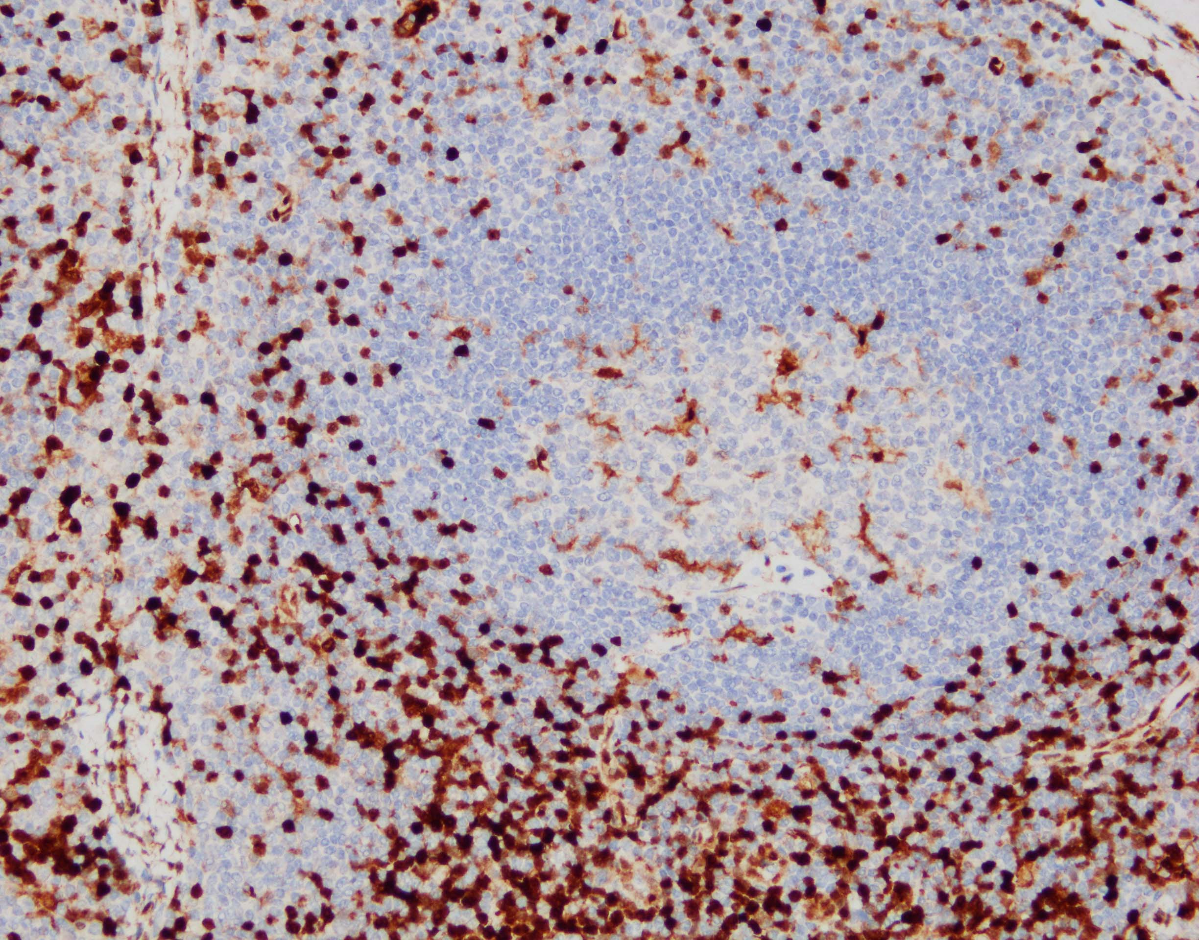
Application: ImmunohistochemistrySample Tested: Lymph node tissueSpecies: HumanVerified Customer | Posted 07/25/2023IHC-P: pH 9 antigen retrieval, primary Ab x1000, HRP-DAB detection system

There are no reviews that match your criteria.
Protocols
Find general support by application which include: protocols, troubleshooting, illustrated assays, videos and webinars.
- Antigen Retrieval Protocol (PIER)
- Antigen Retrieval for Frozen Sections Protocol
- Appropriate Fixation of IHC/ICC Samples
- Cellular Response to Hypoxia Protocols
- Chromogenic IHC Staining of Formalin-Fixed Paraffin-Embedded (FFPE) Tissue Protocol
- Chromogenic Immunohistochemistry Staining of Frozen Tissue
- ClariTSA™ Fluorophore Kits
- Detection & Visualization of Antibody Binding
- Fluorescent IHC Staining of Frozen Tissue Protocol
- Graphic Protocol for Heat-induced Epitope Retrieval
- Graphic Protocol for the Preparation and Fluorescent IHC Staining of Frozen Tissue Sections
- Graphic Protocol for the Preparation and Fluorescent IHC Staining of Paraffin-embedded Tissue Sections
- Graphic Protocol for the Preparation of Gelatin-coated Slides for Histological Tissue Sections
- ICC Cell Smear Protocol for Suspension Cells
- ICC Immunocytochemistry Protocol Videos
- ICC for Adherent Cells
- IHC Sample Preparation (Frozen sections vs Paraffin)
- Immunocytochemistry (ICC) Protocol
- Immunocytochemistry Troubleshooting
- Immunofluorescence of Organoids Embedded in Cultrex Basement Membrane Extract
- Immunofluorescent IHC Staining of Formalin-Fixed Paraffin-Embedded (FFPE) Tissue Protocol
- Immunohistochemistry (IHC) and Immunocytochemistry (ICC) Protocols
- Immunohistochemistry Frozen Troubleshooting
- Immunohistochemistry Paraffin Troubleshooting
- Preparing Samples for IHC/ICC Experiments
- Preventing Non-Specific Staining (Non-Specific Binding)
- Primary Antibody Selection & Optimization
- Protocol for Heat-Induced Epitope Retrieval (HIER)
- Protocol for Making a 4% Formaldehyde Solution in PBS
- Protocol for VisUCyte™ HRP Polymer Detection Reagent
- Protocol for the Fluorescent ICC Staining of Cell Smears - Graphic
- Protocol for the Fluorescent ICC Staining of Cultured Cells on Coverslips - Graphic
- Protocol for the Preparation & Fixation of Cells on Coverslips
- Protocol for the Preparation and Chromogenic IHC Staining of Frozen Tissue Sections
- Protocol for the Preparation and Chromogenic IHC Staining of Frozen Tissue Sections - Graphic
- Protocol for the Preparation and Chromogenic IHC Staining of Paraffin-embedded Tissue Sections
- Protocol for the Preparation and Chromogenic IHC Staining of Paraffin-embedded Tissue Sections - Graphic
- Protocol for the Preparation and Fluorescent ICC Staining of Cells on Coverslips
- Protocol for the Preparation and Fluorescent ICC Staining of Non-adherent Cells
- Protocol for the Preparation and Fluorescent ICC Staining of Stem Cells on Coverslips
- Protocol for the Preparation and Fluorescent IHC Staining of Frozen Tissue Sections
- Protocol for the Preparation and Fluorescent IHC Staining of Paraffin-embedded Tissue Sections
- Protocol for the Preparation of Gelatin-coated Slides for Histological Tissue Sections
- Protocol for the Preparation of a Cell Smear for Non-adherent Cell ICC - Graphic
- TUNEL and Active Caspase-3 Detection by IHC/ICC Protocol
- The Importance of IHC/ICC Controls
- Troubleshooting Guide: Immunohistochemistry
- View all Protocols, Troubleshooting, Illustrated assays and Webinars
Loading...