
Key Product Details
Validated by
Knockout/Knockdown
Species Reactivity
Validated:
Human, Mouse
Cited:
Human, Mouse, Rat
Applications
Validated:
Knockout Validated, Immunohistochemistry, Western Blot, Immunocytochemistry, Simple Western
Cited:
Immunohistochemistry, Immunohistochemistry-Paraffin, Western Blot
Label
Unconjugated
Antibody Source
Polyclonal Rabbit IgG
Loading...
Product Specifications
Immunogen
KLH-coupled human Bax synthetic peptide
Specificity
Detects human and mouse Bax.
Clonality
Polyclonal
Host
Rabbit
Isotype
IgG
Scientific Data Images for Bax Antibody
Detection of Human/Mouse Bax by Western Blot.
Western blot shows lysates of CHP-100 human neuroblastoma cell line, THP-1 human acute monocytic leukemia cell line, L-929 mouse fibroblast cell line, and DA3 mouse myeloma cell line. PVDF membrane was probed with 0.3 µg/mL of Rabbit Anti-Human/Mouse Bax Antigen Affinity-purified Polyclonal Antibody (Catalog # AF820) followed by HRP-conjugated Anti-Rabbit IgG Secondary Antibody (Catalog # HAF008). A specific band was detected for Bax at approximately 21 kDa (as indicated). This experiment was conducted under reducing conditions and using Immunoblot Buffer Group 2.
Bax in A549 Human Cell Line.
Bax was detected in immersion fixed A549 human lung carcinoma cell line using Rabbit Anti-Human/Mouse Bax Antigen Affinity-purified Polyclonal Antibody (Catalog # AF820) at 1.7 µg/mL for 3 hours at room temperature. Cells were stained using the NorthernLights™ 557-conjugated Anti-Rabbit IgG Secondary Antibody (red; Catalog # NL004) and counterstained with DAPI (blue). Specific staining was localized to cytoplasm and nuclei. View our protocol for Fluorescent ICC Staining of Cells on Coverslips.
Bax in Human Breast.
Bax was detected in immersion fixed paraffin-embedded sections of human breast using 15 µg/mL Rabbit Anti-Human/Mouse Bax Antigen Affinity-purified Polyclonal Antibody (Catalog # AF820) overnight at 4 °C. Tissue was stained (red) and counter-stained with hematoxylin (blue). View our protocol for Chromogenic IHC Staining of Paraffin-embedded Tissue Sections.
Bax in Human Liver.
Bax was detected in immersion fixed paraffin-embedded sections of human liver using 10 µg/mL Rabbit Anti-Human/Mouse Bax Antigen Affinity-purified Polyclonal Antibody (Catalog # AF820) overnight at 4 °C. Tissue was stained with the Anti-Rabbit HRP-DAB Cell & Tissue Staining Kit (brown; Catalog # CTS005) and counterstained with hematoxylin (blue). View our protocol for Chromogenic IHC Staining of Paraffin-embedded Tissue Sections.
Western Blot Shows Human Bax Specificity by Using Knockout Cell Line.
Western blot shows lysates of HeLa human cervical epithelial carcinoma parental cell line and Bax knockout HeLa cell line (KO). PVDF membrane was probed with 0.5 µg/mL of Rabbit Anti-Human/Mouse Bax Antigen Affinity-purified Polyclonal Antibody (Catalog # AF820) followed by HRP-conjugated Anti-Rabbit IgG Secondary Antibody (Catalog # HAF008). A specific band was detected for Bax at approximately 20 kDa (as indicated) in the parental HeLa cell line, but is not detectable in knockout HeLa cell line. GAPDH (Catalog # AF5718) is shown as a loading control. This experiment was conducted under reducing conditions and using Immunoblot Buffer Group 1.
Detection of Human Bax by Simple WesternTM.
Simple Western lane view shows lysates of HeLa human cervical epithelial carcinoma parental cell line and Bax knockout HeLa cell line (KO), loaded at 0.2 mg/mL. A specific band was detected for Bax at approximately 26 kDa (as indicated) using 50 µg/mL of Rabbit Anti-Human/Mouse Bax Antigen Affinity-purified Polyclonal Antibody (Catalog # AF820). This experiment was conducted under reducing conditions and using the 12-230 kDa separation system.
Detection of Bax by Western Blot
LPS induced cell pyroptosis, apoptosis and pro-inflammatory cytokines secretion in both IUA mice and cellular models. The expression status of (a) NLRP3, ASC and N-Gasdermin D, and (b) cleaved caspase-3 and Bax were detected by performing Western blot analysis. The pro-inflammatory cytokines generation and secretion were examined by (c) Real-Time qPCR in EPCs cells and (d-g) its supernatants were examined by Real-Time qPCR and ELISA assay. (h) Western blot was used to determine cell pytoptosis in EPCs cells. (i) MTT assay was used to determine cell viability in EPCs cells. (j) Generation and (k, l) secretion of IL-1 beta and IL-18 were respectively examined by Real-Time qPCR and ELISA. Individual experiment repeated 3 times, and *P < 0.05 Image collected and cropped by CiteAb from the following open publication (https://pubmed.ncbi.nlm.nih.gov/34738867), licensed under a CC-BY license. Not internally tested by R&D Systems.
Detection of Bax by Western Blot
Glycyrrhizic acid induces the apoptosis of colorectal cancer cells by inhibiting SIRT3 in vitro. (a,b) 0 or 20 μmol/L glycyrrhizin stimulated colorectal cancer cells for 24 hours, flow cytometry was used to detect the apoptosis of colorectal cancer cells (a) and statistical comparison (b). (c) Western blot analysis of Bax and Bcl2 protein expression in colorectal cancer cells after stimulating with 0 or 20 μmol/L glycyrrhizic acid for 24 hours. (d) Elisa kit was used to determine the activity of caspase 3 in colorectal cancer cells after stimulating with 0 or 20 μmol/L glycyrrhizic acid for 24 hours. Each test is repeated at least 3 times independently. *** P < 0.001 vs WT group, and ### P < 0.001 vs 20 μM GA group. Image collected and cropped by CiteAb from the following open publication (https://pubmed.ncbi.nlm.nih.gov/34747319), licensed under a CC-BY license. Not internally tested by R&D Systems.Applications for Bax Antibody
Application
Recommended Usage
Immunocytochemistry
1-15 µg/mL
Sample: Immersion fixed A549 human lung carcinoma cell line
Sample: Immersion fixed A549 human lung carcinoma cell line
Immunohistochemistry
5-15 µg/mL
Sample: Immersion fixed paraffin-embedded sections of human liver and immersion fixed paraffin-embedded sections of human breast
Sample: Immersion fixed paraffin-embedded sections of human liver and immersion fixed paraffin-embedded sections of human breast
Knockout Validated
Bax is
specifically detected in HeLa human cervical epithelial carcinoma parental cell line but is not detectable in Bax
knockout HeLa cell line.
Simple Western
50 µg/mL
Sample: HeLa human cervical epithelial carcinoma cell line
Sample: HeLa human cervical epithelial carcinoma cell line
Western Blot
0.3 µg/mL
Sample: CHP-100 human neuroblastoma cell line, THP-1 human acute monocytic leukemia cell line, L-929 mouse fibroblast cell line, and DA3 mouse myeloma cell line
Sample: CHP-100 human neuroblastoma cell line, THP-1 human acute monocytic leukemia cell line, L-929 mouse fibroblast cell line, and DA3 mouse myeloma cell line
Reviewed Applications
Read 4 reviews rated 4.3 using AF820 in the following applications:
Formulation, Preparation, and Storage
Purification
Antigen Affinity-purified
Reconstitution
Reconstitute at 0.2 mg/mL in sterile PBS.
Loading...
Formulation
Lyophilized from a 0.2 μm filtered solution in PBS with Trehalose.
Shipping
The product is shipped at ambient temperature. Upon receipt, store it immediately at the temperature recommended below.
Stability & Storage
Use a manual defrost freezer and avoid repeated freeze-thaw cycles.
- 12 months from date of receipt, -20 to -70 °C as supplied.
- 1 month, 2 to 8 °C under sterile conditions after reconstitution.
- 6 months, -20 to -70 °C under sterile conditions after reconstitution.
Calculators
Background: Bax
Long Name
Bcl Associated X Protein
Alternate Names
apoptosis regulator BAX, BCL2-associated X protein, Bcl2-L-4, BCL2L4bcl2-L-4, Bcl-2-like protein 4
Gene Symbol
BAX
Additional Bax Products
Product Documents for Bax Antibody
Certificate of Analysis
To download a Certificate of Analysis, please enter a lot or batch number in the search box below.
Note: Certificate of Analysis not available for kit components.
Product Specific Notices for Bax Antibody
For research use only
Related Research Areas
Citations for Bax Antibody
 Powered by Bioz
Customer Reviews for Bax Antibody (4)
4.3 out of 5
4 Customer Ratings
Have you used Bax Antibody?
Submit a review and receive an Amazon gift card!
$25/€18/£15/$25CAN/¥2500 Yen for a review with an image
$10/€7/£6/$10CAN/¥1110 Yen for a review without an image
Submit a review
Customer Images
Showing
1
-
4 of
4 reviews
Showing All
Filter By:
-
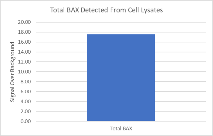
Application: ELISASample Tested: Cancer cell lysatesSpecies: HumanVerified Customer | Posted 07/30/2019

-
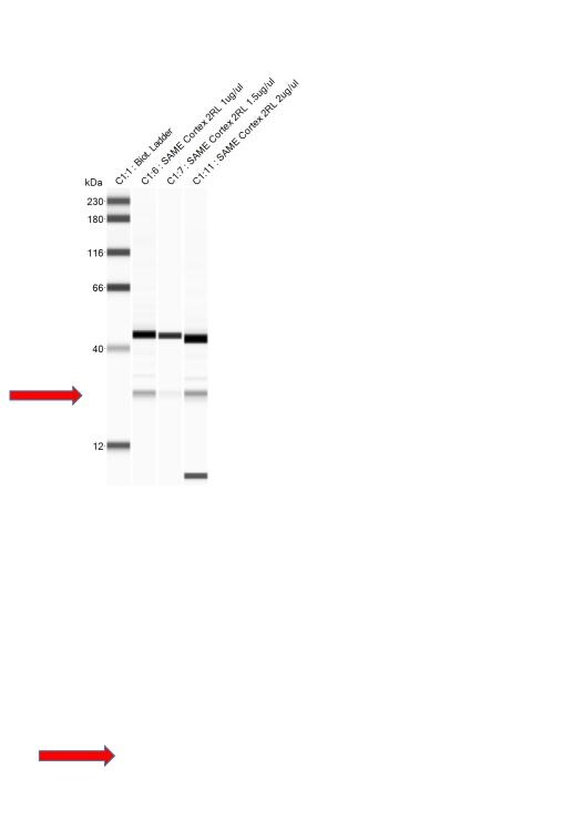
Application: Simple WesternSample Tested: Brain (cortex) tissueSpecies: RatVerified Customer | Posted 01/23/2018Missing band in C1:7 lane.
-
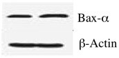
Application: Western BlotSample Tested: MDA-MB-231 human breast cancer cell lineSpecies: HumanVerified Customer | Posted 01/11/2018
-
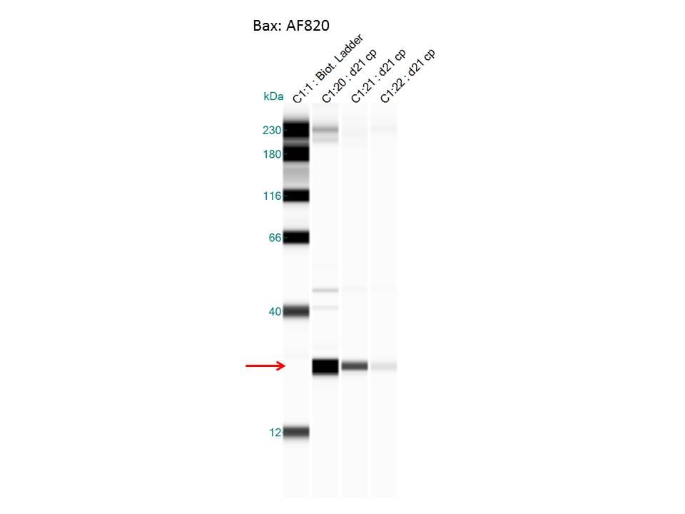
Application: Simple WesternSample Tested:Species: MouseVerified Customer | Posted 09/14/2015Bax AF820
There are no reviews that match your criteria.
Protocols
Find general support by application which include: protocols, troubleshooting, illustrated assays, videos and webinars.
- Antigen Retrieval Protocol (PIER)
- Antigen Retrieval for Frozen Sections Protocol
- Appropriate Fixation of IHC/ICC Samples
- Cellular Response to Hypoxia Protocols
- Chromogenic IHC Staining of Formalin-Fixed Paraffin-Embedded (FFPE) Tissue Protocol
- Chromogenic Immunohistochemistry Staining of Frozen Tissue
- ClariTSA™ Fluorophore Kits
- Detection & Visualization of Antibody Binding
- Fluorescent IHC Staining of Frozen Tissue Protocol
- Graphic Protocol for Heat-induced Epitope Retrieval
- Graphic Protocol for the Preparation and Fluorescent IHC Staining of Frozen Tissue Sections
- Graphic Protocol for the Preparation and Fluorescent IHC Staining of Paraffin-embedded Tissue Sections
- Graphic Protocol for the Preparation of Gelatin-coated Slides for Histological Tissue Sections
- ICC Cell Smear Protocol for Suspension Cells
- ICC Immunocytochemistry Protocol Videos
- ICC for Adherent Cells
- IHC Sample Preparation (Frozen sections vs Paraffin)
- Immunocytochemistry (ICC) Protocol
- Immunocytochemistry Troubleshooting
- Immunofluorescence of Organoids Embedded in Cultrex Basement Membrane Extract
- Immunofluorescent IHC Staining of Formalin-Fixed Paraffin-Embedded (FFPE) Tissue Protocol
- Immunohistochemistry (IHC) and Immunocytochemistry (ICC) Protocols
- Immunohistochemistry Frozen Troubleshooting
- Immunohistochemistry Paraffin Troubleshooting
- Preparing Samples for IHC/ICC Experiments
- Preventing Non-Specific Staining (Non-Specific Binding)
- Primary Antibody Selection & Optimization
- Protocol for Heat-Induced Epitope Retrieval (HIER)
- Protocol for Making a 4% Formaldehyde Solution in PBS
- Protocol for VisUCyte™ HRP Polymer Detection Reagent
- Protocol for the Fluorescent ICC Staining of Cell Smears - Graphic
- Protocol for the Fluorescent ICC Staining of Cultured Cells on Coverslips - Graphic
- Protocol for the Preparation & Fixation of Cells on Coverslips
- Protocol for the Preparation and Chromogenic IHC Staining of Frozen Tissue Sections
- Protocol for the Preparation and Chromogenic IHC Staining of Frozen Tissue Sections - Graphic
- Protocol for the Preparation and Chromogenic IHC Staining of Paraffin-embedded Tissue Sections
- Protocol for the Preparation and Chromogenic IHC Staining of Paraffin-embedded Tissue Sections - Graphic
- Protocol for the Preparation and Fluorescent ICC Staining of Cells on Coverslips
- Protocol for the Preparation and Fluorescent ICC Staining of Non-adherent Cells
- Protocol for the Preparation and Fluorescent ICC Staining of Stem Cells on Coverslips
- Protocol for the Preparation and Fluorescent IHC Staining of Frozen Tissue Sections
- Protocol for the Preparation and Fluorescent IHC Staining of Paraffin-embedded Tissue Sections
- Protocol for the Preparation of Gelatin-coated Slides for Histological Tissue Sections
- Protocol for the Preparation of a Cell Smear for Non-adherent Cell ICC - Graphic
- R&D Systems Quality Control Western Blot Protocol
- TUNEL and Active Caspase-3 Detection by IHC/ICC Protocol
- The Importance of IHC/ICC Controls
- Troubleshooting Guide: Immunohistochemistry
- Troubleshooting Guide: Western Blot Figures
- Western Blot Conditions
- Western Blot Protocol
- Western Blot Protocol for Cell Lysates
- Western Blot Troubleshooting
- Western Blot Troubleshooting Guide
- View all Protocols, Troubleshooting, Illustrated assays and Webinars
Loading...
Associated Pathways

