c-Rel/Rel, the cellular counterpart of the v-Rel oncogene of the avian reticuloendotheliosis retrovirus, is a 69 kDa class II member of the Rel/NK-kappa B family of transcription factors. All Rel family members contain a RHD (Rel homology domain) that is involved in dimerization, DNA and I kappa B binding, and nuclear localization. Class II members contain an additional C-terminal transcriptional activation segment. Rel both homodimerizes and heterodimerizes with multiple family members. Following dimerization, Rel complexes initiate transcription by acting on decameric DNA motifs termed kappa B binding sites. The important role of c-Rel in B-cell development, growth, and survival is well documented. c-Rel is also involved in responses to auto-antigens, allo-antigens, allergens and pathogens and may contribute to the development of certain human lymphoid cell cancers.

Key Product Details
Species Reactivity
Validated:
Mouse
Cited:
Human, Mouse, Transgenic Mouse
Applications
Validated:
Western Blot, Simple Western
Cited:
Immunohistochemistry, Western Blot
Label
Unconjugated
Antibody Source
Polyclonal Goat IgG
Loading...
Product Specifications
Immunogen
E. coli-derived recombinant mouse c-Rel
Met1-Ile588
Accession # NP_033070
Met1-Ile588
Accession # NP_033070
Specificity
Detects mouse c-Rel in Western blots.
Clonality
Polyclonal
Host
Goat
Isotype
IgG
Scientific Data Images for Mouse c-Rel Antibody
Detection of Mouse c-Rel by Western Blot.
Western blot shows lysates of A20 mouse B cell lymphoma cell line, and CH-1 mouse B cell lymphoma cell line. PVDF membrane was probed with 0.5 µg/mL of Goat Anti-Mouse c-Rel Antigen Affinity-purified Polyclonal Antibody (Catalog # AF2699) followed by HRP-conjugated Anti-Goat IgG Secondary Antibody (Catalog # HAF109). A specific band was detected for c-Rel at approximately 69 kDa (as indicated). This experiment was conducted under reducing conditions and using Immunoblot Buffer Group 1.
Detection of Mouse c‑Rel by Simple WesternTM.
Simple Western lane view shows lysates of CH-1 mouse B cell lymphoma cell line, loaded at 0.2 mg/mL. A specific band was detected for c-Rel at approximately 71 kDa (as indicated) using 5 µg/mL of Goat Anti-Mouse c-Rel Antigen Affinity-purified Polyclonal Antibody (Catalog # AF2699) followed by 1:50 dilution of HRP-conjugated Anti-Goat IgG Secondary Antibody (HAF109). This experiment was conducted under reducing conditions and using the 12-230 kDa separation system.Applications for Mouse c-Rel Antibody
Application
Recommended Usage
Simple Western
5 µg/mL
Sample: CH‑1 mouse B cell lymphoma cell line
Sample: CH‑1 mouse B cell lymphoma cell line
Western Blot
0.5 µg/mL
Sample: A20 mouse B cell lymphoma cell line and CH-1 mouse B cell lymphoma cell line
Sample: A20 mouse B cell lymphoma cell line and CH-1 mouse B cell lymphoma cell line
Reviewed Applications
Read 2 reviews rated 5 using AF2699 in the following applications:
Formulation, Preparation, and Storage
Purification
Antigen Affinity-purified
Reconstitution
Reconstitute at 0.2 mg/mL in sterile PBS. For liquid material, refer to CoA for concentration.
Loading...
Formulation
Lyophilized from a 0.2 μm filtered solution in PBS with Trehalose. See Certificate of Analysis for details.
*Small pack size (-SP) is supplied either lyophilized or as a 0.2 µm filtered solution in PBS.
*Small pack size (-SP) is supplied either lyophilized or as a 0.2 µm filtered solution in PBS.
Shipping
Lyophilized product is shipped at ambient temperature. Liquid small pack size (-SP) is shipped with polar packs. Upon receipt, store immediately at the temperature recommended below.
Stability & Storage
Use a manual defrost freezer and avoid repeated freeze-thaw cycles.
- 12 months from date of receipt, -20 to -70 °C as supplied.
- 1 month, 2 to 8 °C under sterile conditions after reconstitution.
- 6 months, -20 to -70 °C under sterile conditions after reconstitution.
Calculators
Background: c-Rel
Long Name
Cellular Repressor of E1A-stimulated Genes
Alternate Names
cRel, REL
Gene Symbol
REL
UniProt
Additional c-Rel Products
Product Documents for Mouse c-Rel Antibody
Certificate of Analysis
To download a Certificate of Analysis, please enter a lot or batch number in the search box below.
Note: Certificate of Analysis not available for kit components.
Product Specific Notices for Mouse c-Rel Antibody
For research use only
Related Research Areas
Citations for Mouse c-Rel Antibody
 Powered by Bioz
Customer Reviews for Mouse c-Rel Antibody (2)
5 out of 5
2 Customer Ratings
Have you used Mouse c-Rel Antibody?
Submit a review and receive an Amazon gift card!
$25/€18/£15/$25CAN/¥2500 Yen for a review with an image
$10/€7/£6/$10CAN/¥1110 Yen for a review without an image
Submit a review
Customer Images

Showing
1
-
2 of
2 reviews
Showing All
Filter By:
-
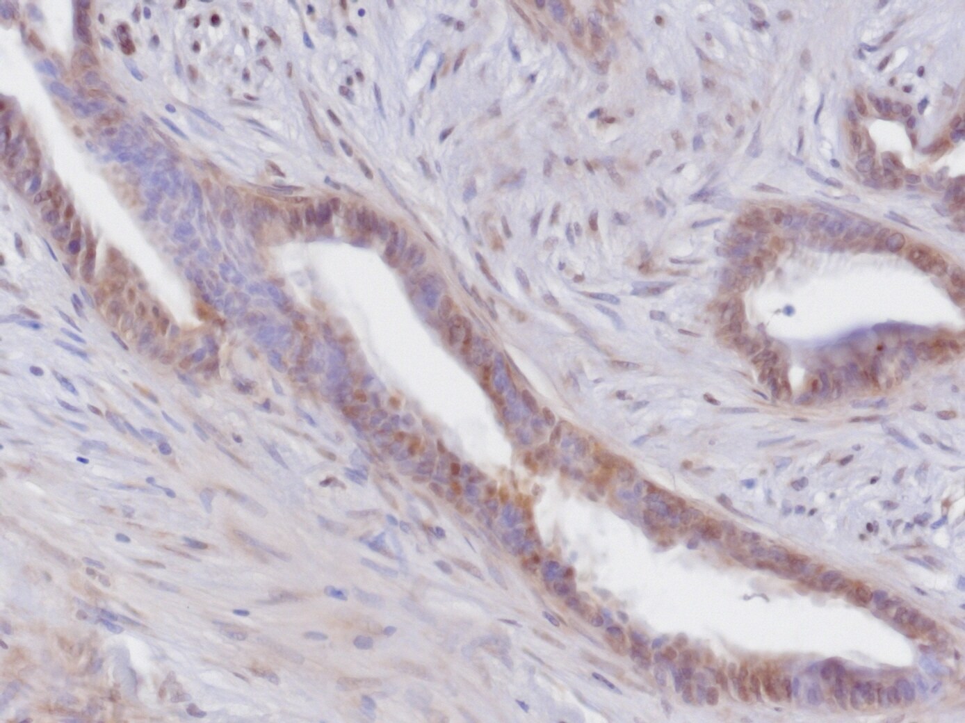
Application: Immunohistochemistry-ParaffinSample Tested: paraffin human pancreatic cancerSpecies: HumanVerified Customer | Posted 07/10/2018Citrate ph:6 based antigen retrieval1:50 primary AB dilution, o/n 4C incubation, dilution factor can be increasedABComplex-DAB developing

-
Application: ImmunohistochemistrySample Tested: Paraffined tumor tissueSpecies: HumanVerified Customer | Posted 06/18/2018Citrate ph:6 based antigen retrieval 1:50 primary AB dilution, o/n 4C incubation, dilution factor can be increased ABComplex-DAB developing
There are no reviews that match your criteria.
Protocols
Find general support by application which include: protocols, troubleshooting, illustrated assays, videos and webinars.
- Cellular Response to Hypoxia Protocols
- R&D Systems Quality Control Western Blot Protocol
- Troubleshooting Guide: Western Blot Figures
- Western Blot Conditions
- Western Blot Protocol
- Western Blot Protocol for Cell Lysates
- Western Blot Troubleshooting
- Western Blot Troubleshooting Guide
- View all Protocols, Troubleshooting, Illustrated assays and Webinars


