
Key Product Details
Validated by
Knockout/Knockdown, Biological Validation
Species Reactivity
Validated:
Human, Mouse
Cited:
Human, Mouse, Rat, Insect - Drosophila melanogaster, Transgenic Mouse
Applications
Validated:
Knockout Validated, Western Blot, Simple Western, Immunoprecipitation
Cited:
Immunohistochemistry, Western Blot, Immunocytochemistry, Proximity Extension Assay
Label
Unconjugated
Antibody Source
Polyclonal Goat IgG
Loading...
Product Specifications
Immunogen
E. coli-derived recombinant human Caspase-3
Met1-His277
Accession # AAA65015
Met1-His277
Accession # AAA65015
Specificity
Detects human Caspase-3.
Clonality
Polyclonal
Host
Goat
Isotype
IgG
Scientific Data Images for Caspase-3 Antibody
Detection of Human and Mouse Caspase‑3 by Western Blot.
Western blot shows lysates of Jurkat human acute T cell leukemia cell line and DA3 mouse myeloma cell line untreated (-) or treated (+) with 1 µg/mL Staurosporine (STS) for 12 hours. PVDF Membrane was probed with 0.5 µg/mL of Goat Anti-Human Caspase-3 Antigen Affinity-purified Polyclonal Antibody (Catalog # AF-605-NA) followed by HRP-conjugated Anti-Goat IgG Secondary Antibody (Catalog # HAF017). Specific bands were detected for Caspase-3 precursor at approximately 34 kDa (as indicated) and Caspase-3 (cleaved) at approximately 18 kDa (as indicated). This experiment was conducted under reducing conditions and using Immunoblot Buffer Group 2.
Immunoprecipitation of Human Caspase‑3.
Jurkat human acute T cell leukemia cell line was treated with indicated concentrations of Staurosporine for 0 or 4 hours. Caspase‑3 was immunoprecipitated from lysates of 1‑2 x 10⁶cells following incubation with 1 or 4 µg Goat Anti-Human Caspase‑3 Antigen Affinity-purified Polyclonal Antibody (Catalog # AF-605-NA) overnight at 4 ºC. Caspase‑3-antibody complexes were absorbed using Protein G expressing Staph cells (Sigma). Immunoprecipitated Caspase‑3 was detected by Western blot using 0.5 µg/mL Goat Anti-Human Caspase‑3 Antigen Affinity-purified Polyclonal Antibody (Catalog # AF‑605‑NA). View our recommended buffer recipes for immunoprecipitation.
Detection of Human Caspase‑3 by Simple WesternTM.
Simple Western lane view shows lysates of HeLa human cervical epithelial carcinoma cell line, HepG2 human hepatocellular carcinoma cell line, and Jurkat human acute T cell leukemia cell line, loaded at 0.2 mg/mL. A specific band was detected for Caspase-3 at approximately 40 kDa (as indicated) using 5 µg/mL of Goat Anti-Human/Mouse Caspase-3 Antigen Affinity-purified Polyclonal Antibody (Catalog # AF-605-NA) 1:50 dilution followed by HRP-conjugated Anti-Goat IgG Secondary Antibody (Catalog # HAF017). This experiment was conducted under reducing conditions and using the 12-230 kDa separation system.
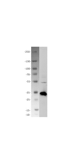
Western Blot Shows Human Caspase‑3 Specificity by Using Knockout Cell Line.
Western blot shows lysates of HeLa human cervical epithelial carcinoma parental cell line and Caspase-3 knockout HeLa cell line (KO). PVDF membrane was probed with 0.2 µg/mL of Goat Anti-Human/Mouse Caspase-3 Antigen Affinity-purified Polyclonal Antibody (Catalog # AF-605-NA) followed by HRP-conjugated Anti-Goat IgG Secondary Antibody (Catalog # HAF017). A specific band was detected for Caspase-3 at approximately 32 kDa (as indicated) in the parental HeLa cell line, but is not detectable in knockout HeLa cell line. GAPDH (Catalog # AF5718) is shown as a loading control. This experiment was conducted under reducing conditions and using Immunoblot Buffer Group 1.Applications for Caspase-3 Antibody
Application
Recommended Usage
Immunoprecipitation
1 µg/106 cells
Sample: Jurkat human acute T cell leukemia cell line treated with Staurosporine, see our available Western blot detection antibodies
Sample: Jurkat human acute T cell leukemia cell line treated with Staurosporine, see our available Western blot detection antibodies
Knockout Validated
Caspase‑3
is specifically detected in HeLa human cervical epithelial carcinoma parental cell line but is not detectable in
Caspase‑3 knockout HeLa cell line.
Simple Western
5 µg/mL
Sample: HeLa human cervical epithelial carcinoma cell line, HepG2 human hepatocellular carcinoma cell line, and Jurkat human acute T cell leukemia cell line
Sample: HeLa human cervical epithelial carcinoma cell line, HepG2 human hepatocellular carcinoma cell line, and Jurkat human acute T cell leukemia cell line
Western Blot
0.5 µg/mL
Sample: Jurkat human acute T cell leukemia cell line and DA3 mouse myeloma cell line treated with Staurosporine
Sample: Jurkat human acute T cell leukemia cell line and DA3 mouse myeloma cell line treated with Staurosporine
Reviewed Applications
Read 1 review rated 4 using AF-605-NA in the following applications:
Formulation, Preparation, and Storage
Purification
Antigen Affinity-purified
Reconstitution
Reconstitute at 0.2 mg/mL in sterile PBS. For liquid material, refer to CoA for concentration.
Loading...
Formulation
Lyophilized from a 0.2 μm filtered solution in PBS with Trehalose. *Small pack size (SP) is supplied either lyophilized or as a 0.2 µm filtered solution in PBS.
Shipping
Lyophilized product is shipped at ambient temperature. Liquid small pack size (-SP) is shipped with polar packs. Upon receipt, store immediately at the temperature recommended below.
Stability & Storage
Use a manual defrost freezer and avoid repeated freeze-thaw cycles.
- 12 months from date of receipt, -20 to -70 °C as supplied.
- 1 month, 2 to 8 °C under sterile conditions after reconstitution.
- 6 months, -20 to -70 °C under sterile conditions after reconstitution.
Calculators
Background: Caspase-3
References
-
Chowdhury, I. et al. (2008) Comp. Biochem. Physiol. B 151:10.
-
Boatright, K.M. & G.S. Salvesen (2003) Curr. Opin. Cell Biol. 15:725.
-
Launay, S. et al. (2005) Oncogene 24:5137.
-
Walsh, J.G. et al. (2008) Proc. Natl. Scad. Sci. USA 105:12815.
-
Nicholson, D.W. et al. (1995) Nature 376:37.
-
Tewari, M. et al. (1995) Cell 81:801.
-
Fernandes-Alnemri, T. et al. (1994) J. Biol. Chem. 269:30761.
-
Milisav, I. et al. (2009) Apoptosis 14:1070.
-
Han, Z. et al. (1997) J. Biol. Chem. 272:13432.
-
Rossig, L. et al. (1999) J. Biol. Chem. 274:6823.
-
Rank, K.B. et al. (2001) Protein Expr. Purif. 22:258.
-
Atkinson, E.A. et al. (1998) J. Biol. Chem. 273:21261.
- Cohen, G.M. (1997) Biochem. J. 326:1.
Alternate Names
Apopain, CASP3, Caspase3, CPP32, LICE-1, YAMA
Gene Symbol
CASP3
UniProt
Additional Caspase-3 Products
Product Documents for Caspase-3 Antibody
Certificate of Analysis
To download a Certificate of Analysis, please enter a lot or batch number in the search box below.
Note: Certificate of Analysis not available for kit components.
Product Specific Notices for Caspase-3 Antibody
For research use only
Related Research Areas
Citations for Caspase-3 Antibody
 Powered by Bioz
Customer Reviews for Caspase-3 Antibody (1)
4 out of 5
1 Customer Rating
Have you used Caspase-3 Antibody?
Submit a review and receive an Amazon gift card!
$25/€18/£15/$25CAN/¥2500 Yen for a review with an image
$10/€7/£6/$10CAN/¥1110 Yen for a review without an image
Submit a review
Customer Images
Showing
1
-
1 of
1 review
Showing All
Filter By:
-
Application: Western BlotSample Tested: A549 human lung carcinoma cell lineSpecies: HumanVerified Customer | Posted 04/08/2022

There are no reviews that match your criteria.
Protocols
Find general support by application which include: protocols, troubleshooting, illustrated assays, videos and webinars.
- Cellular Response to Hypoxia Protocols
- Immunoprecipitation Protocol
- R&D Systems Quality Control Western Blot Protocol
- Troubleshooting Guide: Western Blot Figures
- Western Blot Conditions
- Western Blot Protocol
- Western Blot Protocol for Cell Lysates
- Western Blot Troubleshooting
- Western Blot Troubleshooting Guide
- View all Protocols, Troubleshooting, Illustrated assays and Webinars
Loading...
Associated Pathways

