cIAP-2 (also known as MIHC and HIAP-1) is a member of the inhibitor of apoptosis (IAP) family of proteins that inhibit the proteolytic activity of mature caspases. Human cIAP-2 is a 604 amino acid protein that contains 3 BIR (baculovirus inhibitor of apoptosis) domains, a RING finger domain, and a caspase recruitment domain (CARD). cIAP-2 inhibits caspases through the direct interaction of its BIR domain with the active caspase. Caspase activity may be restored through interactions with the Reaper like motif on mitochondrial proteins such as SMAC/Diablo or HtrA2/Omi. cIAP-2 is reported to be cleaved by HtrA2/Omi.

Key Product Details
Species Reactivity
Validated:
Human, Mouse
Cited:
Human, Mouse
Applications
Validated:
Immunohistochemistry, Western Blot, Simple Western
Cited:
Immunohistochemistry, Western Blot
Label
Unconjugated
Antibody Source
Monoclonal Mouse IgG1 Clone # 315304
Loading...
Product Specifications
Immunogen
E. coli-derived recombinant human cIAP‑2/HIAP‑1
Asn2-Ser604
Accession # Q13489
Asn2-Ser604
Accession # Q13489
Specificity
Detects human and mouse cIAP‑2/HIAP‑1 in Western blots. Does not cross-react with HEK293 cells transfected with cIAP-1.
Clonality
Monoclonal
Host
Mouse
Isotype
IgG1
Scientific Data Images for cIAP-2/HIAP-1 Antibody (315304)
cIAP‑2/HIAP‑1 in Human Colon Cancer Tissue.
cIAP-2/HIAP-1 was detected in immersion fixed paraffin-embedded sections of human colon cancer tissue using Mouse Anti-Human/Mouse cIAP-2/ HIAP-1 Monoclonal Antibody (Catalog # MAB817) at 25 µg/mL overnight at 4 °C. Tissue was stained using the Anti-Mouse HRP-DAB Cell & Tissue Staining Kit (brown; Catalog # CTS002) and counter-stained with hematoxylin (blue). View our protocol for Chromogenic IHC Staining of Paraffin-embedded Tissue Sections. This application has not been tested in mouse samples.
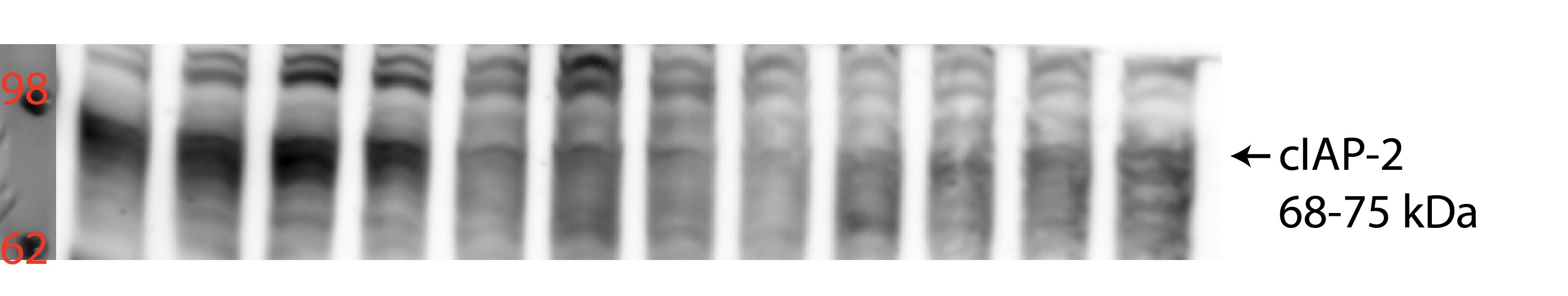
Detection of Human and Mouse cIAP‑2/HIAP‑1 by Western Blot.
Western blot shows lysates of Raji human Burkitt's lymphoma cell line, Daudi human Burkitt's lymphoma cell line, and A20 mouse B cell lymphoma cell line. PVDF membrane was probed with 0.5 µg/mL of Mouse Anti-Human/Mouse cIAP-2/HIAP-1 Monoclonal Antibody (Catalog # MAB817) followed by HRP-conjugated Anti-Mouse IgG Secondary Antibody (Catalog # HAF007). A specific band was detected for cIAP-2/HIAP-1 at approximately 68 kDa (as indicated). This experiment was conducted under reducing conditions and using Immunoblot Buffer Group 5.
cIAP‑2/HIAP‑1 in Human Colon.
cIAP-2/HIAP-1 was detected in immersion fixed paraffin-embedded sections of human colon array using Mouse Anti-Human/Mouse cIAP-2/HIAP-1 Monoclonal Antibody (Catalog # MAB817) at 25 µg/mL overnight at 4 °C. Tissue was stained using the Anti-Mouse HRP-DAB Cell & Tissue Staining Kit (brown; Catalog # CTS002) and counterstained with hematoxylin (blue). Lower panel shows a lack of labeling if primary antibodies are omitted and tissue is stained only with secondary antibody followed by incubation with detection reagents. View our protocol for Chromogenic IHC Staining of Paraffin-embedded Tissue Sections. This application has not been tested in mouse samples.
Detection of Human cIAP‑2/HIAP‑1 by Simple WesternTM.
Simple Western lane view shows lysates of HEK293 human embryonic kidney cell line transfected with either cIAP-1 or cIAP-2 and Raji human Burkitt's lymphoma cell line, loaded at 0.2 mg/mL. A specific band was detected for cIAP‑2/HIAP‑1 at approximately 74 kDa (as indicated) using 10 µg/mL of Mouse Anti-Human/Mouse cIAP‑2/HIAP‑1 Monoclonal Antibody (Catalog # MAB817). This experiment was conducted under reducing conditions and using the 12-230 kDa separation system.Applications for cIAP-2/HIAP-1 Antibody (315304)
Application
Recommended Usage
Immunohistochemistry
8-25 µg/mL
Sample: Immersion fixed paraffin-embedded sections of normal human colon and human colon cancer tissue
Sample: Immersion fixed paraffin-embedded sections of normal human colon and human colon cancer tissue
Simple Western
10 µg/mL
Sample: HEK293 human embryonic kidney cell line transfected with cIAP-2 and Raji human Burkitt's lymphoma cell line
Sample: HEK293 human embryonic kidney cell line transfected with cIAP-2 and Raji human Burkitt's lymphoma cell line
Western Blot
0.5 µg/mL
Sample: Raji human Burkitt's lymphoma cell line, Daudi human Burkitt's lymphoma cell line, and A20 mouse B cell lymphoma cell line
Sample: Raji human Burkitt's lymphoma cell line, Daudi human Burkitt's lymphoma cell line, and A20 mouse B cell lymphoma cell line
Reviewed Applications
Read 1 review rated 4 using MAB817 in the following applications:
Formulation, Preparation, and Storage
Purification
Protein A or G purified from hybridoma culture supernatant
Reconstitution
Reconstitute at 0.5 mg/mL in sterile PBS. For liquid material, refer to CoA for concentration.
Loading...
Formulation
Lyophilized from a 0.2 μm filtered solution in PBS with Trehalose. *Small pack size (SP) is supplied either lyophilized or as a 0.2 µm filtered solution in PBS.
Shipping
Lyophilized product is shipped at ambient temperature. Liquid small pack size (-SP) is shipped with polar packs. Upon receipt, store immediately at the temperature recommended below.
Stability & Storage
Use a manual defrost freezer and avoid repeated freeze-thaw cycles.
- 12 months from date of receipt, -20 to -70 °C as supplied.
- 1 month, 2 to 8 °C under sterile conditions after reconstitution.
- 6 months, -20 to -70 °C under sterile conditions after reconstitution.
Calculators
Background: cIAP-2/HIAP-1
References
- Roy, N. et al. (1997) EMBO J. 23:6914.
- Deveraux, Q. et al. (1997) Nature 388:300.
- Deveraux, Q. and J. Reed (1999) Genes & Develop. 13:239.
- Srinivasula, S.M. et al. (2003) J. Biol. Chem. 278:31469.
- Yang, Q-H. et al. (2003) Genes Dev. 17:1487.
Alternate Names
BIRC3, cIAP2, HIAP-1, MIHC
Entrez Gene IDs
330 (Human)
Gene Symbol
BIRC3
UniProt
Additional cIAP-2/HIAP-1 Products
Product Documents for cIAP-2/HIAP-1 Antibody (315304)
Certificate of Analysis
To download a Certificate of Analysis, please enter a lot or batch number in the search box below.
Note: Certificate of Analysis not available for kit components.
Product Specific Notices for cIAP-2/HIAP-1 Antibody (315304)
For research use only
Related Research Areas
Citations for cIAP-2/HIAP-1 Antibody (315304)
 Powered by Bioz
Customer Reviews for cIAP-2/HIAP-1 Antibody (315304) (1)
4 out of 5
1 Customer Rating
Have you used cIAP-2/HIAP-1 Antibody (315304)?
Submit a review and receive an Amazon gift card!
$25/€18/£15/$25CAN/¥2500 Yen for a review with an image
$10/€7/£6/$10CAN/¥1110 Yen for a review without an image
Submit a review
Customer Images
Showing
1
-
1 of
1 review
Showing All
Filter By:
-
Application: Western BlotSample Tested: Colon tissue and Tissue LysatesSpecies: MouseVerified Customer | Posted 09/23/2021

There are no reviews that match your criteria.
Protocols
Find general support by application which include: protocols, troubleshooting, illustrated assays, videos and webinars.
- Antigen Retrieval Protocol (PIER)
- Antigen Retrieval for Frozen Sections Protocol
- Appropriate Fixation of IHC/ICC Samples
- Cellular Response to Hypoxia Protocols
- Chromogenic IHC Staining of Formalin-Fixed Paraffin-Embedded (FFPE) Tissue Protocol
- Chromogenic Immunohistochemistry Staining of Frozen Tissue
- ClariTSA™ Fluorophore Kits
- Detection & Visualization of Antibody Binding
- Fluorescent IHC Staining of Frozen Tissue Protocol
- Graphic Protocol for Heat-induced Epitope Retrieval
- Graphic Protocol for the Preparation and Fluorescent IHC Staining of Frozen Tissue Sections
- Graphic Protocol for the Preparation and Fluorescent IHC Staining of Paraffin-embedded Tissue Sections
- Graphic Protocol for the Preparation of Gelatin-coated Slides for Histological Tissue Sections
- IHC Sample Preparation (Frozen sections vs Paraffin)
- Immunofluorescent IHC Staining of Formalin-Fixed Paraffin-Embedded (FFPE) Tissue Protocol
- Immunohistochemistry (IHC) and Immunocytochemistry (ICC) Protocols
- Immunohistochemistry Frozen Troubleshooting
- Immunohistochemistry Paraffin Troubleshooting
- Preparing Samples for IHC/ICC Experiments
- Preventing Non-Specific Staining (Non-Specific Binding)
- Primary Antibody Selection & Optimization
- Protocol for Heat-Induced Epitope Retrieval (HIER)
- Protocol for Making a 4% Formaldehyde Solution in PBS
- Protocol for VisUCyte™ HRP Polymer Detection Reagent
- Protocol for the Preparation & Fixation of Cells on Coverslips
- Protocol for the Preparation and Chromogenic IHC Staining of Frozen Tissue Sections
- Protocol for the Preparation and Chromogenic IHC Staining of Frozen Tissue Sections - Graphic
- Protocol for the Preparation and Chromogenic IHC Staining of Paraffin-embedded Tissue Sections
- Protocol for the Preparation and Chromogenic IHC Staining of Paraffin-embedded Tissue Sections - Graphic
- Protocol for the Preparation and Fluorescent IHC Staining of Frozen Tissue Sections
- Protocol for the Preparation and Fluorescent IHC Staining of Paraffin-embedded Tissue Sections
- Protocol for the Preparation of Gelatin-coated Slides for Histological Tissue Sections
- R&D Systems Quality Control Western Blot Protocol
- TUNEL and Active Caspase-3 Detection by IHC/ICC Protocol
- The Importance of IHC/ICC Controls
- Troubleshooting Guide: Immunohistochemistry
- Troubleshooting Guide: Western Blot Figures
- Western Blot Conditions
- Western Blot Protocol
- Western Blot Protocol for Cell Lysates
- Western Blot Troubleshooting
- Western Blot Troubleshooting Guide
- View all Protocols, Troubleshooting, Illustrated assays and Webinars
Loading...
Associated Pathways

