
Key Product Details
Validated by
Biological Validation
Species Reactivity
Validated:
Human, Mouse
Cited:
Human, Mouse, Chicken, Drosophila, Nematode - Caenorhabditis elegans, Transgenic Mouse
Applications
Validated:
Western Blot, Immunocytochemistry, Chromatin Immunoprecipitation (ChIP)
Cited:
Immunohistochemistry, Immunohistochemistry-Paraffin, Western Blot, Immunocytochemistry, Immunoprecipitation, Chromatin Immunoprecipitation (ChIP), Bioassay, CUT&Tag
Label
Unconjugated
Antibody Source
Polyclonal Goat IgG
Loading...
Product Specifications
Immunogen
E. coli-derived recombinant human GLI-3
Met1-Glu479
Accession # P10071
Met1-Glu479
Accession # P10071
Specificity
Detects human and mouse GLI-3 in direct ELISAs and Western blots. In direct ELISAs and Western blots, less than 1% cross-reactivity with recombinant human (rh) GLI-1 and rhGLI-2 is observed.
Clonality
Polyclonal
Host
Goat
Isotype
IgG
Scientific Data Images for GLI-3 Antibody
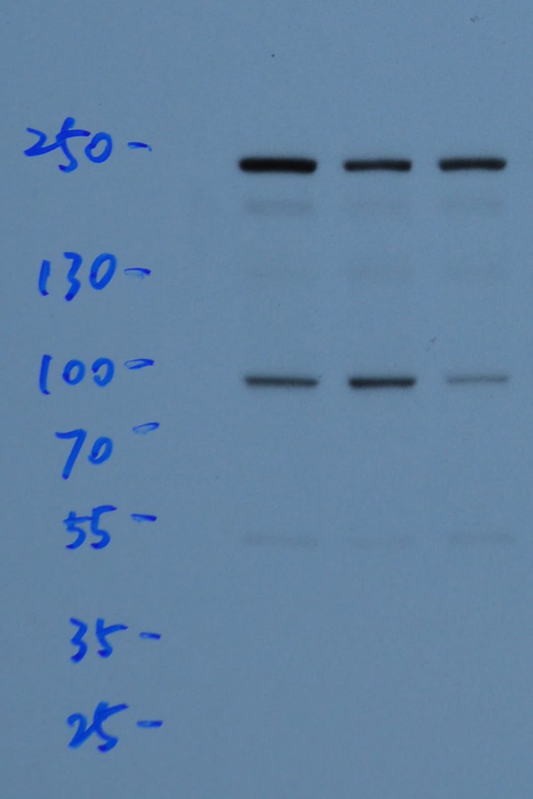
Detection of Mouse GLI‑3 by Western Blot.
Western blot shows lysates of mouse embryo tissue. PVDF membrane was probed with 1 µg/mL of Goat Anti-Human/Mouse GLI-3 Antigen Affinity-purified Polyclonal Antibody (Catalog # AF3690) followed by HRP-conjugated Anti-Goat IgG Secondary Antibody (Catalog # HAF019). A specific band was detected for GLI-3 at approximately 85 kDa (as indicated). This experiment was conducted under reducing conditions and using Immunoblot Buffer Group 8.
Detection of GLI‑3-regulated Genes by Chromatin Immunoprecipitation.
Jurkat human acute T cell leukemia cell line treated with 50 ng/mL PMA and 200 ng/mL calcium ionomycin for 30 minutes was fixed using formaldehyde, resuspended in lysis buffer, and sonicated to shear chromatin. GLI-3/DNA complexes were immunoprecipitated using 5 µg Goat Anti-Human/Mouse GLI-3 Antigen Affinity-purified Polyclonal Antibody (Catalog # AF3690) or control antibody (Catalog # AB-108-C) for 15 minutes in an ultrasonic bath, followed by Biotinylated Anti-Goat IgG Secondary Antibody (Catalog # BAF109). Immunocomplexes were captured using 50 µL of MagCellect Streptavidin Ferrofluid (Catalog # MAG999) and DNA was purified using chelating resin solution. Thegli-1promoter was detected by standard PCR.
GLI‑3 in HeLa Human Cell Line.
GLI-3 was detected in immersion fixed HeLa human cervical epithelial carcinoma cell line using Human/Mouse GLI-3 Antigen Affinity-purified Polyclonal Antibody (Catalog # AF3690) at 10 µg/mL for 3 hours at room temperature. Cells were stained using the NorthernLights™ 557-conjugated Anti-Goat IgG Secondary Antibody (yellow; Catalog # NL001) and counterstained with DAPI (blue). View our protocol for Fluorescent ICC Staining of Cells on Coverslips.
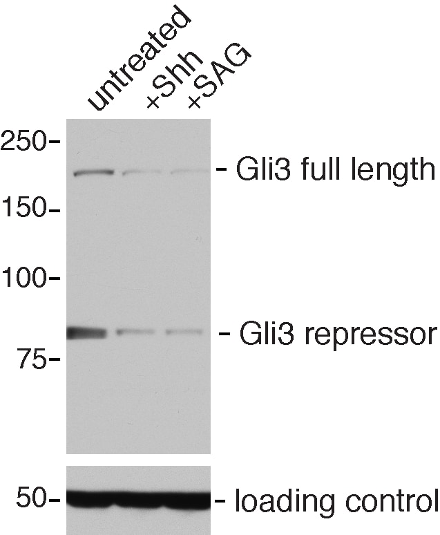
Detection of Mouse GLI-3 by Western Blot
Conditional deletion of GATA6 does not significantly alter Gli3 processing in the hindlimb bud.(A) Western blot analysis of Gli3FL and Gli3R steady state levels in anterior (lanes 1 and 3) or posterior (lanes 2 and 4) halves of E11.5 hindlimb buds isolated from either control (lanes 1 and 2) or Prx1-Cre; GATA6fl/fl (lanes 3 and 4) mouse embryos. alpha –Tubulin is used as a loading control. (B) Quantification of Gli3FL/Gli3R ratios (by densitometry of Western Blots) in anterior or posterior halves of E11.5 hindlimb buds isolated from either littermate controls (lanes 1 and 2) or Prx1-Cre; GATA6fl/fl (lanes 3 and 4) animals, 12 embryos of each genotype were analyzed. Error bar indicates standard deviation, n = 12. Image collected and cropped by CiteAb from the following publication (https://pubmed.ncbi.nlm.nih.gov/24415953), licensed under a CC-BY license. Not internally tested by R&D Systems.
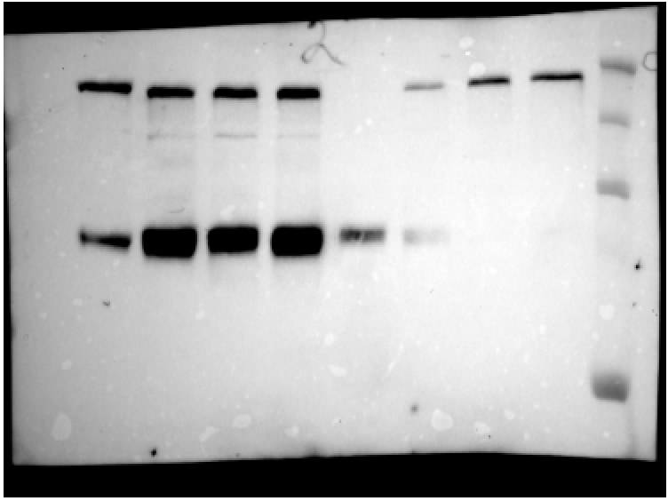
Detection of Mouse GLI-3 by Western Blot
SufuT396I does not stabilize Gli3FL protein and reduces the processing of Gli3FL.(A) Western blotting of lysates prepared from SufuR146X/R146X at E9.5 and SufuT396I/T396I, SufuT396I/+, and wild-type embryos at E10.5 with anti-Gli3, anti-Sufu, and anti-actin antibodies. Each image presented in the Fig. is a representative of independent triplicated experiments. The full gel images are shown in S6A Fig. (B, C) Relative expression of Gli3FL (B) and Gli3REP (B and C). Western blotting was performed two times using lysates prepared from five wild-type and five SufuT396I/T396I embryos at E10.5 (S2A Fig.). Expression levels were quantified from the band intensity shown in S2A Fig. as relative values of the Gli3FL/actin and Gli3REP/actin expression ratios (B) and the direct ratio of Gli3REP/Gli3FL (C). (**) p < 0.01, two-tailed Student’s t-test. Error bars indicate the standard deviations. (D) Western blotting of cell lysates from wild-type and SufuT396I/T396I MEFs with indicated antibodies. The SufuT396I/T396I MEFs were electroporated with 10.0, 1.0, or 0.1 μg of the HA–Sufu construct (lane 3, 4, and 5, respectively), or 10 μg of the HA–SufuT396I construct (lane 6). The mobilities on SDS-PAGE of the wild-type Sufu (lane 3–5) and SufuT396I (lane 6) are identical. The complete gel images are shown in S6B Fig. This image is representative of two independent experiments. (E) Western blotting of cell lysates from Sufu−/− cells with indicated antibodies. The Sufu−/− cells were electroporated with a mixture of the Flag–Gli3 construct (6 μg) and the HA–Sufu construct (4.00, 1.33, 0.44, or 0.15 μg) for the wild-type Sufu cotransfection (lane 2 to 5, respectively), or a mixture of the Flag–Gli3 construct (6 μg) and the HA–SufuT396I construct (4 μg) for mutant Sufu cotransfection (lane 6). The complete gel images are shown in S6C Fig. This image is representative of two independent experiments. (F) Western blotting of immunoprecipitates or lysates from 293T cells transfected with exp
Detection of Mouse GLI-3 by Immunocytochemistry/ Immunofluorescence
Analysis of Ptch receptor-dependent inhibition of Hh signaling. (A,C,E) Immunocytochemistry detection of Gli3 (A), Gli1 (C) and Gli2 (E) in the wild-type, Ptch1−/−, and Ptch2−/− MEFs. Scale bar 50 μm. (B,D,F) Quantification of the intensity of the nuclear GLI3 (B), GLI1 (D), and GLI2 (F) in wild-type, Ptch1−/−, and Ptch2−/− MEFs. **p ≤ 0.005, ***p ≤ 0.0005, ****p < 0.0001. (G) Western blot showing nuclear (N) and cellular (C) expression of Gli2 (FL = full length and RF = repressor form) in wild-type, Ptch1−/− and Ptch2−/− MEFs. Image collected and cropped by CiteAb from the following open publication (https://pubmed.ncbi.nlm.nih.gov/35574464), licensed under a CC-BY license. Not internally tested by R&D Systems.
Detection of Mouse GLI-3 by Immunocytochemistry/ Immunofluorescence
Analysis of Ptch receptor-dependent inhibition of Hh signaling. (A,C,E) Immunocytochemistry detection of Gli3 (A), Gli1 (C) and Gli2 (E) in the wild-type, Ptch1−/−, and Ptch2−/− MEFs. Scale bar 50 μm. (B,D,F) Quantification of the intensity of the nuclear GLI3 (B), GLI1 (D), and GLI2 (F) in wild-type, Ptch1−/−, and Ptch2−/− MEFs. **p ≤ 0.005, ***p ≤ 0.0005, ****p < 0.0001. (G) Western blot showing nuclear (N) and cellular (C) expression of Gli2 (FL = full length and RF = repressor form) in wild-type, Ptch1−/− and Ptch2−/− MEFs. Image collected and cropped by CiteAb from the following open publication (https://pubmed.ncbi.nlm.nih.gov/35574464), licensed under a CC-BY license. Not internally tested by R&D Systems.Applications for GLI-3 Antibody
Application
Recommended Usage
Chromatin Immunoprecipitation (ChIP)
5 µg/106 cells
Sample: PMA and ionomycin treated Jurkat human acute T cell leukemia cell line chromatin, GLI-1 promoter detected by standard PCR
Sample: PMA and ionomycin treated Jurkat human acute T cell leukemia cell line chromatin, GLI-1 promoter detected by standard PCR
Immunocytochemistry
5-15 µg/mL
Sample: Immersion fixed HeLa human cervical epithelial carcinoma cell line
Sample: Immersion fixed HeLa human cervical epithelial carcinoma cell line
Western Blot
1 µg/mL
Sample: Mouse embryo tissue
Sample: Mouse embryo tissue
Reviewed Applications
Read 5 reviews rated 4.8 using AF3690 in the following applications:
Formulation, Preparation, and Storage
Purification
Antigen Affinity-purified
Reconstitution
Reconstitute at 0.2 mg/mL in sterile PBS. For liquid material, refer to CoA for concentration.
Loading...
Formulation
Lyophilized from a 0.2 μm filtered solution in PBS with Trehalose. *Small pack size (SP) is supplied either lyophilized or as a 0.2 µm filtered solution in PBS.
Shipping
Lyophilized product is shipped at ambient temperature. Liquid small pack size (-SP) is shipped with polar packs. Upon receipt, store immediately at the temperature recommended below.
Stability & Storage
Use a manual defrost freezer and avoid repeated freeze-thaw cycles.
- 12 months from date of receipt, -20 to -70 °C as supplied.
- 1 month, 2 to 8 °C under sterile conditions after reconstitution.
- 6 months, -20 to -70 °C under sterile conditions after reconstitution.
Calculators
Background: GLI-3
Long Name
GLI-Kruppel family member GLI3
Alternate Names
ACLS, ADD, Bph, GCPS, GLI3, PAPA1, PAPB, Pdn, PHS, PPDIV, Xt
Gene Symbol
GLI3
UniProt
Additional GLI-3 Products
Product Documents for GLI-3 Antibody
Certificate of Analysis
To download a Certificate of Analysis, please enter a lot or batch number in the search box below.
Note: Certificate of Analysis not available for kit components.
Product Specific Notices for GLI-3 Antibody
For research use only
Related Research Areas
Citations for GLI-3 Antibody
 Powered by Bioz
Customer Reviews for GLI-3 Antibody (5)
4.8 out of 5
5 Customer Ratings
Have you used GLI-3 Antibody?
Submit a review and receive an Amazon gift card!
$25/€18/£15/$25CAN/¥2500 Yen for a review with an image
$10/€7/£6/$10CAN/¥1110 Yen for a review without an image
Submit a review
Customer Images
Showing
1
-
5 of
5 reviews
Showing All
Filter By:
-
Application: Western BlotSample Tested: Cell LysatesSpecies: MouseVerified Customer | Posted 05/22/2019

-
Application: Western BlotSample Tested: NIH-3T3 cellsSpecies: MouseVerified Customer | Posted 11/13/2015Gli3 Western Blot
-
Application: Western BlotSample Tested: Mouse embryo cell lysateSpecies: MouseVerified Customer | Posted 10/26/2015Specificity: Specific<br />Sensitivity: Sensitive<br />Buffer: TBST<br />Dilution: 1/1000
-
Application: Western BlotSample Tested: See PMID 22173325Species: MouseVerified Customer | Posted 01/08/2015
-
Application: Western BlotSample Tested: See PMID 22547067Species: MouseVerified Customer | Posted 01/08/2015
There are no reviews that match your criteria.
Protocols
Find general support by application which include: protocols, troubleshooting, illustrated assays, videos and webinars.
- Appropriate Fixation of IHC/ICC Samples
- Cellular Response to Hypoxia Protocols
- ChIP Protocol Video
- Chromatin Immunoprecipitation (ChIP) Protocol
- Chromatin Immunoprecipitation Protocol
- ClariTSA™ Fluorophore Kits
- Detection & Visualization of Antibody Binding
- ICC Cell Smear Protocol for Suspension Cells
- ICC Immunocytochemistry Protocol Videos
- ICC for Adherent Cells
- Immunocytochemistry (ICC) Protocol
- Immunocytochemistry Troubleshooting
- Immunofluorescence of Organoids Embedded in Cultrex Basement Membrane Extract
- Immunohistochemistry (IHC) and Immunocytochemistry (ICC) Protocols
- Preparing Samples for IHC/ICC Experiments
- Preventing Non-Specific Staining (Non-Specific Binding)
- Primary Antibody Selection & Optimization
- Protocol for VisUCyte™ HRP Polymer Detection Reagent
- Protocol for the Fluorescent ICC Staining of Cell Smears - Graphic
- Protocol for the Fluorescent ICC Staining of Cultured Cells on Coverslips - Graphic
- Protocol for the Preparation and Fluorescent ICC Staining of Cells on Coverslips
- Protocol for the Preparation and Fluorescent ICC Staining of Non-adherent Cells
- Protocol for the Preparation and Fluorescent ICC Staining of Stem Cells on Coverslips
- Protocol for the Preparation of a Cell Smear for Non-adherent Cell ICC - Graphic
- R&D Systems Quality Control Western Blot Protocol
- TUNEL and Active Caspase-3 Detection by IHC/ICC Protocol
- The Importance of IHC/ICC Controls
- Troubleshooting Guide: Western Blot Figures
- Western Blot Conditions
- Western Blot Protocol
- Western Blot Protocol for Cell Lysates
- Western Blot Troubleshooting
- Western Blot Troubleshooting Guide
- View all Protocols, Troubleshooting, Illustrated assays and Webinars
Loading...
Associated Pathways
