
Key Product Details
Species Reactivity
Validated:
Human, Mouse, Rat
Cited:
Human, Mouse, Rat
Applications
Validated:
Western Blot, Flow Cytometry, Immunocytochemistry, Simple Western, CyTOF-ready
Cited:
Immunohistochemistry, Immunohistochemistry-Frozen, Western Blot, Flow Cytometry, Immunocytochemistry, Immunoprecipitation
Label
Unconjugated
Antibody Source
Polyclonal Goat IgG
Loading...
Product Specifications
Immunogen
Mouse myeloma cell line NS0-derived recombinant mouse IGSF8/CD316
Ala25-Thr577
Accession # NP_536344
Ala25-Thr577
Accession # NP_536344
Specificity
Detects mouse IGSF8/CD316 in direct ELISAs. Detects human, mouse, and rat IGSF8/CD316 in Western blots. In direct ELISAs, approximately 15% cross-reactivity with recombinant human IGSF8 is observed and less than 1% cross-reactivity with recombinant mouse IGSF4 is observed.
Clonality
Polyclonal
Host
Goat
Isotype
IgG
Scientific Data Images for IGSF8/CD316 Antibody
Detection of Human and Mouse IGSF8/CD316 by Western Blot.
Western blot shows lysates of SH-SY5Y human neuroblastoma cell line, DU145 human prostate carcinoma cell line, and bEnd.3 mouse endothelioma cell line. PVDF membrane was probed with 1 µg/mL of Goat Anti-Human/Mouse/Rat IGSF8/CD316 Antigen Affinity-purified Polyclonal Antibody (Catalog # AF3117) followed by HRP-conjugated Anti-Goat IgG Secondary Antibody (Catalog # HAF017). Specific bands were detected for IGSF8/CD316 at approximately 70-80 kDa (as indicated). This experiment was conducted under reducing conditions and using Immunoblot Buffer Group 1.
Detection of Human IGSF8/CD316 by Simple WesternTM.
Simple Western lane view shows lysates of Exosome Standards (HEK293) (NBP3-11684) and human cerebellum tissue, loaded at 0.5 mg/ml. A specific band was detected for IGSF8/CD316 at approximately 69 kDa (as indicated) using 20 µg/ml of Goat Anti-Human/Mouse/Rat IGSF8/CD316 Antigen Affinity-purified Polyclonal Antibody (Catalog # AF3117) followed by HRP-conjugated Donkey Anti-Goat Secondary Antibody (Catalog # 042-206). This experiment was conducted under reducing conditions and using the 12-230kDa separation system.
Detection of Rat IGSF8/CD316 by Western Blot.
Western blot shows lysates of rat brain (hippocampus) tissue. PVDF membrane was probed with 1 µg/mL of Goat Anti-Human/Mouse/Rat IGSF8/CD316 Antigen Affinity-purified Polyclonal Antibody (Catalog # AF3117) followed by HRP-conjugated Anti-Goat IgG Secondary Antibody (Catalog # HAF017). A specific band was detected for IGSF8/CD316 at approximately 70 kDa (as indicated). This experiment was conducted under reducing conditions and using Immunoblot Buffer Group 1.
Detection of IGSF8/CD316 in Neuro‑2A Mouse Cell Line by Flow Cytometry.
Neuro-2A mouse neuroblastoma cell line was stained with Goat Anti-Human/Mouse/Rat IGSF8/CD316 Antigen Affinity-purified Polyclonal Antibody (Catalog # AF3117, filled histogram) or isotype control antibody (Catalog # AB-108-C, open histogram), followed by Phycoerythrin-conjugated Anti-Goat IgG Secondary Antibody (Catalog # F0107).
IGSF8/CD316 in Neuro‑2A Mouse Cell Line.
IGSF8/CD316 was detected in immersion fixed Neuro-2A mouse neuroblastoma cell line using Goat Anti-Human/Mouse/Rat IGSF8/CD316 Antigen Affinity-purified Polyclonal Antibody (Catalog # AF3117) at 10 µg/mL for 3 hours at room temperature. Cells were stained using the NorthernLights™ 557-conjugated Anti-Goat IgG Secondary Antibody (red; Catalog # NL001) and counterstained with DAPI (blue). Specific staining was localized to cell surfaces. View our protocol for Fluorescent ICC Staining of Cells on Coverslips.
Detection of Human, Mouse, and Rat IGSF8/CD316 by Simple WesternTM.
Simple Western lane view shows lysates of human cerebellum tissue, human hippocampus tissue, Neuro-2A mouse neuroblastoma cell line, and rat hippocampus tissue, loaded at 0.2 mg/mL. A specific band was detected for IGSF8/CD316 at approximately 71-80 kDa (as indicated) using 20 µg/mL of Goat Anti-Human/Mouse IGSF8/CD316 Antigen Affinity-purified Polyclonal Antibody (Catalog # AF3117) followed by 1:50 dilution of HRP-conjugated Anti-Goat IgG Secondary Antibody (Catalog # HAF109). This experiment was conducted under reducing conditions and using the 12-230 kDa separation system.
Detection of Human IGSF8/CD316 by Western Blot
WGA-HRP identifies a number of EV-specific markers that are present regardless of oncogene status.(A) Matrix depicting samples analyzed during LFQ comparison–Control and Myc cells, as well as Control and Myc EVs. (B) Principle component analysis (PCA) of all four groups analyzed by LFQ. Component 1 (50.4%) and component 2 (15.8%) are graphed. (C) Functional annotation was performed for each gene cluster using DAVID Bioinformatics Resource 6.8 and the highest ranking annotation features for the EV-specific gene cluster are shown. (D) Heatmap of the 50 most upregulated proteins in either RWPE-1 cells or EVs. Proteins are listed in decreasing order of expression with the most highly expressed proteins in EVs on the far left and the most highly expressed proteins in cells on the far right. Averages from all four replicates of each sample type are graphed. Scale indicates intensity, defined as (LFQ Area−Mean LFQ Area)/Standard Deviation. Extracellular proteins with annotated transmembrane domains are bolded and annotated secreted proteins are italicized. (E) Table indicating fold-change of most differentially regulated proteins by LC-MS/MS for RWPE-1 EVs compared to parent cells. (F) Western blot showing the EV-specific marker ITIH4, IGSF8, and MFGE8. Mass spectrometry data is based on two biological and two technical replicates (N=4). Due to limited sample yield, one replicate was performed for the EV western blot. EV, extracellular vesicle; LFQ, label-free quantification.Figure 5—source data 1.Uncropped western blots.Figure 5—source data 2.Mass spectrometry analysis results table.Figure 5—source data 3.List of proteins comparing enriched targets (>2-fold) in Control EVs versus Control cells and Myc EVs versus Myc cells.Uncropped western blots.Mass spectrometry analysis results table.List of proteins comparing enriched targets (>2-fold) in Control EVs versus Control cells and Myc EVs versus Myc cells.Heatmap comparison of biological and technical replicates of RWPE-1 CoApplications for IGSF8/CD316 Antibody
Application
Recommended Usage
CyTOF-ready
Ready to be labeled using established conjugation methods. No BSA or other carrier proteins that could interfere with conjugation.
Flow Cytometry
0.25 µg/106 cells
Sample: Neuro‑2A mouse neuroblastoma cell line
Sample: Neuro‑2A mouse neuroblastoma cell line
Immunocytochemistry
5-15 µg/mL
Sample: Immersion fixed Neuro2A mouse neuroblastoma cell line
Sample: Immersion fixed Neuro2A mouse neuroblastoma cell line
Simple Western
20 µg/mL
Sample: Exosome Standards (HEK293) (Catalog # NBP3-11684), Human cerebellum tissue, Human hippocampus tissue, Neuro‑2A mouse neuroblastoma cell line, and Rat hippocampus tissue
Sample: Exosome Standards (HEK293) (Catalog # NBP3-11684), Human cerebellum tissue, Human hippocampus tissue, Neuro‑2A mouse neuroblastoma cell line, and Rat hippocampus tissue
Western Blot
1 µg/mL
Sample: SH‑SY5Y human neuroblastoma cell line, DU145 human prostate carcinoma cell line, bEnd.3 mouse endothelioma cell line, and Rat brain (hippocampus) tissue
Sample: SH‑SY5Y human neuroblastoma cell line, DU145 human prostate carcinoma cell line, bEnd.3 mouse endothelioma cell line, and Rat brain (hippocampus) tissue
Reviewed Applications
Read 1 review rated 5 using AF3117 in the following applications:
Flow Cytometry Panel Builder
Bio-Techne Knows Flow Cytometry
Save time and reduce costly mistakes by quickly finding compatible reagents using the Panel Builder Tool.
Advanced Features
- Spectra Viewer - Custom analysis of spectra from multiple fluorochromes
- Spillover Popups - Visualize the spectra of individual fluorochromes
- Antigen Density Selector - Match fluorochrome brightness with antigen density

Formulation, Preparation, and Storage
Purification
Antigen Affinity-purified
Reconstitution
Reconstitute at 0.2 mg/mL in sterile PBS. For liquid material, refer to CoA for concentration.
Loading...
Formulation
Lyophilized from a 0.2 μm filtered solution in PBS with Trehalose. See Certificate of Analysis for details.
*Small pack size (-SP) is supplied either lyophilized or as a 0.2 µm filtered solution in PBS.
*Small pack size (-SP) is supplied either lyophilized or as a 0.2 µm filtered solution in PBS.
Shipping
Lyophilized product is shipped at ambient temperature. Liquid small pack size (-SP) is shipped with polar packs. Upon receipt, store immediately at the temperature recommended below.
Stability & Storage
Use a manual defrost freezer and avoid repeated freeze-thaw cycles.
- 12 months from date of receipt, -20 to -70 °C as supplied.
- 1 month, 2 to 8 °C under sterile conditions after reconstitution.
- 6 months, -20 to -70 °C under sterile conditions after reconstitution.
Calculators
Background: IGSF8/CD316
Long Name
Immunoglobulin Superfamily, Member 8
Alternate Names
CD316, CD81P3, EWI2, KCT4, PGRL
Gene Symbol
IGSF8
UniProt
Additional IGSF8/CD316 Products
Product Documents for IGSF8/CD316 Antibody
Certificate of Analysis
To download a Certificate of Analysis, please enter a lot or batch number in the search box below.
Note: Certificate of Analysis not available for kit components.
Product Specific Notices for IGSF8/CD316 Antibody
For research use only
Citations for IGSF8/CD316 Antibody
 Powered by Bioz
Customer Reviews for IGSF8/CD316 Antibody (1)
5 out of 5
1 Customer Rating
Have you used IGSF8/CD316 Antibody?
Submit a review and receive an Amazon gift card!
$25/€18/£15/$25CAN/¥2500 Yen for a review with an image
$10/€7/£6/$10CAN/¥1110 Yen for a review without an image
Submit a review
Customer Images
Showing
1
-
1 of
1 review
Showing All
Filter By:
-
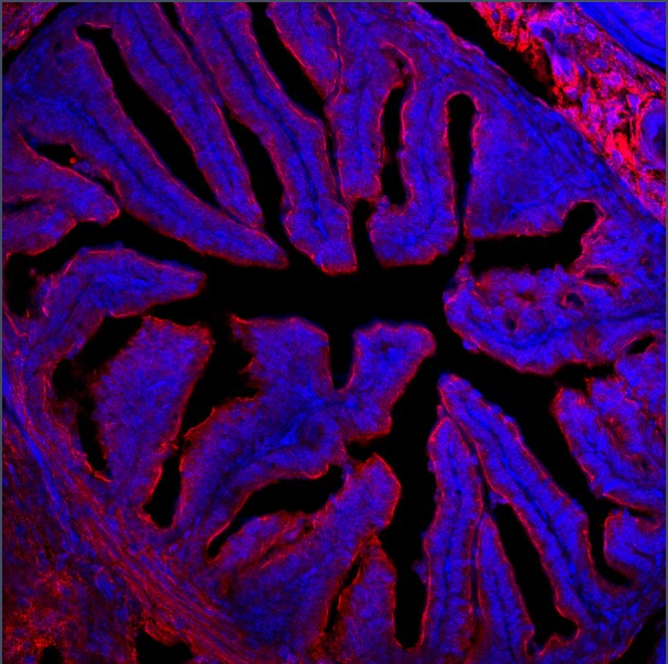
Application: Immunocytochemistry/ImmunofluorescenceSample Tested: fixed frozen tissueSpecies: MouseVerified Customer | Posted 01/30/2020Immunofluorescence experiment on mouse ovary. Was incubated with the primary antibody overnight at room temperature

There are no reviews that match your criteria.
Protocols
Find general support by application which include: protocols, troubleshooting, illustrated assays, videos and webinars.
- 7-Amino Actinomycin D (7-AAD) Cell Viability Flow Cytometry Protocol
- Appropriate Fixation of IHC/ICC Samples
- Cellular Response to Hypoxia Protocols
- ClariTSA™ Fluorophore Kits
- Detection & Visualization of Antibody Binding
- Extracellular Membrane Flow Cytometry Protocol
- Flow Cytometry Protocol for Cell Surface Markers
- Flow Cytometry Protocol for Staining Membrane Associated Proteins
- Flow Cytometry Staining Protocols
- Flow Cytometry Troubleshooting Guide
- ICC Cell Smear Protocol for Suspension Cells
- ICC Immunocytochemistry Protocol Videos
- ICC for Adherent Cells
- Immunocytochemistry (ICC) Protocol
- Immunocytochemistry Troubleshooting
- Immunofluorescence of Organoids Embedded in Cultrex Basement Membrane Extract
- Immunohistochemistry (IHC) and Immunocytochemistry (ICC) Protocols
- Intracellular Flow Cytometry Protocol Using Alcohol (Methanol)
- Intracellular Flow Cytometry Protocol Using Detergents
- Intracellular Nuclear Staining Flow Cytometry Protocol Using Detergents
- Intracellular Staining Flow Cytometry Protocol Using Alcohol Permeabilization
- Intracellular Staining Flow Cytometry Protocol Using Detergents to Permeabilize Cells
- Preparing Samples for IHC/ICC Experiments
- Preventing Non-Specific Staining (Non-Specific Binding)
- Primary Antibody Selection & Optimization
- Propidium Iodide Cell Viability Flow Cytometry Protocol
- Protocol for Liperfluo
- Protocol for VisUCyte™ HRP Polymer Detection Reagent
- Protocol for the Characterization of Human Th22 Cells
- Protocol for the Characterization of Human Th9 Cells
- Protocol for the Fluorescent ICC Staining of Cell Smears - Graphic
- Protocol for the Fluorescent ICC Staining of Cultured Cells on Coverslips - Graphic
- Protocol for the Preparation and Fluorescent ICC Staining of Cells on Coverslips
- Protocol for the Preparation and Fluorescent ICC Staining of Non-adherent Cells
- Protocol for the Preparation and Fluorescent ICC Staining of Stem Cells on Coverslips
- Protocol for the Preparation of a Cell Smear for Non-adherent Cell ICC - Graphic
- Protocol: Annexin V and PI Staining by Flow Cytometry
- Protocol: Annexin V and PI Staining for Apoptosis by Flow Cytometry
- R&D Systems Quality Control Western Blot Protocol
- TUNEL and Active Caspase-3 Detection by IHC/ICC Protocol
- The Importance of IHC/ICC Controls
- Troubleshooting Guide: Fluorokine Flow Cytometry Kits
- Troubleshooting Guide: Western Blot Figures
- Western Blot Conditions
- Western Blot Protocol
- Western Blot Protocol for Cell Lysates
- Western Blot Troubleshooting
- Western Blot Troubleshooting Guide
- View all Protocols, Troubleshooting, Illustrated assays and Webinars
Loading...