 by Western Blot.")
Loading...
Key Product Details
Validated by
Biological Validation
Species Reactivity
Validated:
Human, Mouse
Cited:
Human, Mouse, Hamster
Applications
Validated:
Western Blot, Intracellular Staining by Flow Cytometry, Simple Western, CyTOF-ready
Cited:
Immunohistochemistry, Western Blot, Immunocytochemistry, Capillary Immunoblot
Label
Unconjugated
Antibody Source
Polyclonal Rabbit IgG
Loading...
Product Specifications
Immunogen
Phosphopeptide containing human STAT1 Y701 site
Specificity
Detects human STAT1 when phosphorylated at Y701 in Western blots.
Clonality
Polyclonal
Host
Rabbit
Isotype
IgG
Scientific Data Images for phospho-STAT1 (Y701) Antibody
Detection of Human Phospho-STAT1 (Y701) by Western Blot.
Western blot shows lysates of Daudi human Burkitt's lymphoma cell line untreated (-) or treated (+) with Recombinant Human IFN-a2a for 20 minutes. PVDF membrane was probed with 0.5 µg/mL of Rabbit Anti-Human/Mouse Phospho-STAT1 (Y701) Antigen Affinity-purified Polyclonal Antibody (Catalog # AF2894), followed by HRP-conjugated Anti-Rabbit IgG Secondary Antibody (Catalog # HAF008). A specific band was detected for Phospho-STAT1 (Y701) at approximately 94 kDa (as indicated). This experiment was conducted under reducing conditions and using Immunoblot Buffer Group 2.
Detection of Phospho-STAT1 in IFN-alpha -treated Daudi Human Cell Line by Flow Cytometry.
Daudi human Burkitt's lymphoma cell line was unstimulated (light orange filled histogram) or treated with 500 units/mL Recombinant Human IFN-a (Catalog # 11100-1) for 20 minutes (dark orange filled histogram), then stained with Rabbit Anti-Human/Mouse Phospho-STAT1 (Y701) Antigen Affinity-purified Polyclonal Antibody (Catalog # AF2894) or control antibody (Catalog # AB-105-C, open histogram), followed by Allophycocyanin-conjugated Anti-Rabbit IgG Secondary Antibody (Catalog # F0111). To facilitate intracellular staining, cells were fixed with paraformaldehyde and permeabilized with methanol. by Simple Western<SUP>TM</SUP>.")
Detection of Human Phospho-STAT1 (Y701) by Simple WesternTM.
Simple Western lane view shows lysates of Daudi human Burkitt's lymphoma cell line untreated (-) or treated (+) with Recombinant Human IFN-aA (Catalog # 11100-1) for 20 minutes, loaded at 0.2 mg/mL. A specific band was detected for Phospho-STAT1 (Y701) at approximately 86 kDa (as indicated) using 5 µg/mL of Rabbit Anti-Human/Mouse Phospho-STAT1 (Y701) Antigen Affinity-purified Polyclonal Antibody (Catalog # AF2894). This experiment was conducted under reducing conditions and using the 12-230 kDa separation system.
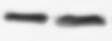
Detection of Mouse STAT1 by Western Blot
Effects of kurarinone on the phosphorylation of p-STAT1 and p-STAT3 in lymph nodes. Single cell suspensions were collected from ILNs on day 42, the protein expression levels of p-STAT1, STAT1, p-STAT3, and STAT3 were measured using Western blots. (A) Representative images of Western blot and (B) Densitometric analysis for protein expressions was performed using ImageJ software. Data are presented as mean ± SEM of 6 mice from one of three experiments. (*) p < 0.05, (**) p < 0.01, (***) p < 0.001 versus vehicle-treated CIA mice group (One Way ANOVA followed by Tukey’s multiple comparison test). Image collected and cropped by CiteAb from the following open publication (https://pubmed.ncbi.nlm.nih.gov/33924467), licensed under a CC-BY license. Not internally tested by R&D Systems. by Western Blot")
Detection of Phospho-STAT1 (Y701) by Western Blot
The loss of C9ORF72 leads to JAK-STAT activation(A) RT-qPCR analysis of Sting levels in control and C9orf72−/− RAW264.7 cells untreated or treated with DMXAA for 16h. Data represent the mean ± SEM. Statistical significance was analyzed by two-way ANOVA (n = 3), ns = not significant, ∗p < 0.05, ∗∗p < 0.01.(B) Protein levels of STAT1 in spleen lysates from 6-month-old WT and C9orf72−/− mice were analyzed using western blot. Mixed male and female mice were used. Data represent the mean ± SEM. Statistical significance was analyzed by unpaired one-tail Student’s t test (n = 4), ns = not significant, ∗p < 0.05.(C) Protein levels of STAT1 and p-STAT1 in control or C9orf72−/− RAW264.7 cells untreated or treated with DMXAA for 2h were analyzed using western blot and normalized to GAPDH. Data represent the mean ± SEM. Statistical significance was analyzed by unpaired two-tail Student’s t test (n = 3), ns = not significant, ∗p < 0.05.(D) RT-qPCR analysis of Stat1 levels in control and C9orf72−/− RAW264.7 cells untreated or treated with DMXAA for 16h. Data represent the mean ± SEM. Statistical significance was analyzed by two-way ANOVA (n = 3), ns = not significant, ∗p < 0.05, ∗∗∗∗p < 0.0001.(E) IFN-beta levels in control and C9orf72−/− RAW264.7 cells untreated or treated with DMXAA for 16h were measured using ELISA. Data represent the mean ± SEM. Statistical significance was analyzed by two-way ANOVA (n = 3), ns = not significant, ∗∗p < 0.01. Image collected and cropped by CiteAb from the following open publication (https://pubmed.ncbi.nlm.nih.gov/37250330), licensed under a CC-BY license. Not internally tested by R&D Systems. by Western Blot")
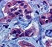
Detection of Phospho-STAT1 (Y701) by Western Blot
The loss of C9ORF72 leads to JAK-STAT activation(A) RT-qPCR analysis of Sting levels in control and C9orf72−/− RAW264.7 cells untreated or treated with DMXAA for 16h. Data represent the mean ± SEM. Statistical significance was analyzed by two-way ANOVA (n = 3), ns = not significant, ∗p < 0.05, ∗∗p < 0.01.(B) Protein levels of STAT1 in spleen lysates from 6-month-old WT and C9orf72−/− mice were analyzed using western blot. Mixed male and female mice were used. Data represent the mean ± SEM. Statistical significance was analyzed by unpaired one-tail Student’s t test (n = 4), ns = not significant, ∗p < 0.05.(C) Protein levels of STAT1 and p-STAT1 in control or C9orf72−/− RAW264.7 cells untreated or treated with DMXAA for 2h were analyzed using western blot and normalized to GAPDH. Data represent the mean ± SEM. Statistical significance was analyzed by unpaired two-tail Student’s t test (n = 3), ns = not significant, ∗p < 0.05.(D) RT-qPCR analysis of Stat1 levels in control and C9orf72−/− RAW264.7 cells untreated or treated with DMXAA for 16h. Data represent the mean ± SEM. Statistical significance was analyzed by two-way ANOVA (n = 3), ns = not significant, ∗p < 0.05, ∗∗∗∗p < 0.0001.(E) IFN-beta levels in control and C9orf72−/− RAW264.7 cells untreated or treated with DMXAA for 16h were measured using ELISA. Data represent the mean ± SEM. Statistical significance was analyzed by two-way ANOVA (n = 3), ns = not significant, ∗∗p < 0.01. Image collected and cropped by CiteAb from the following open publication (https://pubmed.ncbi.nlm.nih.gov/37250330), licensed under a CC-BY license. Not internally tested by R&D Systems.Applications for phospho-STAT1 (Y701) Antibody
Application
Recommended Usage
CyTOF-ready
Ready to be labeled using established conjugation methods. No BSA or other carrier proteins that could interfere with conjugation.
Intracellular Staining by Flow Cytometry
2.5 µg/106 cells
Sample: IFN-alpha -treated Daudi human Burkitt's lymphoma cell line fixed with paraformaldehyde and permeabilized with methanol
Sample: IFN-alpha -treated Daudi human Burkitt's lymphoma cell line fixed with paraformaldehyde and permeabilized with methanol
Simple Western
5 µg/mL
Sample: Daudi human Burkitt's lymphoma cell line treated with Recombinant Human IFN‑ alpha A (Catalog # 11100-1)
Sample: Daudi human Burkitt's lymphoma cell line treated with Recombinant Human IFN‑ alpha A (Catalog # 11100-1)
Western Blot
0.5 µg/mL
Sample: Daudi human Burkitt's lymphoma cell line treated with Recombinant Human IFN-alpha 2a
Sample: Daudi human Burkitt's lymphoma cell line treated with Recombinant Human IFN-alpha 2a
Reviewed Applications
Read 9 reviews rated 4.4 using AF2894 in the following applications:
Flow Cytometry Panel Builder
Bio-Techne Knows Flow Cytometry
Save time and reduce costly mistakes by quickly finding compatible reagents using the Panel Builder Tool.
Advanced Features
- Spectra Viewer - Custom analysis of spectra from multiple fluorochromes
- Spillover Popups - Visualize the spectra of individual fluorochromes
- Antigen Density Selector - Match fluorochrome brightness with antigen density

Formulation, Preparation, and Storage
Purification
Antigen Affinity-purified
Reconstitution
Reconstitute at 0.2 mg/mL in sterile PBS. For liquid material, refer to CoA for concentration.
Loading...
Formulation
Lyophilized from a 0.2 μm filtered solution in PBS with Trehalose. *Small pack size (SP) is supplied either lyophilized or as a 0.2 µm filtered solution in PBS.
Shipping
Lyophilized product is shipped at ambient temperature. Liquid small pack size (-SP) is shipped with polar packs. Upon receipt, store immediately at the temperature recommended below.
Stability & Storage
Use a manual defrost freezer and avoid repeated freeze-thaw cycles.
- 12 months from date of receipt, -20 to -70 °C as supplied.
- 1 month, 2 to 8 °C under sterile conditions after reconstitution.
- 6 months, -20 to -70 °C under sterile conditions after reconstitution.
Calculators
Background: STAT1
Long Name
Signal Transducer and Activator of Transcription 1
Alternate Names
CANDF7, ISGF-3, STAT91
Gene Symbol
STAT1
Additional STAT1 Products
Product Documents for phospho-STAT1 (Y701) Antibody
Certificate of Analysis
To download a Certificate of Analysis, please enter a lot or batch number in the search box below.
Note: Certificate of Analysis not available for kit components.
Product Specific Notices for phospho-STAT1 (Y701) Antibody
For research use only
Citations for phospho-STAT1 (Y701) Antibody
 Powered by Bioz
Customer Reviews for phospho-STAT1 (Y701) Antibody (9)
4.4 out of 5
9 Customer Ratings
Have you used phospho-STAT1 (Y701) Antibody?
Submit a review and receive an Amazon gift card!
$25/€18/£15/$25CAN/¥2500 Yen for a review with an image
$10/€7/£6/$10CAN/¥1110 Yen for a review without an image
Submit a review
Customer Images

Showing
1
-
5 of
9 reviews
Showing All
Filter By:
-
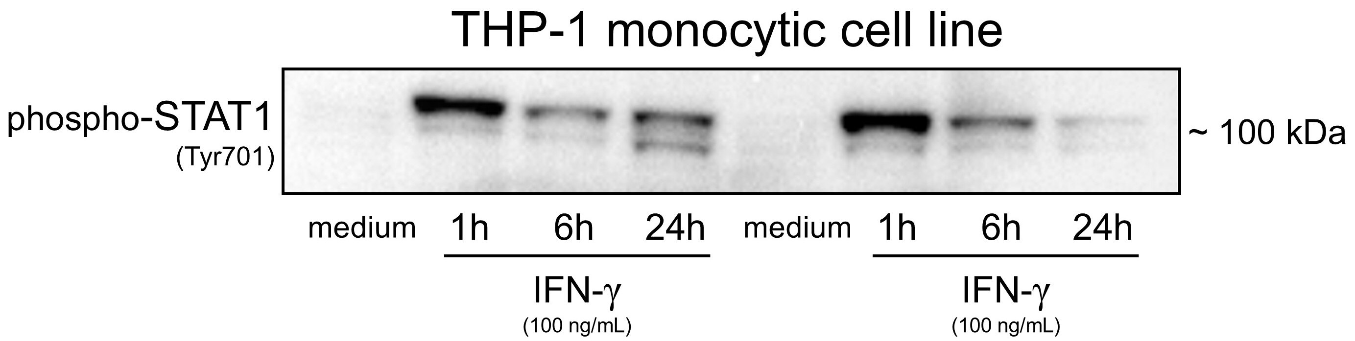
Application: Western BlotSample Tested: THP-1 human acute monocytic leukemia cell lineSpecies: HumanVerified Customer | Posted 10/12/2025

-
Application: Western BlotSample Tested: RAW 264.7 mouse monocyte/macrophage cell lineSpecies: MouseVerified Customer | Posted 03/28/2023First antibody: 1:2000, 4 oC, overnight Second antibody: 1:5000, room temperature, 1 h
-
Application: Western BlotSample Tested: MDA-MB-231 human breast cancer cell lineSpecies: HumanVerified Customer | Posted 04/18/2022
-
Application: Western BlotSample Tested: Human lung macrophages and JurkatSpecies: HumanVerified Customer | Posted 04/04/2020
-
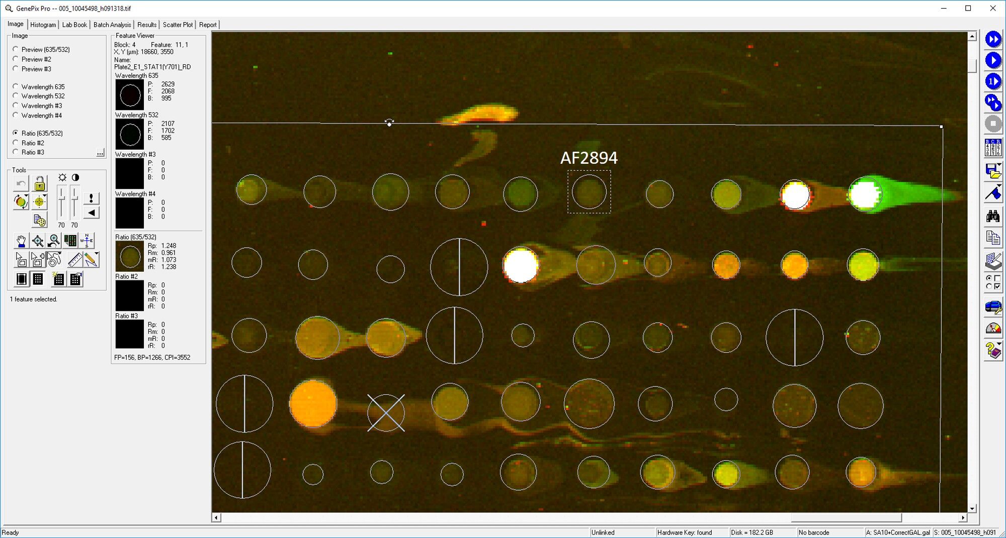
Application: MicroarraysSample Tested: EDTA PlasmaSpecies: HumanVerified Customer | Posted 03/11/2019
-
Application: ImmunohistochemistrySample Tested: Breast cancer tissueSpecies: HumanVerified Customer | Posted 02/19/2019
-
Application: ELISASample Tested: Serum and PlasmaSpecies: Human and MouseVerified Customer | Posted 11/08/2018
-
Application: MicroarraySample Tested: EDTA PlasmaSpecies: HumanVerified Customer | Posted 11/02/2018
-
Application: Western BlotSample Tested: EMT6 cells and CT26Species: MouseVerified Customer | Posted 10/17/2018
There are no reviews that match your criteria.
Protocols
Find general support by application which include: protocols, troubleshooting, illustrated assays, videos and webinars.
- 7-Amino Actinomycin D (7-AAD) Cell Viability Flow Cytometry Protocol
- Cellular Response to Hypoxia Protocols
- Extracellular Membrane Flow Cytometry Protocol
- Flow Cytometry Protocol for Cell Surface Markers
- Flow Cytometry Protocol for Staining Membrane Associated Proteins
- Flow Cytometry Staining Protocols
- Flow Cytometry Troubleshooting Guide
- Intracellular Flow Cytometry Protocol Using Alcohol (Methanol)
- Intracellular Flow Cytometry Protocol Using Detergents
- Intracellular Nuclear Staining Flow Cytometry Protocol Using Detergents
- Intracellular Staining Flow Cytometry Protocol Using Alcohol Permeabilization
- Intracellular Staining Flow Cytometry Protocol Using Detergents to Permeabilize Cells
- Propidium Iodide Cell Viability Flow Cytometry Protocol
- Protocol for Liperfluo
- Protocol for the Characterization of Human Th22 Cells
- Protocol for the Characterization of Human Th9 Cells
- Protocol: Annexin V and PI Staining by Flow Cytometry
- Protocol: Annexin V and PI Staining for Apoptosis by Flow Cytometry
- R&D Systems Quality Control Western Blot Protocol
- Troubleshooting Guide: Fluorokine Flow Cytometry Kits
- Troubleshooting Guide: Western Blot Figures
- Western Blot Conditions
- Western Blot Protocol
- Western Blot Protocol for Cell Lysates
- Western Blot Troubleshooting
- Western Blot Troubleshooting Guide
- View all Protocols, Troubleshooting, Illustrated assays and Webinars


