Human/Mouse/Rat Cytochrome c Antibody
R&D Systems | Catalog # MAB897

Key Product Details
Species Reactivity
Validated:
Human, Mouse, Rat
Cited:
Human
Applications
Validated:
Western Blot, Simple Western
Cited:
Western Blot
Label
Unconjugated
Antibody Source
Monoclonal Mouse IgG2B Clone # 7H8.2C12
Loading...
Product Specifications
Immunogen
Pigeon Cytochrome c
Specificity
Reacts with denatured human, mouse and rat Cytochrome c, but not native Cytochrome c in Western blots.
Clonality
Monoclonal
Host
Mouse
Isotype
IgG2B
Scientific Data Images for Human/Mouse/Rat Cytochrome c Antibody
Detection of Human and Mouse Cytochrome c by Western Blot.
Western blot shows lysates of Jurkat human acute T cell leukemia cell line and CTLL-2 mouse cytotoxic T cell line. PVDF membrane was probed with 0.5 µg/mL of Mouse Anti-Human/Mouse/Rat Cytochrome c Monoclonal Antibody (Catalog # MAB897) followed by HRP-conjugated Anti-Mouse IgG Secondary Antibody (Catalog # HAF007). A specific band was detected for Cytochrome c at approximately 12 kDa (as indicated). This experiment was conducted under reducing conditions and using Immunoblot Buffer Group 4.
Detection of Human Cytochrome c by Simple WesternTM.
Simple Western lane view shows lysates of human heart tissue, loaded at 0.2 mg/mL. A specific band was detected for Cytochrome c at approximately 23 kDa (as indicated) using 2.5 µg/mL of Mouse Anti-Human/Mouse/Rat Cytochrome c Monoclonal Antibody (Catalog # MAB897). This experiment was conducted under reducing conditions and using the 12-230 kDa separation system.
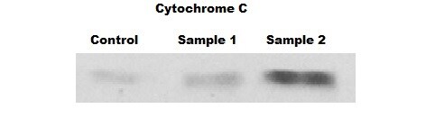
Detection of Human Cytochrome c by Western Blot
FAM210B is transported into and is localized in the mitochondria. (a) Schematic representation of FAM210B domains based on the primary structure of FAM210B. (b) Confocal microscopy of HeLa cells transfected with GFP-tagged human FAM210B and RFP-tagged mitochondria. FAM210B-GFP, GFP sequence introduced at the C terminus of FAM210B; FAM210B (MTS)-GFP, the MTS (aa 1–47) of FAM210B was added to the GFP N terminus; FAM210B ( delta MTS)-GFP, GFP was added to the C terminus of MTS (1–47)-deleted CRIF1. Scale bar, 20 mm. (c) Confocal microscopy of HeLa cells transfected with GFP-tagged human FAM210B and RFP-tagged endoplasmic reticulum. (d) Western blotting analysis following subcellular fractionation of GFP-tagged human FAM210B HeLa cells. (e) Western blotting analysis following subcellular fractionation of endogenous in HeLa cells. (f) The mitochondria of HeLa cells were swollen and sonicated to disrupt membranes, washed with alkali buffer (pH 11.5) to detach loosely associated proteins from membranes, and then re-isolated by ultracentrifugation. The supernatant (Supe) and membrane fractions (Pellet) were subjected to western blotting for FAM210B, TOM20, or MnSOD. (g) Mitochondria isolated from HeLa cells were subjected to proteinase K (PK) proteolysis to digest exposed proteins, and detergent (SDS) was used to disrupt both IMMs (inner membrane of mitochondria) and OMMs (outer membrane of mitochondria). The lysates were resolved and subjected to immunoblot analyses. The submitochondrial markers used are Tom20 (OMM), Cyt C (Cytochrome c, intermembrane space), NDUFS1 (IMM), and MnSOD (mitochondrial matrix) Image collected and cropped by CiteAb from the following publication (https://pubmed.ncbi.nlm.nih.gov/28594398), licensed under a CC-BY license. Not internally tested by R&D Systems.Applications for Human/Mouse/Rat Cytochrome c Antibody
Application
Recommended Usage
Simple Western
2.5 µg/mL
Sample: Human heart tissue
Sample: Human heart tissue
Western Blot
0.5 µg/mL
Sample: Jurkat human acute T cell leukemia cell line and CTLL-2 mouse cytotoxic T cell line
Sample: Jurkat human acute T cell leukemia cell line and CTLL-2 mouse cytotoxic T cell line
Reviewed Applications
Read 1 review rated 5 using MAB897 in the following applications:
Formulation, Preparation, and Storage
Purification
Protein A or G purified from ascites
Reconstitution
Reconstitute at 0.5 mg/mL in sterile PBS. For liquid material, refer to CoA for concentration.
Loading...
Formulation
Lyophilized from a 0.2 μm filtered solution in PBS with Trehalose. *Small pack size (SP) is supplied either lyophilized or as a 0.2 µm filtered solution in PBS.
Shipping
Lyophilized product is shipped at ambient temperature. Liquid small pack size (-SP) is shipped with polar packs. Upon receipt, store immediately at the temperature recommended below.
Stability & Storage
Use a manual defrost freezer and avoid repeated freeze-thaw cycles.
- 12 months from date of receipt, -20 to -70 °C as supplied.
- 1 month, 2 to 8 °C under sterile conditions after reconstitution.
- 6 months, -20 to -70 °C under sterile conditions after reconstitution.
Calculators
Background: Cytochrome c
Additional Cytochrome c Products
Product Documents for Human/Mouse/Rat Cytochrome c Antibody
Certificate of Analysis
To download a Certificate of Analysis, please enter a lot or batch number in the search box below.
Note: Certificate of Analysis not available for kit components.
Product Specific Notices for Human/Mouse/Rat Cytochrome c Antibody
For research use only
Related Research Areas
Citations for Human/Mouse/Rat Cytochrome c Antibody
 Powered by Bioz
Customer Reviews for Human/Mouse/Rat Cytochrome c Antibody (1)
5 out of 5
1 Customer Rating
Have you used Human/Mouse/Rat Cytochrome c Antibody?
Submit a review and receive an Amazon gift card!
$25/€18/£15/$25CAN/¥2500 Yen for a review with an image
$10/€7/£6/$10CAN/¥1110 Yen for a review without an image
Submit a review
Customer Images
Showing
1
-
1 of
1 review
Showing All
Filter By:
-
Application: Western BlotSample Tested: Cartilage tissueSpecies: MouseVerified Customer | Posted 08/06/2019Antibody Dilution 1:100. We used this on mouse meniscus lysates. Antibody was incubated overnight and developed with ECL kit. It worked very well and we would use it again.

There are no reviews that match your criteria.
Protocols
Find general support by application which include: protocols, troubleshooting, illustrated assays, videos and webinars.
- Cellular Response to Hypoxia Protocols
- R&D Systems Quality Control Western Blot Protocol
- Troubleshooting Guide: Western Blot Figures
- Western Blot Conditions
- Western Blot Protocol
- Western Blot Protocol for Cell Lysates
- Western Blot Troubleshooting
- Western Blot Troubleshooting Guide
- View all Protocols, Troubleshooting, Illustrated assays and Webinars
Loading...
Associated Pathways

