
Key Product Details
Validated by
Biological Validation
Species Reactivity
Validated:
Human, Mouse, Rat
Cited:
Human, Mouse, Rat, Chinese Hamster, Rabbit, Transgenic Mouse, Xenograft, Zebrafish
Applications
Validated:
Immunohistochemistry, Western Blot, Simple Western
Cited:
Immunohistochemistry, Immunohistochemistry-Paraffin, Western Blot, Flow Cytometry, Immunocytochemistry
Label
Unconjugated
Antibody Source
Monoclonal Mouse IgG1 Clone # 2D2-B2
Loading...
Product Specifications
Immunogen
Recombinant human iNOS
Pro781-His798
Accession # P35228
Pro781-His798
Accession # P35228
Specificity
Detects human iNOS. By using synthetic peptides, the epitope recognized by this antibody has been mapped to aa 781-798 of human iNOS. The corresponding sequence of mouse iNOS is identical.
Clonality
Monoclonal
Host
Mouse
Isotype
IgG1
Scientific Data Images for iNOS Antibody (2D2-B2)
Detection of Human iNOS by Western Blot.
Western blot shows lysates of DLD clone 2C2 human colon adenocarcinoma cell line untreated (-) or treated (+) with Recombinant Human IL-1 beta, Recombinant Human TNF-alpha, and Recombinant Human IFN-gamma (Catalog # HAF007). A specific band was detected for iNOS at approximately 130 kDa (as indicated). This experiment was conducted under reducing conditions and using Immunoblot Buffer Group 6.
Detection of Mouse and Rat iNOS by Western Blot.
Western blot shows lysates of RAW 264.7 mouse monocyte/macrophage cell line and NR8383 rat alveolar macrophage cell line untreated (-) or treated (+) with 10 µg/mL LPS for 4 hours. PVDF membrane was probed with 1 µg/mL of Mouse Anti-Human/Mouse/Rat iNOS Monoclonal Antibody (Catalog # MAB9502) followed by HRP-conjugated Anti-Mouse IgG Secondary Antibody (Catalog # HAF018). A specific band was detected for iNOS at approximately 125 kDa (as indicated). This experiment was conducted under reducing conditions and using Immunoblot Buffer Group 1.
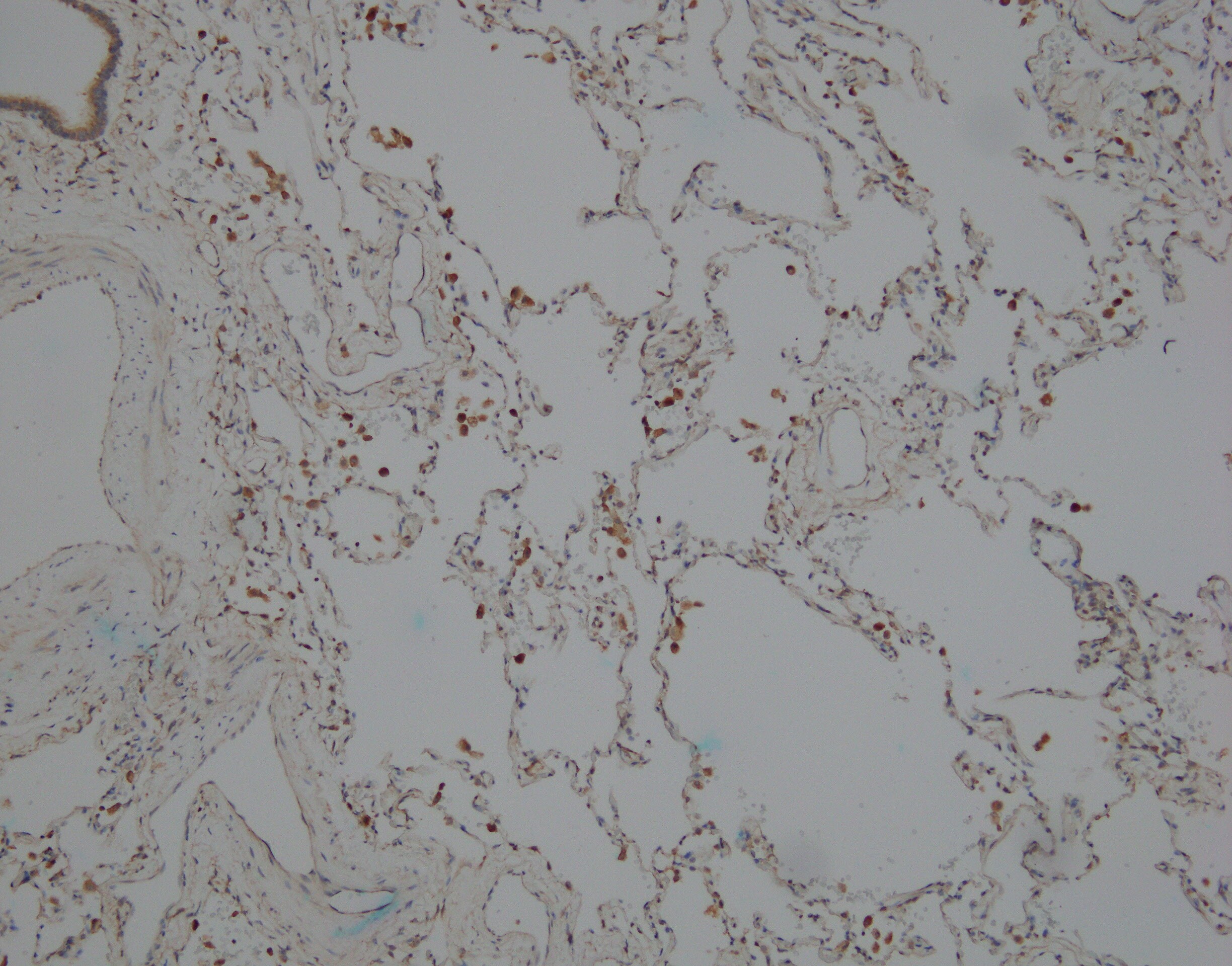
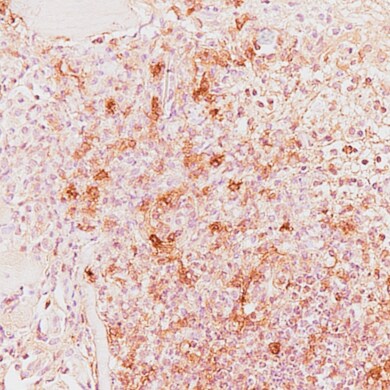
iNOS in Human Brain.
iNOS was detected in immersion fixed paraffin-embedded sections of human brain (medulla) using Mouse Anti-Human/Mouse/Rat iNOS Monoclonal Antibody (Catalog # MAB9502) at 5 µg/mL overnight at 4 °C. Tissue was stained using the Anti-Mouse HRP-DAB Cell & Tissue Staining Kit (brown; Catalog # CTS002) and counterstained with hematoxylin (blue). Specific staining was localized to neuronal cell bodies. View our protocol for Chromogenic IHC Staining of Paraffin-embedded Tissue Sections.
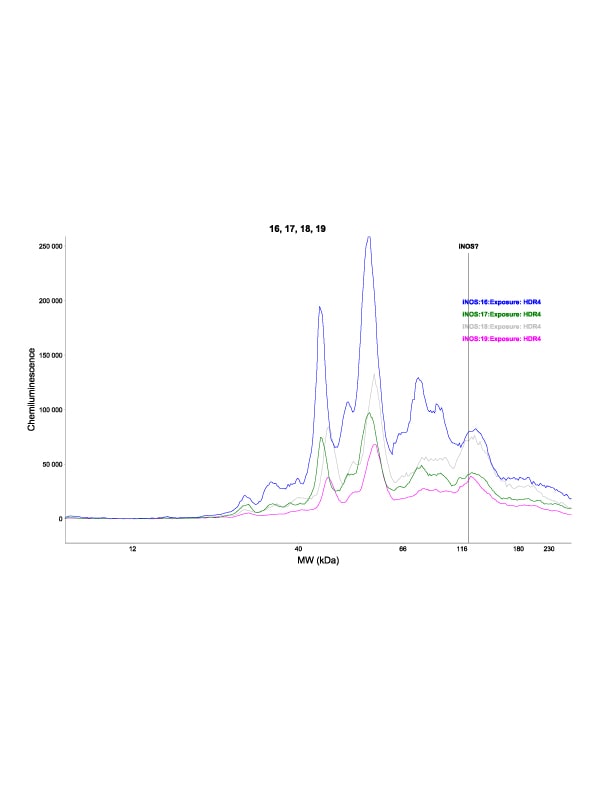
Detection of Mouse iNOS by Simple WesternTM.
Simple Western lane view shows lysates of RAW 264.7 mouse monocyte/macrophage cell line untreated (-) or treated (+) with LPS, loaded at 0.2 mg/mL. A specific band was detected for iNOS at approximately 136 kDa (as indicated) using 10 µg/mL of Mouse Anti-Human/Mouse/Rat iNOS Monoclonal Antibody (Catalog # MAB9502). This experiment was conducted under reducing conditions and using the 12-230 kDa separation system.
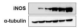
Detection of Rat iNOS by Western Blot
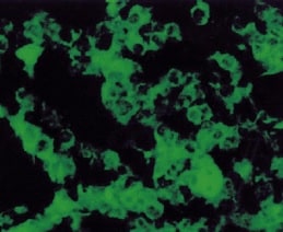
Ultrasound (US) stimulation reduces ED-1, iNOS, and TNF-alpha expression in the acute spinal cord injury (SCI) phase. (A–C) ED-1 (macrophage/microglia, green) and iNOS (inducible nitric oxide synthase, red) were evaluated in the sham (A), SCIU40 (B), and SCIU5 (c) experimental groups at 7 days post-SCI (five sections per rat, n = 3). (D–F) Cross-sections were counterstained with anti-TNF-alpha (red) and ED-1 (green) antibodies (five sections per rat, n = 3). (G–I) Quantification of fluorescence intensity of ED-1, iNOS, and TNF-alpha. Two-way ANOVA with Turkey's test for multiple comparisons was used for analyses. *P < 0.05, ****P < 0.0001 compared with the sham control, #P < 0.05, ###P < 0.001, ####P < 0.0001 compared with the SCIU5-treated group. (J) Representative western blotting results for ED-1, iNOS, TNF-alpha, and GAPDH at 7 days post-SCI (Lane 1, sham; lane 2, SCIU40; lane 3, SCIU5;). Relative expression levels of ED-1, iNOS, and TNF-alpha were calculated after normalization to the level of GAPDH. Full-length blots are presented in Supplementary Fig. 8. Data are presented as means ± SEM. Two-tailed Student’s t-tests were used for comparison. *P < 0.05 compared with the sham control. Image collected and cropped by CiteAb from the following open publication (https://pubmed.ncbi.nlm.nih.gov/35256617), licensed under a CC-BY license. Not internally tested by R&D Systems.Applications for iNOS Antibody (2D2-B2)
Application
Recommended Usage
Immunohistochemistry
8-25 µg/mL
Sample: Immersion fixed paraffin-embedded sections of human brain (medulla).
Sample: Immersion fixed paraffin-embedded sections of human brain (medulla).
Simple Western
10 µg/mL
Sample: RAW 264.7 mouse monocyte/macrophage cell line treated with LPS
Sample: RAW 264.7 mouse monocyte/macrophage cell line treated with LPS
Western Blot
1 µg/mL
Sample: DLD clone 2C2 human colon adenocarcinoma cell line treated with Recombinant Human IL-1 beta, Recombinant Human TNF-alpha, and Recombinant Human IFN-gamma (Catalog # 201-LB, 210-TA, 285-IF), RAW 264.7 mouse monocyte/macrophage cell line treated with LPS, and NR8383 rat alveolar macrophage cell line treated with LPS
Sample: DLD clone 2C2 human colon adenocarcinoma cell line treated with Recombinant Human IL-1 beta, Recombinant Human TNF-alpha, and Recombinant Human IFN-gamma (Catalog # 201-LB, 210-TA, 285-IF), RAW 264.7 mouse monocyte/macrophage cell line treated with LPS, and NR8383 rat alveolar macrophage cell line treated with LPS
Reviewed Applications
Read 9 reviews rated 3.9 using MAB9502 in the following applications:
Formulation, Preparation, and Storage
Purification
Protein A or G purified from ascites
Reconstitution
Reconstitute at 0.5 mg/mL in sterile PBS. For liquid material, refer to CoA for concentration.
Loading...
Formulation
Lyophilized from a 0.2 μm filtered solution in PBS with Trehalose. *Small pack size (SP) is supplied either lyophilized or as a 0.2 µm filtered solution in PBS.
Shipping
Lyophilized product is shipped at ambient temperature. Liquid small pack size (-SP) is shipped with polar packs. Upon receipt, store immediately at the temperature recommended below.
Stability & Storage
Use a manual defrost freezer and avoid repeated freeze-thaw cycles.
- 12 months from date of receipt, -20 to -70 °C as supplied.
- 1 month, 2 to 8 °C under sterile conditions after reconstitution.
- 6 months, -20 to -70 °C under sterile conditions after reconstitution.
Calculators
Background: iNOS
Long Name
Inducible Nitic Oxide Synthase
Alternate Names
NOS2, NOS2A
Gene Symbol
NOS2
UniProt
Additional iNOS Products
Product Documents for iNOS Antibody (2D2-B2)
Certificate of Analysis
To download a Certificate of Analysis, please enter a lot or batch number in the search box below.
Note: Certificate of Analysis not available for kit components.
Product Specific Notices for iNOS Antibody (2D2-B2)
For research use only
Citations for iNOS Antibody (2D2-B2)
 Powered by Bioz
Customer Reviews for iNOS Antibody (2D2-B2) (9)
3.9 out of 5
9 Customer Ratings
Have you used iNOS Antibody (2D2-B2)?
Submit a review and receive an Amazon gift card!
$25/€18/£15/$25CAN/¥2500 Yen for a review with an image
$10/€7/£6/$10CAN/¥1110 Yen for a review without an image
Submit a review
Customer Images
Showing
1
-
5 of
9 reviews
Showing All
Filter By:
-
Application: Western BlotSample Tested: Lung homogenatesSpecies: MouseVerified Customer | Posted 10/30/2025Didn't work well. Somehow I couldn't get the inos signal.
-
Application: ImmunohistochemistrySample Tested: Lung tissueSpecies: HumanVerified Customer | Posted 04/11/2023Leica Bond ER2 heat retrieval primary Ab MAB9502 at 5ug/mL in BSA-TBS Leica standard IHC protocol on Leica Rx stainer

-
Application: ImmunohistochemistrySample Tested: Bone marrow tissueSpecies: MouseVerified Customer | Posted 08/13/2021
-
Application: Immunocytochemistry/ImmunofluorescenceSample Tested: Connective tissueSpecies: MouseVerified Customer | Posted 07/06/2021
-
Application: Western BlotSample Tested: RAW 264.7 mouse monocyte/macrophage cell lineSpecies: Human and MouseVerified Customer | Posted 04/03/2021Antibody also works very well for immunohistochemistry
-
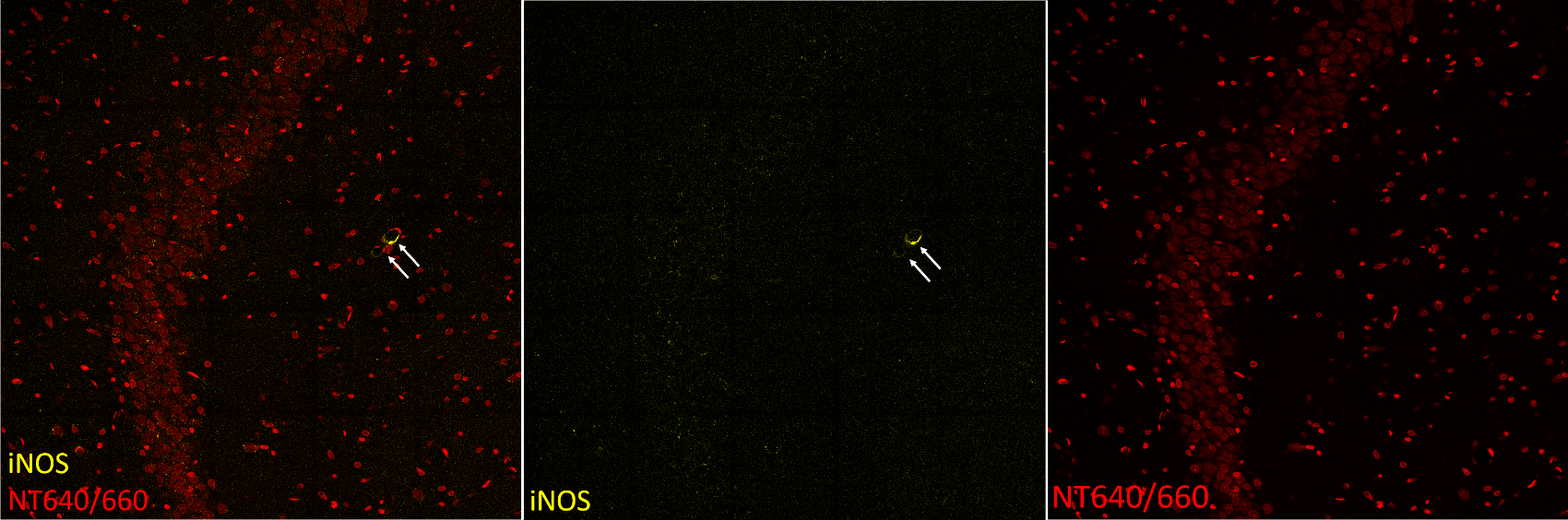
Application: ImmunohistochemistrySample Tested: Adult brainSpecies: RatVerified Customer | Posted 07/28/2020Rats were perfused with 4% paraformaldehyde in PBS (pH=7.6). The brain was removed and immersed in PFA then in 30% sucrose. The brain was sliced with a freezing microtome. A mixture of 1% Triton-X and 10% donkey serum (in TRIS) was used for antigen retrieval and blocking. Tmem119 antibody was used in 1:100 dilution and was visualized by a Cy3 conjugated anti-mouse secondary antibody (1:500, yellow). Cell bodies were stained with NeuroTrace 640/660 (1:250, red). Pictures show a part of the rat hippocampus. The product was assumed to label activated microglial cells. It seems, it did not. However, it labelled some cerebral blood vessels, which is also meaningful, thus, it may be useful for this application.
-
Application: Simple WesternSample Tested: skin wound - diabeticSpecies: RatVerified Customer | Posted 02/12/2020
Bio-Techne ResponseThis review was submitted through the legacy Novus Innovators Program, reflecting a new species or application tested on a primary antibody.
-
Application: Western BlotSample Tested: See PMID 21075192Species: MouseVerified Customer | Posted 02/25/2015
-
Application: Immunohistochemistry-ParaffinSample Tested: See PMID 23147558Species: HumanVerified Customer | Posted 02/25/2015
There are no reviews that match your criteria.
Protocols
Find general support by application which include: protocols, troubleshooting, illustrated assays, videos and webinars.
- Antigen Retrieval Protocol (PIER)
- Antigen Retrieval for Frozen Sections Protocol
- Appropriate Fixation of IHC/ICC Samples
- Cellular Response to Hypoxia Protocols
- Chromogenic IHC Staining of Formalin-Fixed Paraffin-Embedded (FFPE) Tissue Protocol
- Chromogenic Immunohistochemistry Staining of Frozen Tissue
- ClariTSA™ Fluorophore Kits
- Detection & Visualization of Antibody Binding
- Fluorescent IHC Staining of Frozen Tissue Protocol
- Graphic Protocol for Heat-induced Epitope Retrieval
- Graphic Protocol for the Preparation and Fluorescent IHC Staining of Frozen Tissue Sections
- Graphic Protocol for the Preparation and Fluorescent IHC Staining of Paraffin-embedded Tissue Sections
- Graphic Protocol for the Preparation of Gelatin-coated Slides for Histological Tissue Sections
- IHC Sample Preparation (Frozen sections vs Paraffin)
- Immunofluorescent IHC Staining of Formalin-Fixed Paraffin-Embedded (FFPE) Tissue Protocol
- Immunohistochemistry (IHC) and Immunocytochemistry (ICC) Protocols
- Immunohistochemistry Frozen Troubleshooting
- Immunohistochemistry Paraffin Troubleshooting
- Preparing Samples for IHC/ICC Experiments
- Preventing Non-Specific Staining (Non-Specific Binding)
- Primary Antibody Selection & Optimization
- Protocol for Heat-Induced Epitope Retrieval (HIER)
- Protocol for Making a 4% Formaldehyde Solution in PBS
- Protocol for VisUCyte™ HRP Polymer Detection Reagent
- Protocol for the Preparation & Fixation of Cells on Coverslips
- Protocol for the Preparation and Chromogenic IHC Staining of Frozen Tissue Sections
- Protocol for the Preparation and Chromogenic IHC Staining of Frozen Tissue Sections - Graphic
- Protocol for the Preparation and Chromogenic IHC Staining of Paraffin-embedded Tissue Sections
- Protocol for the Preparation and Chromogenic IHC Staining of Paraffin-embedded Tissue Sections - Graphic
- Protocol for the Preparation and Fluorescent IHC Staining of Frozen Tissue Sections
- Protocol for the Preparation and Fluorescent IHC Staining of Paraffin-embedded Tissue Sections
- Protocol for the Preparation of Gelatin-coated Slides for Histological Tissue Sections
- R&D Systems Quality Control Western Blot Protocol
- TUNEL and Active Caspase-3 Detection by IHC/ICC Protocol
- The Importance of IHC/ICC Controls
- Troubleshooting Guide: Immunohistochemistry
- Troubleshooting Guide: Western Blot Figures
- Western Blot Conditions
- Western Blot Protocol
- Western Blot Protocol for Cell Lysates
- Western Blot Troubleshooting
- Western Blot Troubleshooting Guide
- View all Protocols, Troubleshooting, Illustrated assays and Webinars
Loading...