Plexin A2 (also OCT) is a 220-230 kDa member of the plexin-A subfamily, plexin family of proteins. It is found on cerebellar granule cells, sensory neurons, and cardiac neural crest cells. It participates in cell migration and axon guidance, and does so by serving as a receptor for Sema6A and B, and as a coreceptor with neuropilin-1 for Sema3A and C. Mature mouse Plexin A2 is an 1860 amino acid (aa) type I transmembrane glycoprotein. It contains a 1203 aa extracellular domain (ECD) and a 636 aa cytoplasmic region. The ECD contains one Sema domain (aa 35-508), three PSI domains (aa 510-855) and four IPT regions (aa 858-1228). There is one alternate start site 10 aa upstream of the standard start site. Over aa 35-1237, mouse Plexin A2 shares 96% aa sequence identity with human Plexin A2.

Key Product Details
Species Reactivity
Validated:
Human, Mouse, Rat
Cited:
Mouse, Rat, Transgenic Mouse
Applications
Validated:
Western Blot, Flow Cytometry, Immunocytochemistry, Simple Western, CyTOF-ready
Cited:
Immunohistochemistry, Western Blot, Neutralization, Immunocytochemistry
Label
Unconjugated
Antibody Source
Polyclonal Goat IgG
Loading...
Product Specifications
Immunogen
Chinese hamster ovary cell line CHO-derived recombinant mouse Plexin A2
Met35-Pro1237
Accession # P70207
Met35-Pro1237
Accession # P70207
Specificity
Detects mouse and rat Plexin A2 in direct ELISAs and Western blots. In direct ELISAs, less than 1% cross-reactivity with recombinant mouse (rm) Plexin A1, rmPlexin A3, and recombinant human Plexin B1 is observed.
Clonality
Polyclonal
Host
Goat
Isotype
IgG
Scientific Data Images for Plexin A2 Antibody
Detection of Mouse and Rat Plexin A2 by Western Blot.
Western blot shows lysates of bEnd.3 mouse endothelioma cell line and rat embryonic cortical neuron cells. PVDF Membrane was probed with 1 µg/mL of Goat Anti-Human/Mouse/Rat Plexin A2 Antigen Affinity-purified Polyclonal Antibody (Catalog # AF5486) followed by HRP-conjugated Anti-Goat IgG Secondary Antibody (Catalog # HAF019). A specific band was detected for Plexin A2 at approximately 210 kDa (as indicated). This experiment was conducted under reducing conditions and using Immunoblot Buffer Group 8.
Detection of Plexin A2 in bEnd.3 Mouse Cell Line by Flow Cytometry.
bEnd.3 mouse endothelioma cell line was stained with Goat Anti-Human/Mouse/Rat Plexin A2 Antigen Affinity-purified Polyclonal Antibody (Catalog # AF5486, filled histogram) or isotype control antibody (Catalog # AB-108-C, open histogram), followed by Allophycocyanin-conjugated Anti-Goat IgG Secondary Antibody (Catalog # F0108).
Plexin A2 in bEnd.3 Mouse Cell Line.
Plexin A2 was detected in immersion fixed bEnd.3 mouse endothelioma cell line using Goat Anti-Human/Mouse/Rat Plexin A2 Antigen Affinity-purified Polyclonal Antibody (Catalog # AF5486) at 10 µg/mL for 3 hours at room temperature. Cells were stained using the NorthernLights™ 557-conjugated Anti-Goat IgG Secondary Antibody (red; Catalog # NL001) and counterstained with DAPI (blue). Specific staining was localized to cell surfaces and cytoplasm. View our protocol for Fluorescent ICC Staining of Cells on Coverslips.
Plexin A2 in HUVEC Human Cells.
Plexin A2 was detected in immersion fixed HUVEC human umbilical vein endothelial cells using Goat Anti-Human/Mouse/Rat Plexin A2 Antigen Affinity-purified Polyclonal Antibody (Catalog # AF5486) at 10 µg/mL for 3 hours at room temperature. Cells were stained using the NorthernLights™ 557-conjugated Anti-Goat IgG Secondary Antibody (red; Catalog # NL001) and counterstained with DAPI (blue). Specific staining was localized to cell surfaces and cytoplasm. View our protocol for Fluorescent ICC Staining of Cells on Coverslips.
Detection of Mouse Plexin A2 by Simple WesternTM.
Simple Western lane view shows lysates of bEnd.3 mouse endothelioma cell line, loaded at 0.2 mg/mL. A specific band was detected for Plexin A2 at approximately 246 kDa (as indicated) using 10 µg/mL of Goat Anti-Human/Mouse/Rat Plexin A2 Antigen Affinity-purified Polyclonal Antibody (Catalog # AF5486) followed by 1:50 dilution of HRP-conjugated Anti-Goat IgG Secondary Antibody (Catalog # HAF109). This experiment was conducted under reducing conditions and using the 66-440 kDa separation system.Applications for Plexin A2 Antibody
Application
Recommended Usage
CyTOF-ready
Ready to be labeled using established conjugation methods. No BSA or other carrier proteins that could interfere with conjugation.
Flow Cytometry
2.5 µg/106 cells
Sample: bEnd.3 mouse endothelioma cell line
Sample: bEnd.3 mouse endothelioma cell line
Immunocytochemistry
10 µg/mL
Sample: bEnd.3 mouse endothelioma cell line, HUVEC human endothelial cell line, and rat embryonic cortical neuron cells
Sample: bEnd.3 mouse endothelioma cell line, HUVEC human endothelial cell line, and rat embryonic cortical neuron cells
Simple Western
10 µg/mL
Sample: bEnd.3 mouse endothelioma cell line
Sample: bEnd.3 mouse endothelioma cell line
Western Blot
1 µg/mL
Sample: bEnd.3 mouse endothelioma cell line and rat embryonic cortical neuron cells
Sample: bEnd.3 mouse endothelioma cell line and rat embryonic cortical neuron cells
Reviewed Applications
Read 1 review rated 1 using AF5486 in the following applications:
Flow Cytometry Panel Builder
Bio-Techne Knows Flow Cytometry
Save time and reduce costly mistakes by quickly finding compatible reagents using the Panel Builder Tool.
Advanced Features
- Spectra Viewer - Custom analysis of spectra from multiple fluorochromes
- Spillover Popups - Visualize the spectra of individual fluorochromes
- Antigen Density Selector - Match fluorochrome brightness with antigen density

Formulation, Preparation, and Storage
Purification
Antigen Affinity-purified
Reconstitution
Reconstitute at 0.2 mg/mL in sterile PBS. For liquid material, refer to CoA for concentration.
Loading...
Formulation
Lyophilized from a 0.2 μm filtered solution in PBS with Trehalose. *Small pack size (SP) is supplied either lyophilized or as a 0.2 µm filtered solution in PBS.
Shipping
Lyophilized product is shipped at ambient temperature. Liquid small pack size (-SP) is shipped with polar packs. Upon receipt, store immediately at the temperature recommended below.
Stability & Storage
Use a manual defrost freezer and avoid repeated freeze-thaw cycles.
- 12 months from date of receipt, -20 to -70 °C as supplied.
- 1 month, 2 to 8 °C under sterile conditions after reconstitution.
- 6 months, -20 to -70 °C under sterile conditions after reconstitution.
Calculators
Background: Plexin A2
Alternate Names
OCT, PLXN2, PLXNA2
Gene Symbol
PLXNA2
UniProt
Additional Plexin A2 Products
Product Documents for Plexin A2 Antibody
Certificate of Analysis
To download a Certificate of Analysis, please enter a lot or batch number in the search box below.
Note: Certificate of Analysis not available for kit components.
Product Specific Notices for Plexin A2 Antibody
For research use only
Citations for Plexin A2 Antibody
 Powered by Bioz
Customer Reviews for Plexin A2 Antibody (1)
1 out of 5
1 Customer Rating
Have you used Plexin A2 Antibody?
Submit a review and receive an Amazon gift card!
$25/€18/£15/$25CAN/¥2500 Yen for a review with an image
$10/€7/£6/$10CAN/¥1110 Yen for a review without an image
Submit a review
Customer Images
Showing
1
-
1 of
1 review
Showing All
Filter By:
-
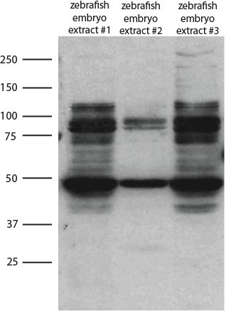
Application: Western BlotSample Tested: ZebrafishSpecies: ZebrafishVerified Customer | Posted 03/29/2018Western blot on zebrafish embryo extracts Customer is aware this antibody is not validated with Zebrafish. Provided review as help to others.

There are no reviews that match your criteria.
Protocols
Find general support by application which include: protocols, troubleshooting, illustrated assays, videos and webinars.
- 7-Amino Actinomycin D (7-AAD) Cell Viability Flow Cytometry Protocol
- Appropriate Fixation of IHC/ICC Samples
- Cellular Response to Hypoxia Protocols
- ClariTSA™ Fluorophore Kits
- Detection & Visualization of Antibody Binding
- Extracellular Membrane Flow Cytometry Protocol
- Flow Cytometry Protocol for Cell Surface Markers
- Flow Cytometry Protocol for Staining Membrane Associated Proteins
- Flow Cytometry Staining Protocols
- Flow Cytometry Troubleshooting Guide
- ICC Cell Smear Protocol for Suspension Cells
- ICC Immunocytochemistry Protocol Videos
- ICC for Adherent Cells
- Immunocytochemistry (ICC) Protocol
- Immunocytochemistry Troubleshooting
- Immunofluorescence of Organoids Embedded in Cultrex Basement Membrane Extract
- Immunohistochemistry (IHC) and Immunocytochemistry (ICC) Protocols
- Intracellular Flow Cytometry Protocol Using Alcohol (Methanol)
- Intracellular Flow Cytometry Protocol Using Detergents
- Intracellular Nuclear Staining Flow Cytometry Protocol Using Detergents
- Intracellular Staining Flow Cytometry Protocol Using Alcohol Permeabilization
- Intracellular Staining Flow Cytometry Protocol Using Detergents to Permeabilize Cells
- Preparing Samples for IHC/ICC Experiments
- Preventing Non-Specific Staining (Non-Specific Binding)
- Primary Antibody Selection & Optimization
- Propidium Iodide Cell Viability Flow Cytometry Protocol
- Protocol for Liperfluo
- Protocol for VisUCyte™ HRP Polymer Detection Reagent
- Protocol for the Characterization of Human Th22 Cells
- Protocol for the Characterization of Human Th9 Cells
- Protocol for the Fluorescent ICC Staining of Cell Smears - Graphic
- Protocol for the Fluorescent ICC Staining of Cultured Cells on Coverslips - Graphic
- Protocol for the Preparation and Fluorescent ICC Staining of Cells on Coverslips
- Protocol for the Preparation and Fluorescent ICC Staining of Non-adherent Cells
- Protocol for the Preparation and Fluorescent ICC Staining of Stem Cells on Coverslips
- Protocol for the Preparation of a Cell Smear for Non-adherent Cell ICC - Graphic
- Protocol: Annexin V and PI Staining by Flow Cytometry
- Protocol: Annexin V and PI Staining for Apoptosis by Flow Cytometry
- R&D Systems Quality Control Western Blot Protocol
- TUNEL and Active Caspase-3 Detection by IHC/ICC Protocol
- The Importance of IHC/ICC Controls
- Troubleshooting Guide: Fluorokine Flow Cytometry Kits
- Troubleshooting Guide: Western Blot Figures
- Western Blot Conditions
- Western Blot Protocol
- Western Blot Protocol for Cell Lysates
- Western Blot Troubleshooting
- Western Blot Troubleshooting Guide
- View all Protocols, Troubleshooting, Illustrated assays and Webinars
Loading...