
Phosphatase of Regenerating Liver-3 (PRL-3), also known as PTP4A3, is an enzyme that removes phosphate modifications from tyrosine residues on proteins. Prenylation at the C-terminal end targets PRL-3 to membrane compartments in the cell, especially to early endosomes and the plasma membrane. Elevated levels of PRL-3 are observed in metastatic colorectal, ovarian, liver and skin cancers. PRL-3 levels may correlate with cell migration rates and tumor invasiveness.


Scientific images may be resized, cropped, or adjusted for brightness or contrast for presentation purposes. These updates are limited to presentation and do not affect the underlying scientific interpretation of the data. Where available, additional source data may be provided upon request.
Key Product Details
Species Reactivity
Validated:
Human, Mouse, Rat
Cited:
Human, Zebrafish
Applications
Validated:
Western Blot
Cited:
Immunohistochemistry-Paraffin, Western Blot, Immunoprecipitation
Label
Unconjugated
Antibody Source
Monoclonal Mouse IgG2B Clone # 334402
Loading...
Product Specifications
Immunogen
E. coli-derived recombinant human PRL-3
Ala2-Met173
Accession # O75365
Ala2-Met173
Accession # O75365
Specificity
Detects endogenous human, mouse and rat PRL-3 in Western blots. In Western blots, this antibody does not cross‑react with recombinant human PRL-1 or PRL‑2.
Clonality
Monoclonal
Host
Mouse
Isotype
IgG2B
Scientific Data Images for PRL-3/PTP4A3 Antibody (334402)
Detection of Human/Mouse/Rat PRL‑3 by Western Blot.
Western blot shows lysates of HeLa human cervical epithelial carcinoma cell line, Neuro-2A mouse neuroblastoma cell line, and Nb2-11 rat lymphoma cell line. PVDF membrane was probed with 0.3 µg/mL of Human/Mouse/Rat PRL-3 Monoclonal Antibody (Catalog # MAB3219) followed by HRP-conjugated Anti-Mouse IgG Secondary Antibody (Catalog # HAF007). A specific band was detected for PRL-3 at approximately 22 kDa (as indicated). This experiment was conducted under reducing conditions and using Immunoblot Buffer Group 1.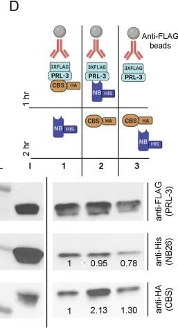
Detection of PRL-3/PTP4A3 by Western Blot
Anti-PRL-3 nanobodies partially interact with the PRL-3 active site and site of CNNM3 CBS-domain binding.(A) Phosphatase activity of PRL-3 alone or in complex with nanobody 91, against a generic diFMUP substrate. DiFMUP fluoresces when phosphate is released. The graph shows normalized fluorescence to PRL-3 alone, n = 12 ± standard deviation. Assays were completed with six technical replicates and repeated in two biological replicates. Error bars represent standard deviation. ns = not significant, **p < 0.01, ***p<0.001 ****p < 0.0001 by one-way ANOVA with Dunnett’s multiple comparisons test. (B) PDB:5TSR where the CNNM CBS domain is colored in orange, and PRL-3 in blue. Specifically, the dark blue is the footprint for nanobody 91 binding (Fig 5), and cyan is the rest of the PRL-3 surface filling structure. (C) Controls for competition assays between CNNM3 CBS domain and nanobody 26. 3XFLAG-PRL-3 pulldown and co-immunoprecipitation controls include His-tagged nanobody 26 pulldown alone or HA-tagged CNNM CBS domain pulldown alone, to show both proteins Co-IP with PRL-3. (D) Immunoprecipitation competition assays 1–3. 3XFLAG-tagged PRL-3 was complexed with anti-FLAG beads and either the HA-tagged CBS domain of CNNM3 (1), histidine-tagged nanobody 26 (2), or neither for 1 hour. After 1 hr incubation, nanobody 26-His (1) CBS-HA (2), or both proteins (3) were added to the complex for the second hour. L, ladder; I, input. Antibodies used for western blot are shown. Quantification is of CBS-HA and nanobody 26-His pulldown normalized to 3XFLAG-PRL-3 immunoprecipitation lane by ImageLab normalization analysis. Image collected and cropped by CiteAb from the following open publication (https://pubmed.ncbi.nlm.nih.gov/37220097), licensed under a CC-BY license. Not internally tested by R&D Systems.Applications for PRL-3/PTP4A3 Antibody (334402)
Application
Recommended Usage
Western Blot
0.3 µg/mL
Sample: HeLa human cervical epithelial carcinoma cell line, Neuro-2A mouse neuroblastoma cell line, and Nb2-11 rat lymphoma cell line
Sample: HeLa human cervical epithelial carcinoma cell line, Neuro-2A mouse neuroblastoma cell line, and Nb2-11 rat lymphoma cell line
Reviewed Applications
Read 3 reviews rated 3.7 using MAB3219 in the following applications:
Formulation, Preparation, and Storage
Purification
Protein A or G purified from hybridoma culture supernatant
Reconstitution
Reconstitute at 0.5 mg/mL in sterile PBS. For liquid material, refer to CoA for concentration.
Loading...
Formulation
Lyophilized from a 0.2 μm filtered solution in PBS with Trehalose. *Small pack size (SP) is supplied either lyophilized or as a 0.2 µm filtered solution in PBS.
Shipping
Lyophilized product is shipped at ambient temperature. Liquid small pack size (-SP) is shipped with polar packs. Upon receipt, store immediately at the temperature recommended below.
Stability & Storage
Use a manual defrost freezer and avoid repeated freeze-thaw cycles.
- 12 months from date of receipt, -20 to -70 °C as supplied.
- 1 month, 2 to 8 °C under sterile conditions after reconstitution.
- 6 months, -20 to -70 °C under sterile conditions after reconstitution.
Calculators
Background: PRL-3/PTP4A3
Long Name
Phosphatase of Regenerating Liver 3
Alternate Names
PRL3, PTP4A3
Gene Symbol
PTP4A3
UniProt
Additional PRL-3/PTP4A3 Products
Product Documents for PRL-3/PTP4A3 Antibody (334402)
Certificate of Analysis
To download a Certificate of Analysis, please enter a lot or batch number in the search box below.
Note: Certificate of Analysis not available for kit components.
Product Specific Notices for PRL-3/PTP4A3 Antibody (334402)
For research use only
Related Research Areas
Citations for PRL-3/PTP4A3 Antibody (334402)
 Powered by Bioz
Customer Reviews for PRL-3/PTP4A3 Antibody (334402) (3)
3.7 out of 5
3 Customer Ratings
Have you used PRL-3/PTP4A3 Antibody (334402)?
Submit a review and receive an Amazon gift card!
$25/€18/£15/$25CAN/¥2500 for a review with an image
$10/€7/£6/$10CAN/¥1110 for a review without an image
Submit a review
Customer Images

Showing
1
-
3 of
3 reviews
Showing All
Filter By:
-
Application: MicroarraysSample Tested: EDTA PlasmaSpecies: HumanVerified Customer | Posted 01/03/2020

-
Application: MicroarraySample Tested: EDTA PlasmaSpecies: HumanVerified Customer | Posted 02/21/2019
-
Application: ELISASample Tested: Serum and PlasmaSpecies: HumanVerified Customer | Posted 11/11/2018
There are no reviews that match your criteria.
Protocols
Find general support by application which include: protocols, troubleshooting, illustrated assays, videos and webinars.
- Cellular Response to Hypoxia Protocols
- R&D Systems Quality Control Western Blot Protocol
- Troubleshooting Guide: Western Blot Figures
- Western Blot Conditions
- Western Blot Protocol
- Western Blot Protocol for Cell Lysates
- Western Blot Troubleshooting
- Western Blot Troubleshooting Guide
- View all Protocols, Troubleshooting, Illustrated assays and Webinars
Loading...