
Key Product Details
Species Reactivity
Validated:
Human, Mouse
Cited:
Human, Mouse
Applications
Validated:
Western Blot
Cited:
Immunohistochemistry, Immunohistochemistry-Frozen, Western Blot, Neutralization
Label
Unconjugated
Antibody Source
Monoclonal Mouse IgG2B Clone # 259820
Loading...
Product Specifications
Immunogen
Mouse myeloma cell line NS0-derived recombinant human TSG-6
Trp18-Leu277
Accession # P98066
Trp18-Leu277
Accession # P98066
Specificity
Detects human and mouse TSG-6 in direct ELISAs and Western blots.
Clonality
Monoclonal
Host
Mouse
Isotype
IgG2B
Scientific Data Images for TSG-6 Antibody (259820)
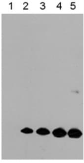
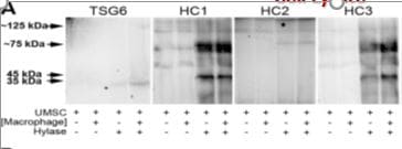
Detection of TSG-6 by Western Blot
Phosphorylated beta -catenin at S311 by PLK1 induced metastasis in an in vivo model. A549 cells expressing RFP-tagged WT of beta -catenin and WT at S311, S311D, or S311A of beta -cateninmtGSK3 beta were injected intravenously into the tail veins of four-week-old BALB/c nude mice, and the tumorigenic and metastatic properties were evaluated after 15 weeks. A, Representative lung tumors from the mouse model. B, The number of metastatic lung tumors was counted and plotted (n = 5). Data are presented as mean ± SD. C-F, Representative H&E staining (C-D) and Ki-67 staining (E-F) were performed using lung tissue from the mice. The relative density of H&E staining (D) and Ki-67 staining (F) was analyzed and plotted. *p <0.05; **p <0.01; ***p <0.001. Data are presented as mean ± SD. G, Immunoblotting was performed using lung tissue lysates from each mouse model. beta -catenin, RFP, N-cadherin, vimentin, CD44, c-Jun, TSG6, laminin gamma 2, and beta -actin were detected using specific antibodies. The band intensity values were quantified using densitometry of Photoshop software, normalized, and plotted (left panel). *p <0.05; **p <0.01; ***p <0.001. H, qRT-PCR was performed for RFP, CDH1, CDH2, CD44, JUN, LAMC2, and TNFAIP6 using lung tissue lysates from each mouse model. The relative mRNA expression was plotted. *p <0.05; **p <0.01; ***p <0.001. Image collected and cropped by CiteAb from the following open publication (https://pubmed.ncbi.nlm.nih.gov/36793862), licensed under a CC-BY license. Not internally tested by R&D Systems.
Detection of TSG-6 by Western Blot
Clinical relevance of PLK1 and beta -catenin in metastatic NSCLC. A-C, The overall survival (OS) times in NSCLC whole patients (n=1292) (A), NSCLC patients with stage 1 (n=522) (B), and stages 3-4 (n=48) (C) were analyzed according to their PLK1 and CTNNB1 expression levels using KM PLOTTER. High (Hi) and low (Lo) were generated by dividing patients according to their expression at the median cut-off. D, A heatmap analysis was performed from the TCGA dataset of lung adenocarcinoma patients for CTNNB1, PLK1, TNFAIP6, LAMC2, CD44, epithelial marker (CDH1), and mesenchymal markers (CDH2, SNAI1, SNAI2, TWIST1, TWIST2, ZEB1, and ZEB2) in paired normal (left) and tumor tissues (right) depending on stages. E-F, A549 and NCI-H460 (H460) non-small cell lung cancer cells were treated with 2.5 ng/ml of TGF-beta for 48 hours. E, Immunoblotting was performed to measure the protein levels of beta -catenin, laminin gamma 2, TSG6, CD44, c-Jun, c-Fos, PLK1, p-PLK1T210, p-Smad2S465/S467, Smad2/3, E-cadherin, N-cadherin, vimentin, SNAI1, SNAI2, and GAPDH in A549 (left panel) and NCI-H460 (H460) cells (right panel). F, The relative band intensities for beta -catenin, laminin gamma 2, CD44, TSG6, c-Jun, c-Fos, PLK1, p-PLK1T210, E-cadherin, N-cadherin, vimentin, SNAI1, and SNAI2 were quantified using densitometry of Photoshop software. G, The relative band intensities for beta -catenin/GAPDH, p-PLK1T210/PLK1, and p-Smad2S465/S467/Smad2 were quantified using densitometry of Photoshop software. H-I, qRT-PCR was performed for CTNNB1, CDH1, CDH2, PLK1, TNFAIP6, LAMC2, and CD44 expression in A549 (H) and NCI-H460 (I) cells. Data are presented as mean ± SD of at least three independent experiments (significantly different as compared with experimental control). *p <0.05; **p <0.01; ***p <0.001. Image collected and cropped by CiteAb from the following open publication (https://pubmed.ncbi.nlm.nih.gov/36793862), licensed under a CC-BY license. Not internally tested by R&D Systems.
Detection of TSG-6 by Western Blot
Phosphorylated beta -catenin at S311 by PLK1 induced metastasis in an in vivo model. A549 cells expressing RFP-tagged WT of beta -catenin and WT at S311, S311D, or S311A of beta -cateninmtGSK3 beta were injected intravenously into the tail veins of four-week-old BALB/c nude mice, and the tumorigenic and metastatic properties were evaluated after 15 weeks. A, Representative lung tumors from the mouse model. B, The number of metastatic lung tumors was counted and plotted (n = 5). Data are presented as mean ± SD. C-F, Representative H&E staining (C-D) and Ki-67 staining (E-F) were performed using lung tissue from the mice. The relative density of H&E staining (D) and Ki-67 staining (F) was analyzed and plotted. *p <0.05; **p <0.01; ***p <0.001. Data are presented as mean ± SD. G, Immunoblotting was performed using lung tissue lysates from each mouse model. beta -catenin, RFP, N-cadherin, vimentin, CD44, c-Jun, TSG6, laminin gamma 2, and beta -actin were detected using specific antibodies. The band intensity values were quantified using densitometry of Photoshop software, normalized, and plotted (left panel). *p <0.05; **p <0.01; ***p <0.001. H, qRT-PCR was performed for RFP, CDH1, CDH2, CD44, JUN, LAMC2, and TNFAIP6 using lung tissue lysates from each mouse model. The relative mRNA expression was plotted. *p <0.05; **p <0.01; ***p <0.001. Image collected and cropped by CiteAb from the following open publication (https://pubmed.ncbi.nlm.nih.gov/36793862), licensed under a CC-BY license. Not internally tested by R&D Systems.
Detection of TSG-6 by Western Blot
Clinical relevance of PLK1 and beta -catenin in metastatic NSCLC. A-C, The overall survival (OS) times in NSCLC whole patients (n=1292) (A), NSCLC patients with stage 1 (n=522) (B), and stages 3-4 (n=48) (C) were analyzed according to their PLK1 and CTNNB1 expression levels using KM PLOTTER. High (Hi) and low (Lo) were generated by dividing patients according to their expression at the median cut-off. D, A heatmap analysis was performed from the TCGA dataset of lung adenocarcinoma patients for CTNNB1, PLK1, TNFAIP6, LAMC2, CD44, epithelial marker (CDH1), and mesenchymal markers (CDH2, SNAI1, SNAI2, TWIST1, TWIST2, ZEB1, and ZEB2) in paired normal (left) and tumor tissues (right) depending on stages. E-F, A549 and NCI-H460 (H460) non-small cell lung cancer cells were treated with 2.5 ng/ml of TGF-beta for 48 hours. E, Immunoblotting was performed to measure the protein levels of beta -catenin, laminin gamma 2, TSG6, CD44, c-Jun, c-Fos, PLK1, p-PLK1T210, p-Smad2S465/S467, Smad2/3, E-cadherin, N-cadherin, vimentin, SNAI1, SNAI2, and GAPDH in A549 (left panel) and NCI-H460 (H460) cells (right panel). F, The relative band intensities for beta -catenin, laminin gamma 2, CD44, TSG6, c-Jun, c-Fos, PLK1, p-PLK1T210, E-cadherin, N-cadherin, vimentin, SNAI1, and SNAI2 were quantified using densitometry of Photoshop software. G, The relative band intensities for beta -catenin/GAPDH, p-PLK1T210/PLK1, and p-Smad2S465/S467/Smad2 were quantified using densitometry of Photoshop software. H-I, qRT-PCR was performed for CTNNB1, CDH1, CDH2, PLK1, TNFAIP6, LAMC2, and CD44 expression in A549 (H) and NCI-H460 (I) cells. Data are presented as mean ± SD of at least three independent experiments (significantly different as compared with experimental control). *p <0.05; **p <0.01; ***p <0.001. Image collected and cropped by CiteAb from the following open publication (https://pubmed.ncbi.nlm.nih.gov/36793862), licensed under a CC-BY license. Not internally tested by R&D Systems.
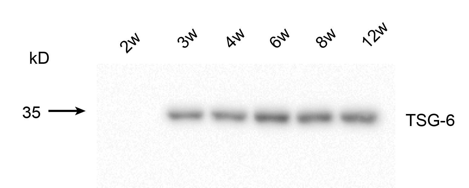
Detection of TSG-6 by Western Blot
Reduced reproductive performance in old mice. A) Comparison of litter size in young and old mice. B) Decreased proliferation of granulosa cells in older mice. C–E) Cell cycle analysis in young and old mice. F–H) The mRNA (F) and protein (G-H) expression levels of CDK1, HAS2, TNFAIP6 and PTX3 in GCs. (n = 3). *P < 0.05, **P < 0.01 Image collected and cropped by CiteAb from the following open publication (https://pubmed.ncbi.nlm.nih.gov/39497025), licensed under a CC-BY license. Not internally tested by R&D Systems.Applications for TSG-6 Antibody (259820)
Application
Recommended Usage
Western Blot
1 µg/mL
Sample: Recombinant Human TSG‑6 (Catalog # 2104-TS)
Recombinant Mouse TSG‑6 (Catalog # 2326-TS)
Sample: Recombinant Human TSG‑6 (Catalog # 2104-TS)
Recombinant Mouse TSG‑6 (Catalog # 2326-TS)
Reviewed Applications
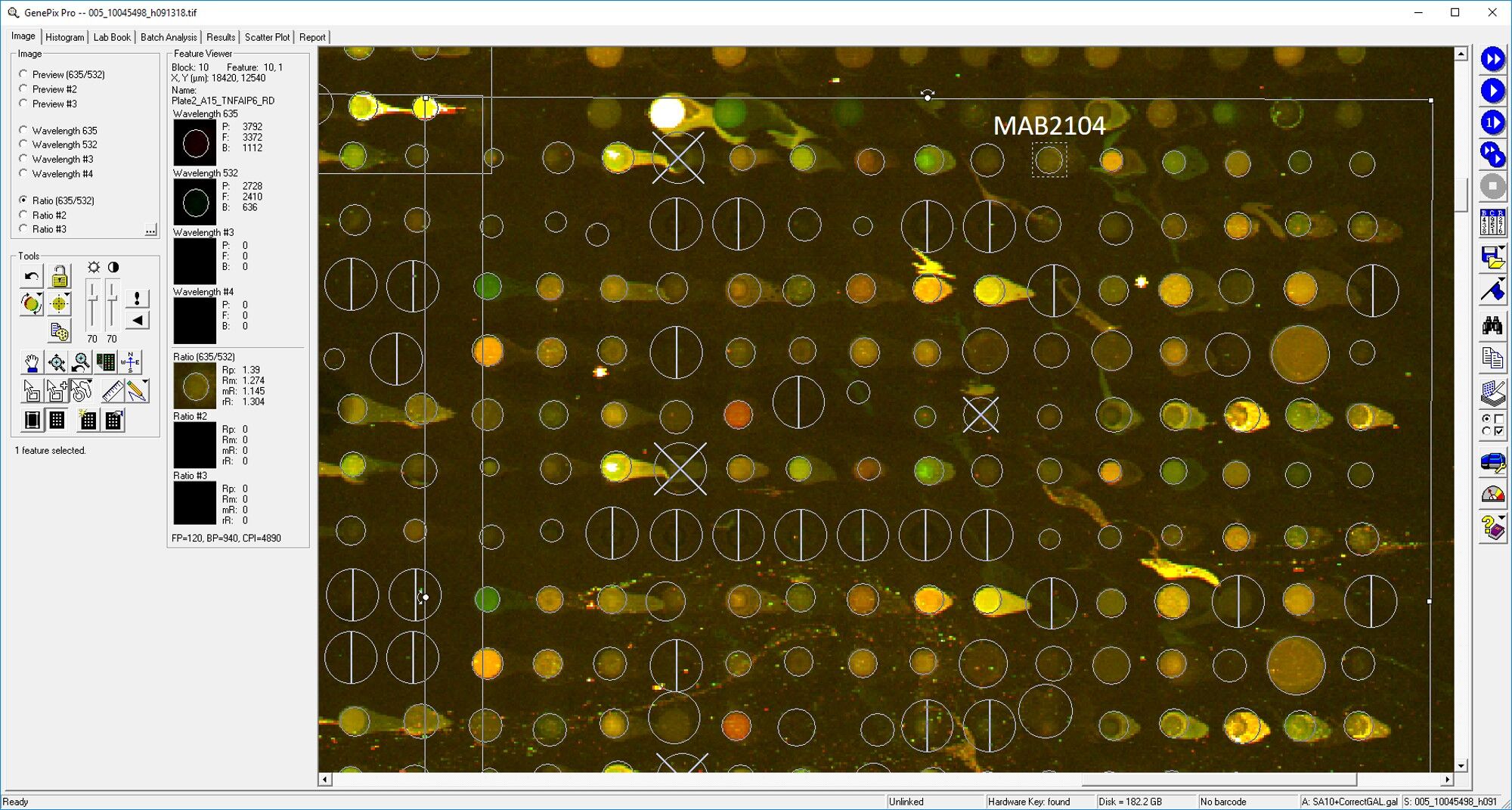
Read 10 reviews rated 4.1 using MAB2104 in the following applications:
Formulation, Preparation, and Storage
Purification
Protein A or G purified from hybridoma culture supernatant
Reconstitution
Reconstitute at 0.5 mg/mL in sterile PBS. For liquid material, refer to CoA for concentration.
Loading...
Formulation
Lyophilized from a 0.2 μm filtered solution in PBS with Trehalose. *Small pack size (SP) is supplied either lyophilized or as a 0.2 µm filtered solution in PBS.
Shipping
Lyophilized product is shipped at ambient temperature. Liquid small pack size (-SP) is shipped with polar packs. Upon receipt, store immediately at the temperature recommended below.
Stability & Storage
Use a manual defrost freezer and avoid repeated freeze-thaw cycles.
- 12 months from date of receipt, -20 to -70 °C as supplied.
- 1 month, 2 to 8 °C under sterile conditions after reconstitution.
- 6 months, -20 to -70 °C under sterile conditions after reconstitution.
Calculators
Background: TSG-6
Long Name
Tumor Necrosis Factor-stimulated Gene Sequence 6
Alternate Names
TNFAIP6, TSG6
Gene Symbol
TNFAIP6
UniProt
Additional TSG-6 Products
Product Documents for TSG-6 Antibody (259820)
Certificate of Analysis
To download a Certificate of Analysis, please enter a lot or batch number in the search box below.
Note: Certificate of Analysis not available for kit components.
Product Specific Notices for TSG-6 Antibody (259820)
For research use only
Related Research Areas
Citations for TSG-6 Antibody (259820)
 Powered by Bioz
Customer Reviews for TSG-6 Antibody (259820) (10)
4.1 out of 5
10 Customer Ratings
Have you used TSG-6 Antibody (259820)?
Submit a review and receive an Amazon gift card!
$25/€18/£15/$25CAN/¥2500 Yen for a review with an image
$10/€7/£6/$10CAN/¥1110 Yen for a review without an image
Submit a review
Customer Images
Showing
1
-
5 of
10 reviews
Showing All
Filter By:
-
Application: Western BlotSample Tested: extracellular vesiclesSpecies: HumanVerified Customer | Posted 09/10/2021

-
Application: Western BlotSample Tested: PlasmaSpecies: MouseVerified Customer | Posted 09/01/2021
-
Application: Western BlotSample Tested: Brain (cerebral cortex)Species: MouseVerified Customer | Posted 08/06/2021this antibody was detected in the mouse cortex at different timepoint. It works very good with very clear band.
-
Application: MicroarraysSample Tested: EDTA PlasmaSpecies: HumanVerified Customer | Posted 03/11/2019
-
Application: MicroarraySample Tested: EDTA PlasmaSpecies: HumanVerified Customer | Posted 02/21/2019
-
Application: Western BlotSample Tested: HUVEC human umbilical vein endothelial cellsSpecies: HumanVerified Customer | Posted 01/19/2019Western Blot with diff concentration
-
Application: ImmunohistochemistrySample Tested: HUVEC human umbilical vein endothelial cellsSpecies: HumanVerified Customer | Posted 12/08/2018
-
Application: ELISASample Tested: Serum and PlasmaSpecies: HumanVerified Customer | Posted 11/11/2018
-
Application: Western BlotSample Tested: See PMID 22351758Species: HumanVerified Customer | Posted 02/19/2015
-
Application: Immunohistochemistry-FrozenSample Tested: See PMID 22351758Species: HumanVerified Customer | Posted 02/19/2015
There are no reviews that match your criteria.
Protocols
Find general support by application which include: protocols, troubleshooting, illustrated assays, videos and webinars.
- Cellular Response to Hypoxia Protocols
- R&D Systems Quality Control Western Blot Protocol
- Troubleshooting Guide: Western Blot Figures
- Western Blot Conditions
- Western Blot Protocol
- Western Blot Protocol for Cell Lysates
- Western Blot Troubleshooting
- Western Blot Troubleshooting Guide
- View all Protocols, Troubleshooting, Illustrated assays and Webinars
Loading...