Notch-1 (so named for "notches" in fly wings; also TAN-1) is a 300 kDa member of the Notch family of glycoproteins. It is associated with gene activation in both embryo and adult. Human Notch-1 is a 2538 amino acid (aa) type I transmembrane glycoprotein. It undergoes Golgi processing to generate a heterodimer composed of a 180‑200 kDa disulfide-linked extracellular domain (aa 18‑1664) and a 120 kDa membrane-bound segment (aa 1665‑2556). Upon ligand binding, the 110 kDa segment undergoes two cleavages which generate an NICD (notch intracellular domain) (aa 1754‑2556), a nuclear transcription factor. One isoform shows a deletion of aa 248‑288. Over aa 2428‑2556, human Notch 1 is 83% and 89% aa identical to canine and mouse Notch-1, respectively.
Human Notch-1 Intracellular Domain Antibody
R&D Systems | Catalog # AF3647

Key Product Details
Validated by
Biological Validation
Species Reactivity
Validated:
Human
Cited:
Human
Applications
Validated:
Western Blot, Flow Cytometry, Immunocytochemistry, Chromatin Immunoprecipitation (ChIP), CyTOF-ready
Cited:
Immunohistochemistry, Western Blot, Flow Cytometry, Immunocytochemistry
Label
Unconjugated
Antibody Source
Polyclonal Sheep IgG
Loading...
Product Specifications
Immunogen
E. coli-derived recombinant human Notch‑1
Gly2428-Lys2556
Accession # P46531
Gly2428-Lys2556
Accession # P46531
Specificity
Detects the intracellular domain (ICD) of human Notch-1 in direct ELISAs and Western blots. In direct ELISAs and Western blots, less than 1% cross-reactivity with recombinant human (rh) Notch-2 ICD, rhNotch-3 ICD, and rhNotch-4 ICD is observed.
Clonality
Polyclonal
Host
Sheep
Isotype
IgG
Scientific Data Images for Human Notch-1 Intracellular Domain Antibody
Detection of Human Notch-1.
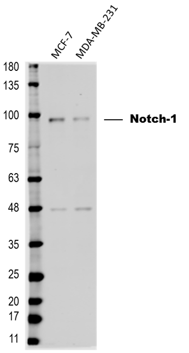
Western blot shows lysates of MOLT-4 human acute lymphoblastic leukemia cell line, Jurkat human acute T cell leukemia cell line, RPMI 8226 human multiple myeloma cell line, 293T human embryonic kidney cell line, and 293T human embryonic kidney cell line (1 µg per lane), transfected with full length human Notch-1. PVDF membrane was probed with 1 µg/mL of Sheep Anti-Human Notch-1 Intracellular Domain Antigen Affinity-purified Polyclonal Antibody (Catalog # AF3647) followed by HRP-conjugated Anti-Sheep IgG Secondary Antibody (Catalog # HAF016). Specific bands were detected for Notch-1 intracellular domain (NICD) and full length Notch-1 (Notch-1 FL) at approximately 115 and 300 kDa (as indicated). This experiment was conducted under reducing conditions and using Immunoblot Buffer Group 1.
Notch‑1 in Saos-2 Human Cell Line.
Notch-1 was detected in immersion fixed Saos-2 human osteosarcoma cell line using 10 µg/mL Sheep Anti-Human Notch-1 Intracellular Domain Antigen Affinity-purified Polyclonal Antibody (Catalog # AF3647) for 3 hours at room temperature. Cells were stained with the NorthernLights™ 557-conjugated Anti-Sheep IgG Secondary Antibody (red; Catalog # NL010) and counterstained with DAPI (blue). View our protocol for Fluorescent ICC Staining of Cells on Coverslips.
Detection of Notch‑1-regulated Genes by Chromatin Immunoprecipitation.
Jurkat human acute T cell leukemia cell line treated with 50 ng/mL PMA and 200 ng/mL calcium ionomycin for 30 minutes was fixed using formaldehyde, resuspended in lysis buffer, and sonicated to shear chromatin. Notch-1/DNA complexes were immunoprecipitated using 5 µg Sheep Anti-Human Notch-1 Intracellular Domain Antigen Affinity-purified Polyclonal Antibody (Catalog # AF3647) or control antibody (Catalog # 5-001-A) for 15 minutes in an ultrasonic bath, followed by Biotinylated Anti-Sheep IgG Secondary Antibody (Catalog # BAF016). Immunocomplexes were captured using 50 µL of MagCellect Streptavidin Ferrofluid (Catalog # MAG999) and DNA was purified using chelating resin solution. Thec-mycpromoter was detected by standard PCR.Applications for Human Notch-1 Intracellular Domain Antibody
Application
Recommended Usage
Chromatin Immunoprecipitation (ChIP)
5 µg/5 x 106 cells
Sample: PMA and calcium ionomycin treated Jurkat human acute T cell leukemia cell line chromatin, c-myc promoter detected by standard PCR
Sample: PMA and calcium ionomycin treated Jurkat human acute T cell leukemia cell line chromatin, c-myc promoter detected by standard PCR
CyTOF-ready
Ready to be labeled using established conjugation methods. No BSA or other carrier proteins that could interfere with conjugation.
Flow Cytometry
2.5 µg/106 cells
Sample: U2OS human osteosarcoma cell line
Sample: U2OS human osteosarcoma cell line
Immunocytochemistry
5-15 µg/mL
Sample: Immersion fixed Saos-2 human osteosarcoma cell line
Sample: Immersion fixed Saos-2 human osteosarcoma cell line
Western Blot
1 µg/mL
Sample: MOLT‑4 human acute lymphoblastic leukemia cell line, Jurkat human acute T cell leukemia cell line, RPMI 8226 human multiple myeloma cell line, 293T human embryonic kidney cell line, and 293T human embryonic kidney cell line, transfected with full length human Notch-1
Sample: MOLT‑4 human acute lymphoblastic leukemia cell line, Jurkat human acute T cell leukemia cell line, RPMI 8226 human multiple myeloma cell line, 293T human embryonic kidney cell line, and 293T human embryonic kidney cell line, transfected with full length human Notch-1
Reviewed Applications
Read 4 reviews rated 3.8 using AF3647 in the following applications:
Flow Cytometry Panel Builder
Bio-Techne Knows Flow Cytometry
Save time and reduce costly mistakes by quickly finding compatible reagents using the Panel Builder Tool.
Advanced Features
- Spectra Viewer - Custom analysis of spectra from multiple fluorochromes
- Spillover Popups - Visualize the spectra of individual fluorochromes
- Antigen Density Selector - Match fluorochrome brightness with antigen density

Formulation, Preparation, and Storage
Purification
Antigen Affinity-purified
Reconstitution
Reconstitute at 0.2 mg/mL in sterile PBS. For liquid material, refer to CoA for concentration.
Loading...
Formulation
Lyophilized from a 0.2 μm filtered solution in PBS with Trehalose. *Small pack size (SP) is supplied either lyophilized or as a 0.2 µm filtered solution in PBS.
Shipping
Lyophilized product is shipped at ambient temperature. Liquid small pack size (-SP) is shipped with polar packs. Upon receipt, store immediately at the temperature recommended below.
Stability & Storage
Use a manual defrost freezer and avoid repeated freeze-thaw cycles.
- 12 months from date of receipt, -20 to -70 °C as supplied.
- 1 month, 2 to 8 °C under sterile conditions after reconstitution.
- 6 months, -20 to -70 °C under sterile conditions after reconstitution.
Calculators
Background: Notch-1
Alternate Names
Notch1, TAN1
Gene Symbol
NOTCH1
UniProt
Additional Notch-1 Products
Product Documents for Human Notch-1 Intracellular Domain Antibody
Certificate of Analysis
To download a Certificate of Analysis, please enter a lot or batch number in the search box below.
Note: Certificate of Analysis not available for kit components.
Product Specific Notices for Human Notch-1 Intracellular Domain Antibody
For research use only
Related Research Areas
Citations for Human Notch-1 Intracellular Domain Antibody
 Powered by Bioz
Customer Reviews for Human Notch-1 Intracellular Domain Antibody (4)
3.8 out of 5
4 Customer Ratings
Have you used Human Notch-1 Intracellular Domain Antibody?
Submit a review and receive an Amazon gift card!
$25/€18/£15/$25CAN/¥2500 Yen for a review with an image
$10/€7/£6/$10CAN/¥1110 Yen for a review without an image
Submit a review
Customer Images
Showing
1
-
4 of
4 reviews
Showing All
Filter By:
-
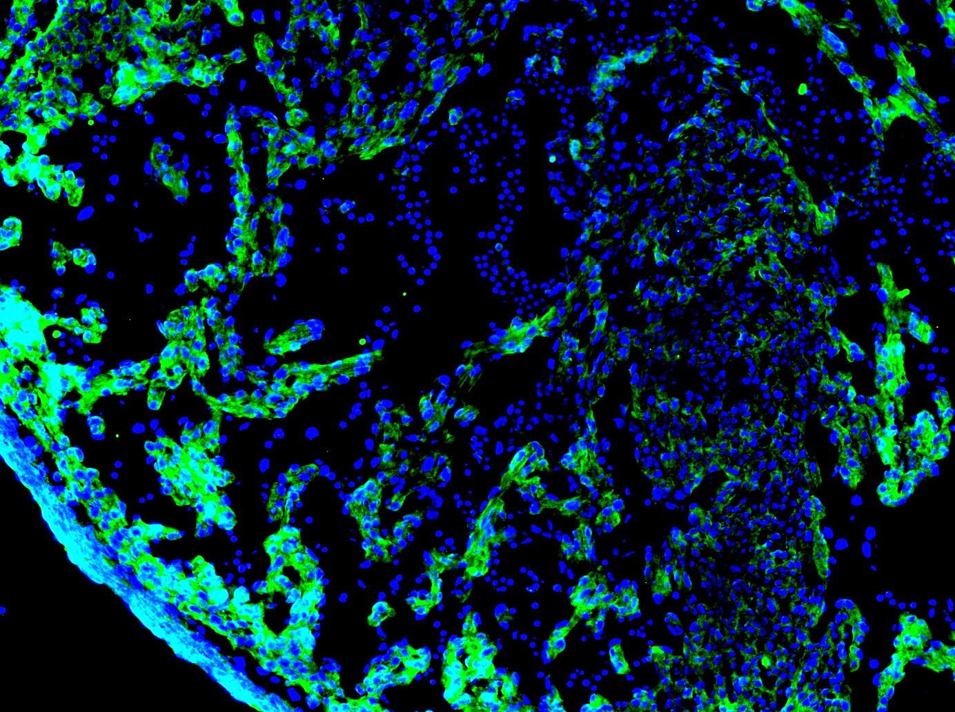
Application: Immunocytochemistry/ImmunofluorescenceSample Tested: E12.5 mouse embryo fixed in 4% PFASpecies: MouseVerified Customer | Posted 06/23/2021Fixed 4% PFA overnight. Blocked with 1% BSA Primary antibody dilution - 1:20 Secondary antibody - Invitrogen Alexa Fluor 488 Secondary antibody dilution - 1:1000 Stained on an E12.5 mouse right ventricle heart section

-
Application: Western BlotSample Tested: iPS2 human induced pluripotent stem cellsSpecies: HumanVerified Customer | Posted 01/25/2018
-
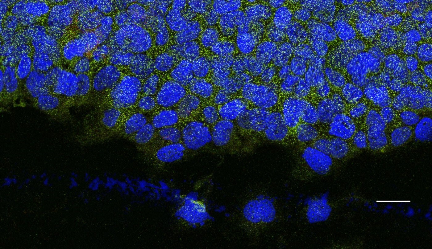
Application: Immunocytochemistry/ImmunofluorescenceSample Tested: Bladder tissue and Bladder cancer tissueSpecies: HumanVerified Customer | Posted 12/10/2017
-
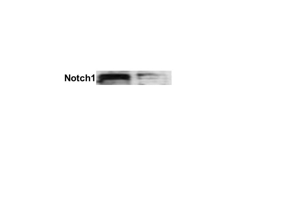
Application: Western BlotSample Tested: Breast cancer cellsSpecies: HumanVerified Customer | Posted 03/01/2017Expression of cleaved Notch-1 (Intracellular Domain) in mammary tumor cell lines. Dilution: 1:200 in PBS with 5% BSA. Secondary Ab: anti-Sheep IgG 1:5,000.
There are no reviews that match your criteria.
Protocols
Find general support by application which include: protocols, troubleshooting, illustrated assays, videos and webinars.
- 7-Amino Actinomycin D (7-AAD) Cell Viability Flow Cytometry Protocol
- Appropriate Fixation of IHC/ICC Samples
- Cellular Response to Hypoxia Protocols
- ChIP Protocol Video
- Chromatin Immunoprecipitation (ChIP) Protocol
- Chromatin Immunoprecipitation Protocol
- ClariTSA™ Fluorophore Kits
- Detection & Visualization of Antibody Binding
- Extracellular Membrane Flow Cytometry Protocol
- Flow Cytometry Protocol for Cell Surface Markers
- Flow Cytometry Protocol for Staining Membrane Associated Proteins
- Flow Cytometry Staining Protocols
- Flow Cytometry Troubleshooting Guide
- ICC Cell Smear Protocol for Suspension Cells
- ICC Immunocytochemistry Protocol Videos
- ICC for Adherent Cells
- Immunocytochemistry (ICC) Protocol
- Immunocytochemistry Troubleshooting
- Immunofluorescence of Organoids Embedded in Cultrex Basement Membrane Extract
- Immunohistochemistry (IHC) and Immunocytochemistry (ICC) Protocols
- Intracellular Flow Cytometry Protocol Using Alcohol (Methanol)
- Intracellular Flow Cytometry Protocol Using Detergents
- Intracellular Nuclear Staining Flow Cytometry Protocol Using Detergents
- Intracellular Staining Flow Cytometry Protocol Using Alcohol Permeabilization
- Intracellular Staining Flow Cytometry Protocol Using Detergents to Permeabilize Cells
- Preparing Samples for IHC/ICC Experiments
- Preventing Non-Specific Staining (Non-Specific Binding)
- Primary Antibody Selection & Optimization
- Propidium Iodide Cell Viability Flow Cytometry Protocol
- Protocol for Liperfluo
- Protocol for VisUCyte™ HRP Polymer Detection Reagent
- Protocol for the Characterization of Human Th22 Cells
- Protocol for the Characterization of Human Th9 Cells
- Protocol for the Fluorescent ICC Staining of Cell Smears - Graphic
- Protocol for the Fluorescent ICC Staining of Cultured Cells on Coverslips - Graphic
- Protocol for the Preparation and Fluorescent ICC Staining of Cells on Coverslips
- Protocol for the Preparation and Fluorescent ICC Staining of Non-adherent Cells
- Protocol for the Preparation and Fluorescent ICC Staining of Stem Cells on Coverslips
- Protocol for the Preparation of a Cell Smear for Non-adherent Cell ICC - Graphic
- Protocol: Annexin V and PI Staining by Flow Cytometry
- Protocol: Annexin V and PI Staining for Apoptosis by Flow Cytometry
- R&D Systems Quality Control Western Blot Protocol
- TUNEL and Active Caspase-3 Detection by IHC/ICC Protocol
- The Importance of IHC/ICC Controls
- Troubleshooting Guide: Fluorokine Flow Cytometry Kits
- Troubleshooting Guide: Western Blot Figures
- Western Blot Conditions
- Western Blot Protocol
- Western Blot Protocol for Cell Lysates
- Western Blot Troubleshooting
- Western Blot Troubleshooting Guide
- View all Protocols, Troubleshooting, Illustrated assays and Webinars
Loading...
Associated Pathways


