Human p16INK4a / CDKN2A Antibody
R&D Systems | Catalog # AF5779

Key Product Details
Species Reactivity
Validated:
Human
Cited:
Human
Applications
Validated:
Western Blot, Immunocytochemistry, Simple Western
Cited:
Immunohistochemistry, Western Blot, Immunocytochemistry, Simple Western
Label
Unconjugated
Antibody Source
Polyclonal Goat IgG
Loading...
Product Specifications
Immunogen
E. coli-derived recombinant human p16INK4a/CDKN2A
Glu2-Asp156
Accession # P42771
Glu2-Asp156
Accession # P42771
Specificity
Detects human p16INK4a/CDKN2A in Western blots.
Clonality
Polyclonal
Host
Goat
Isotype
IgG
Scientific Data Images for Human p16INK4a / CDKN2A Antibody
Detection of Human p16INK4a/CDKN2A by Western Blot.
Western blot shows lysates of HEK293 human embryonic kidney cell line, HepG2 human hepatocellular carcinoma cell line, and Saos-2 human osteosarcoma cell line. PVDF membrane was probed with 1 µg/mL of Goat Anti-Human p16INK4a/CDKN2A Antigen Affinity-purified Polyclonal Antibody (Catalog # AF5779) followed by HRP-conjugated Anti-Goat IgG Secondary Antibody (Catalog # HAF109). A specific band was detected for p16INK4a/CDKN2A at approximately 16 kDa (as indicated). This experiment was conducted under reducing conditions and using Immunoblot Buffer Group 1.
p16INK4a / CDKN2A in HeLa Human Cell Line.
p16INK4a / CDKN2A was detected in immersion fixed HeLa human cervical epithelial carcinoma cell line using Goat Anti-Human p16INK4a / CDKN2A Antigen Affinity-purified Polyclonal Antibody (Catalog # AF5779) at 0.3 µg/mL for 3 hours at room temperature. Cells were stained using the NorthernLights™ 557-conjugated Anti-Goat IgG Secondary Antibody (red; Catalog # NL001) and counterstained with DAPI (blue). Specific staining was localized to cytoplasm and nuclei. View our protocol for Fluorescent ICC Staining of Cells on Coverslips.
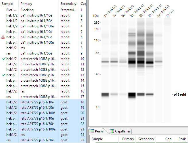
Detection of Human p16INK4a / CDKN2A by Simple WesternTM.
Simple Western lane view shows lysates of HEK293 human embryonic kidney cell line, loaded at 0.2 mg/mL. A specific band was detected for p16INK4a / CDKN2A at approximately 24 kDa (as indicated) using 10 µg/mL of Goat Anti-Human p16INK4a / CDKN2A Antigen Affinity-purified Polyclonal Antibody (Catalog # AF5779) followed by 1:50 dilution of HRP-conjugated Anti-Goat IgG Secondary Antibody (Catalog # HAF109). This experiment was conducted under reducing conditions and using the 12-230 kDa separation system.
Detection of p16INK4a/ CDKN2A by Western Blot
Protein markers of cellular senescence. Western blot analysis of (A) NHEM transduced with mutated BRAFV600E (all n = 7, p16 n = 6) and (B) melanoma cell line Mel Juso treated with 100 µM etoposide (n = 3). (C,D) Immunofluorescent stainings of PML and DAPI. Scale bars equal 20 µm. Bars are shown as mean ± SEM (Student’s t-test). (*: p < 0.05, **: p < 0.01, ***: p < 0.001, ****: p < 0.0001). Image collected and cropped by CiteAb from the following open publication (https://pubmed.ncbi.nlm.nih.gov/35563794), licensed under a CC-BY license. Not internally tested by R&D Systems.
Detection of p16INK4a/ CDKN2A by Western Blot
Protein markers of cellular senescence. Western blot analysis of (A) NHEM transduced with mutated BRAFV600E (all n = 7, p16 n = 6) and (B) melanoma cell line Mel Juso treated with 100 µM etoposide (n = 3). (C,D) Immunofluorescent stainings of PML and DAPI. Scale bars equal 20 µm. Bars are shown as mean ± SEM (Student’s t-test). (*: p < 0.05, **: p < 0.01, ***: p < 0.001, ****: p < 0.0001). Image collected and cropped by CiteAb from the following open publication (https://pubmed.ncbi.nlm.nih.gov/35563794), licensed under a CC-BY license. Not internally tested by R&D Systems.Applications for Human p16INK4a / CDKN2A Antibody
Application
Recommended Usage
Immunocytochemistry
0.3-15 µg/mL
Sample: Immersion fixed HeLa human cervical epithelial carcinoma cell line
Sample: Immersion fixed HeLa human cervical epithelial carcinoma cell line
Simple Western
10 µg/mL
Sample: HEK293 human embryonic kidney cell line
Sample: HEK293 human embryonic kidney cell line
Western Blot
1 µg/mL
Sample: HEK293 human embryonic kidney cell line, HepG2 human hepatocellular carcinoma cell line, and Saos-2 human osteosarcoma cell line
Sample: HEK293 human embryonic kidney cell line, HepG2 human hepatocellular carcinoma cell line, and Saos-2 human osteosarcoma cell line
Reviewed Applications
Read 2 reviews rated 4.5 using AF5779 in the following applications:
Formulation, Preparation, and Storage
Purification
Antigen Affinity-purified
Reconstitution
Reconstitute at 0.2 mg/mL in sterile PBS. For liquid material, refer to CoA for concentration.
Loading...
Formulation
Lyophilized from a 0.2 μm filtered solution in PBS with Trehalose. *Small pack size (SP) is supplied either lyophilized or as a 0.2 µm filtered solution in PBS.
Shipping
Lyophilized product is shipped at ambient temperature. Liquid small pack size (-SP) is shipped with polar packs. Upon receipt, store immediately at the temperature recommended below.
Stability & Storage
Use a manual defrost freezer and avoid repeated freeze-thaw cycles.
- 12 months from date of receipt, -20 to -70 °C as supplied.
- 1 month, 2 to 8 °C under sterile conditions after reconstitution.
- 6 months, -20 to -70 °C under sterile conditions after reconstitution.
Calculators
Background: p16INK4a / CDKN2A
(aa 11‑139) that interact with cyclin. There are at least two splice variants for p16INK4a. One is termed p12 and shows a 65 aa substitution for aa 52‑156; the other simply shows an alternate start site at Met52. Full‑length human p16INK4a shares 63% aa identity with mouse p16INK4a.
Long Name
p16 Cyclin Dependent Kinase 4 Inhibitor 2A
Alternate Names
ARF, CDK4I, CDKN2A, INK4a, MLM, p14ARF, p16, p19ARF
Gene Symbol
CDKN2A
UniProt
Additional p16INK4a / CDKN2A Products
Product Documents for Human p16INK4a / CDKN2A Antibody
Certificate of Analysis
To download a Certificate of Analysis, please enter a lot or batch number in the search box below.
Note: Certificate of Analysis not available for kit components.
Product Specific Notices for Human p16INK4a / CDKN2A Antibody
For research use only
Related Research Areas
Citations for Human p16INK4a / CDKN2A Antibody
 Powered by Bioz
Customer Reviews for Human p16INK4a / CDKN2A Antibody (2)
4.5 out of 5
2 Customer Ratings
Have you used Human p16INK4a / CDKN2A Antibody?
Submit a review and receive an Amazon gift card!
$25/€18/£15/$25CAN/¥2500 Yen for a review with an image
$10/€7/£6/$10CAN/¥1110 Yen for a review without an image
Submit a review
Customer Images
Showing
1
-
2 of
2 reviews
Showing All
Filter By:
-
Application: Simple WesternSample Tested: HEK293 human embryonic kidney cell lineSpecies: HumanVerified Customer | Posted 08/12/2021Human p16INK4a/CDKN2A Antibody Ref:AF5779 R et D Systems Efficacité validée dans conditions suivantes: Dilution anticorps p16= 1/10è (0,2mg/ml solution stock) [HEK]= 0,65mg/ml Anticorps 2aire goat prêt à l’emploi

-
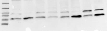
Application: Western BlotSample Tested: Mesenchymal stem cellsSpecies: HumanVerified Customer | Posted 05/20/20165% non fat milk as blocking agent for 1 hour and overnight incubation with primary antibody at 1:3000. Specific band at 16kDa as expected, but also band at 25 KDa. Cells at passage 2 had low p16 expression, while at passage 6 expression was increased as expected. Loading: p2,p6-p2,p6-p2,p6-p2,p6 of MSCs from 4 different individuals.
There are no reviews that match your criteria.
Protocols
Find general support by application which include: protocols, troubleshooting, illustrated assays, videos and webinars.
- Appropriate Fixation of IHC/ICC Samples
- Cellular Response to Hypoxia Protocols
- ClariTSA™ Fluorophore Kits
- Detection & Visualization of Antibody Binding
- ICC Cell Smear Protocol for Suspension Cells
- ICC Immunocytochemistry Protocol Videos
- ICC for Adherent Cells
- Immunocytochemistry (ICC) Protocol
- Immunocytochemistry Troubleshooting
- Immunofluorescence of Organoids Embedded in Cultrex Basement Membrane Extract
- Immunohistochemistry (IHC) and Immunocytochemistry (ICC) Protocols
- Preparing Samples for IHC/ICC Experiments
- Preventing Non-Specific Staining (Non-Specific Binding)
- Primary Antibody Selection & Optimization
- Protocol for VisUCyte™ HRP Polymer Detection Reagent
- Protocol for the Fluorescent ICC Staining of Cell Smears - Graphic
- Protocol for the Fluorescent ICC Staining of Cultured Cells on Coverslips - Graphic
- Protocol for the Preparation and Fluorescent ICC Staining of Cells on Coverslips
- Protocol for the Preparation and Fluorescent ICC Staining of Non-adherent Cells
- Protocol for the Preparation and Fluorescent ICC Staining of Stem Cells on Coverslips
- Protocol for the Preparation of a Cell Smear for Non-adherent Cell ICC - Graphic
- R&D Systems Quality Control Western Blot Protocol
- TUNEL and Active Caspase-3 Detection by IHC/ICC Protocol
- The Importance of IHC/ICC Controls
- Troubleshooting Guide: Western Blot Figures
- Western Blot Conditions
- Western Blot Protocol
- Western Blot Protocol for Cell Lysates
- Western Blot Troubleshooting
- Western Blot Troubleshooting Guide
- View all Protocols, Troubleshooting, Illustrated assays and Webinars
Loading...