Pepsins are aspartic proteases that are synthesized in the gastric mucosa and secreted into the stomach. They are released as zymogens called pepsinogens which are then converted to active pepsins by the acidic pH of gastric juices (1-3). PGA3, PGA4, and PGA5 are human Pepsinogen A isozymogens that differ in sequence by 24 amino acid (aa) residues (4, 5). This recombinant human Pepsinogen A corresponds to PGA5. Human Pepsinogen A isozymogens share approximately 56% aa sequence identity with mouse and rat Pepsinogen A isozymogens. Pepsins have optimal activity under conditions of acidic pH and are inhibited by pepstatin (6, 7). Pepsin A has broad substrate specificity, but preferentially cleaves peptide bonds involving aromatic and aliphatic amino acids.

Key Product Details
Species Reactivity
Human
Applications
Immunohistochemistry, Western Blot, Simple Western
Label
Unconjugated
Antibody Source
Monoclonal Mouse IgG1 Clone # 974731
Loading...
Product Specifications
Immunogen
Human embryonic kidney cell, HEK293-derived human Pepsinogen A5/PGA5 protein
Ile16-Ala388
Accession # P0DJD9
Ile16-Ala388
Accession # P0DJD9
Specificity
Detects human Pepsinogen A5/PGA5 protein in direct ELISAs.
Clonality
Monoclonal
Host
Mouse
Isotype
IgG1
Scientific Data Images for Human Pepsinogen A5/PGA5 Antibody (974731)
Detection of Human Pepsinogen A5/PGA5 by Western Blot.
Western blot shows lysates of human stomach cancer tissue. PVDF membrane was probed with 2 µg/mL of Mouse Anti-Human Pepsinogen A5/PGA5 Monoclonal Antibody (Catalog # MAB8457) followed by HRP-conjugated Anti-Mouse IgG Secondary Antibody (Catalog # HAF018). A specific band was detected for Pepsinogen A5/PGA5 at approximately 45 kDa (as indicated). This experiment was conducted under reducing conditions and using Immunoblot Buffer Group 1.
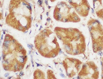
Pepsinogen A5/PGA5 in Human Stomach.
Pepsinogen A5/PGA5 was detected in immersion fixed paraffin-embedded sections of human stomach using Mouse Anti-Human Pepsinogen A5/PGA5 Monoclonal Antibody (Catalog # MAB8457) at 0.5 µg/mL for 1 hour at room temperature followed by incubation with the Anti-Mouse IgG VisUCyte™ HRP Polymer Antibody (Catalog # VC001). Before incubation with the primary antibody, tissue was subjected to heat-induced epitope retrieval using Antigen Retrieval Reagent-Basic (Catalog # CTS013). Tissue was stained using DAB (brown) and counterstained with hematoxylin (blue). Specific staining was localized to intestinal glands. View our protocol for IHC Staining with VisUCyte HRP Polymer Detection Reagents.
Detection of Human Pepsinogen A5/PGA5 by Simple WesternTM.
Simple Western lane view shows lysates of human stomach tissue and human stomach cancer tissue, loaded at 0.2 mg/mL. A specific band was detected for Pepsinogen A5/PGA5 at approximately 48 kDa (as indicated) using 20 µg/mL of Mouse Anti-Human Pepsinogen A5/PGA5 Monoclonal Antibody (Catalog # MAB8457). This experiment was conducted under reducing conditions and using the 12-230 kDa separation system. Non-specific interaction with the 230 kDa Simple Western standard may be seen with this antibody.Applications for Human Pepsinogen A5/PGA5 Antibody (974731)
Application
Recommended Usage
Immunohistochemistry
0.5-25 µg/mL
Sample: Immersion fixed paraffin-embedded sections of human stomach
Sample: Immersion fixed paraffin-embedded sections of human stomach
Simple Western
20 µg/mL
Sample: Human stomach tissue and Human stomach cancer tissue
Sample: Human stomach tissue and Human stomach cancer tissue
Western Blot
2 µg/mL
Sample: Human stomach cancer tissue
Sample: Human stomach cancer tissue
Reviewed Applications
Read 1 review rated 5 using MAB8457 in the following applications:
Formulation, Preparation, and Storage
Purification
Protein A or G purified from hybridoma culture supernatant
Reconstitution
Reconstitute at 0.5 mg/mL in sterile PBS. For liquid material, refer to CoA for concentration.
Loading...
Formulation
Lyophilized from a 0.2 μm filtered solution in PBS with Trehalose. *Small pack size (SP) is supplied either lyophilized or as a 0.2 µm filtered solution in PBS.
Shipping
Lyophilized product is shipped at ambient temperature. Liquid small pack size (-SP) is shipped with polar packs. Upon receipt, store immediately at the temperature recommended below.
Stability & Storage
Use a manual defrost freezer and avoid repeated freeze-thaw cycles.
- 12 months from date of receipt, -20 to -70 °C as supplied.
- 1 month, 2 to 8 °C under sterile conditions after reconstitution.
- 6 months, -20 to -70 °C under sterile conditions after reconstitution.
Calculators
Background: Pepsinogen A5/PGA5
References
- Athauda, S.B. et al. (1989) J. Biochem. 106:920.
- Kageyama, T. et al. (1989) J. Biochem. 105:15.
- Kageyama, T. (2002) Cell. Mol. Life Sci. 59:288.
- Zwiers, A. et al. (1994) Clin. Nephrol. 41:153.
- Nakai, H. et al. (1986) Cytogenet. Cell Genet. 43:215.
- Marciniszyn, J., Jr. et al. (1976) J. Biol. Chem. 251:7088.
- Kageyama, T. and K. Takahashi (1980) J. Biochem. 88:571.
Alternate Names
PGA5
Entrez Gene IDs
5222 (Human)
Gene Symbol
PGA5
UniProt
Additional Pepsinogen A5/PGA5 Products
Product Documents for Human Pepsinogen A5/PGA5 Antibody (974731)
Certificate of Analysis
To download a Certificate of Analysis, please enter a lot or batch number in the search box below.
Note: Certificate of Analysis not available for kit components.
Product Specific Notices for Human Pepsinogen A5/PGA5 Antibody (974731)
For research use only
Related Research Areas
Customer Reviews for Human Pepsinogen A5/PGA5 Antibody (974731) (1)
5 out of 5
1 Customer Rating
Have you used Human Pepsinogen A5/PGA5 Antibody (974731)?
Submit a review and receive an Amazon gift card!
$25/€18/£15/$25CAN/¥2500 Yen for a review with an image
$10/€7/£6/$10CAN/¥1110 Yen for a review without an image
Submit a review
Customer Images
Showing
1
-
1 of
1 review
Showing All
Filter By:
-
Application: ImmunohistochemistrySample Tested: Stomach tissueSpecies: HumanVerified Customer | Posted 06/28/2022

There are no reviews that match your criteria.
Protocols
Find general support by application which include: protocols, troubleshooting, illustrated assays, videos and webinars.
- Antigen Retrieval Protocol (PIER)
- Antigen Retrieval for Frozen Sections Protocol
- Appropriate Fixation of IHC/ICC Samples
- Cellular Response to Hypoxia Protocols
- Chromogenic IHC Staining of Formalin-Fixed Paraffin-Embedded (FFPE) Tissue Protocol
- Chromogenic Immunohistochemistry Staining of Frozen Tissue
- ClariTSA™ Fluorophore Kits
- Detection & Visualization of Antibody Binding
- Fluorescent IHC Staining of Frozen Tissue Protocol
- Graphic Protocol for Heat-induced Epitope Retrieval
- Graphic Protocol for the Preparation and Fluorescent IHC Staining of Frozen Tissue Sections
- Graphic Protocol for the Preparation and Fluorescent IHC Staining of Paraffin-embedded Tissue Sections
- Graphic Protocol for the Preparation of Gelatin-coated Slides for Histological Tissue Sections
- IHC Sample Preparation (Frozen sections vs Paraffin)
- Immunofluorescent IHC Staining of Formalin-Fixed Paraffin-Embedded (FFPE) Tissue Protocol
- Immunohistochemistry (IHC) and Immunocytochemistry (ICC) Protocols
- Immunohistochemistry Frozen Troubleshooting
- Immunohistochemistry Paraffin Troubleshooting
- Preparing Samples for IHC/ICC Experiments
- Preventing Non-Specific Staining (Non-Specific Binding)
- Primary Antibody Selection & Optimization
- Protocol for Heat-Induced Epitope Retrieval (HIER)
- Protocol for Making a 4% Formaldehyde Solution in PBS
- Protocol for VisUCyte™ HRP Polymer Detection Reagent
- Protocol for the Preparation & Fixation of Cells on Coverslips
- Protocol for the Preparation and Chromogenic IHC Staining of Frozen Tissue Sections
- Protocol for the Preparation and Chromogenic IHC Staining of Frozen Tissue Sections - Graphic
- Protocol for the Preparation and Chromogenic IHC Staining of Paraffin-embedded Tissue Sections
- Protocol for the Preparation and Chromogenic IHC Staining of Paraffin-embedded Tissue Sections - Graphic
- Protocol for the Preparation and Fluorescent IHC Staining of Frozen Tissue Sections
- Protocol for the Preparation and Fluorescent IHC Staining of Paraffin-embedded Tissue Sections
- Protocol for the Preparation of Gelatin-coated Slides for Histological Tissue Sections
- R&D Systems Quality Control Western Blot Protocol
- TUNEL and Active Caspase-3 Detection by IHC/ICC Protocol
- The Importance of IHC/ICC Controls
- Troubleshooting Guide: Immunohistochemistry
- Troubleshooting Guide: Western Blot Figures
- Western Blot Conditions
- Western Blot Protocol
- Western Blot Protocol for Cell Lysates
- Western Blot Troubleshooting
- Western Blot Troubleshooting Guide
- View all Protocols, Troubleshooting, Illustrated assays and Webinars
Loading...