The cAMP response element binding protein (CREB) belongs to the bZIP superfamily of transcription factors, containing a basic domain that mediates DNA binding and a leucine zipper domain that facilitates dimerization. Within the promoter of target genes, CREB dimers bind cAMP response elements, defined by the palindromic consensus sequence TGACGTCA. When phosphorylated at Ser133, CREB also binds the coactivator CREB binding protein (CBP), which enhances transcription by acetylating histones to facilitate chromatin unraveling.
Human phospho-CREB (S133) Antibody (702710R)
R&D Systems | Catalog # MAB6906R
Recombinant Monoclonal Antibody.
 by Western Blot.")
Key Product Details
Validated by
Biological Validation
Species Reactivity
Human
Applications
Western Blot
Label
Unconjugated
Antibody Source
Recombinant Monoclonal Mouse IgG2A Clone # 702710R
Loading...
Product Specifications
Immunogen
Phosphopeptide containing the human CREB S133 site
Specificity
Detects human CREB when phosphorylated at S133.
Clonality
Monoclonal
Host
Mouse
Isotype
IgG2A
Scientific Data Images for Human phospho-CREB (S133) Antibody (702710R)
Detection of Human Phospho-CREB (S133) by Western Blot.
Western blot shows lysates of HeLa human cervical epithelial carcinoma cell line and HEK293T human embryonic kidney cell line untreated (-) or treated (+) with 20 mJ/cm2 ultraviolet light (UV) with a 30 minute recovery. PVDF membrane was probed with 0.5 µg/mL of Mouse Anti-Human Phospho-CREB (S133) Monoclonal Antibody (Catalog # MAB6906R) followed by HRP-conjugated Anti-Mouse IgG Secondary Antibody (Catalog # HAF018). A specific band was detected for Phospho-CREB (S133) at approximately 45 kDa (as indicated). This experiment was conducted under reducing conditions and using Immunoblot Buffer Group 1.Applications for Human phospho-CREB (S133) Antibody (702710R)
Application
Recommended Usage
Western Blot
0.5 µg/mL
Sample: Hela human cervical epithelial carcinoma cells and HEK293 human embryonic kidney cells stimulated with ultraviolet light (UV)
Sample: Hela human cervical epithelial carcinoma cells and HEK293 human embryonic kidney cells stimulated with ultraviolet light (UV)
Reviewed Applications
Read 1 review rated 3 using MAB6906R in the following applications:
Formulation, Preparation, and Storage
Purification
Protein A or G purified from cell culture supernatant
Reconstitution
Reconstitute at 0.5 mg/mL in sterile PBS. For liquid material, refer to CoA for concentration.
Loading...
Formulation
Lyophilized from a 0.2 μm filtered solution in PBS with Trehalose. *Small pack size (SP) is supplied either lyophilized or as a 0.2 µm filtered solution in PBS.
Shipping
Lyophilized product is shipped at ambient temperature. Liquid small pack size (-SP) is shipped with polar packs. Upon receipt, store immediately at the temperature recommended below.
Stability & Storage
Use a manual defrost freezer and avoid repeated freeze-thaw cycles.
- 12 months from date of receipt, -20 to -70 °C as supplied.
- 1 month, 2 to 8 °C under sterile conditions after reconstitution.
- 6 months, -20 to -70 °C under sterile conditions after reconstitution.
Calculators
Background: CREB
Long Name
cAMP Response Element-binding Protein
Alternate Names
CREB1
Gene Symbol
CREB1
Additional CREB Products
Product Documents for Human phospho-CREB (S133) Antibody (702710R)
Certificate of Analysis
To download a Certificate of Analysis, please enter a lot or batch number in the search box below.
Note: Certificate of Analysis not available for kit components.
Product Specific Notices for Human phospho-CREB (S133) Antibody (702710R)
For research use only
Customer Reviews for Human phospho-CREB (S133) Antibody (702710R) (1)
3 out of 5
1 Customer Rating
Have you used Human phospho-CREB (S133) Antibody (702710R)?
Submit a review and receive an Amazon gift card!
$25/€18/£15/$25CAN/¥2500 Yen for a review with an image
$10/€7/£6/$10CAN/¥1110 Yen for a review without an image
Submit a review
Customer Images
Showing
1
-
1 of
1 review
Showing All
Filter By:
-
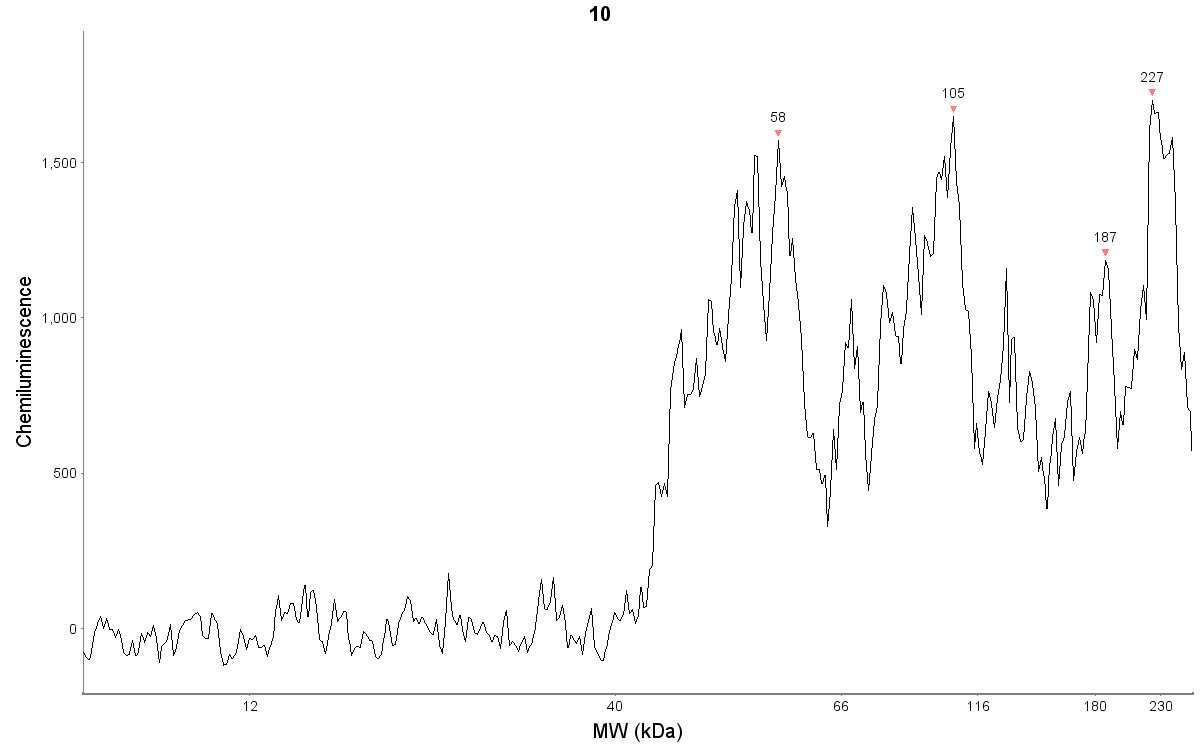
Application: Simple WesternSample Tested: NHEK human normal epidermal keratinocytesSpecies: HumanVerified Customer | Posted 05/24/2019Protein concentration of 750 ug/mL. Antibody dilution of 1:20. Detection is chemiluminescence.Standard JESS run settings.

There are no reviews that match your criteria.
Protocols
Find general support by application which include: protocols, troubleshooting, illustrated assays, videos and webinars.
- Cellular Response to Hypoxia Protocols
- R&D Systems Quality Control Western Blot Protocol
- Troubleshooting Guide: Western Blot Figures
- Western Blot Conditions
- Western Blot Protocol
- Western Blot Protocol for Cell Lysates
- Western Blot Troubleshooting
- Western Blot Troubleshooting Guide
- View all Protocols, Troubleshooting, Illustrated assays and Webinars
Loading...
Associated Pathways


