

Scientific images may be resized, cropped, or adjusted for brightness or contrast for presentation purposes. These updates are limited to presentation and do not affect the underlying scientific interpretation of the data. Where available, additional source data may be provided upon request.
Key Product Details
Species Reactivity
Validated:
Cited:
Applications
Validated:
Cited:
Label
Antibody Source
Product Specifications
Immunogen
Gln20-Val256
Accession # Q9NP55
Specificity
Clonality
Host
Isotype
Scientific Data Images for Human PLUNC Antibody (251512)
Detection of PLUNC by Western Blot
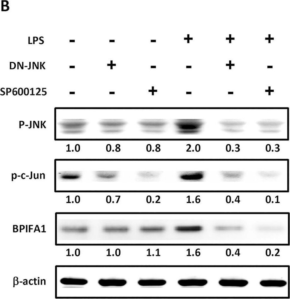
JNK/c-Jun pathway is involved in LPS-mediated up-regulation of BPIFA1 expression in nasal epithelial cells.(A) Cells were pretreated for 1 h with 20 μM PD98059 (ERK inhibitor), 10 μM SP600125 (JNK inhibitor), or 20 μM SB203580 (p38 inhibitor) and incubated with 10 μg/ml LPS for 2 h. (B) Cells were transfected with a JNK-dominant negative (DN-JNK) mutant for 24 h or pretreated with SP600125 for 30 min prior to incubation with LPS for 1 h. The protein expression levels were determined using western blot. beta -actin was used as the loading control. The western blots were carried out independently in triplicate and results were representative of one of three independent experiments. The expression level of each protein was quantified by signal intensity and was indicated at the bottom of each lane. The quantitative analysis of western blot for three independent experiments was shown in S2 Fig. ANOVA with Tukey’s test was used to compare the overall difference between the groups. P < 0.05 was considered statistically significant. Image collected and cropped by CiteAb from the following open publication (https://pubmed.ncbi.nlm.nih.gov/26646664), licensed under a CC-BY license. Not internally tested by R&D Systems.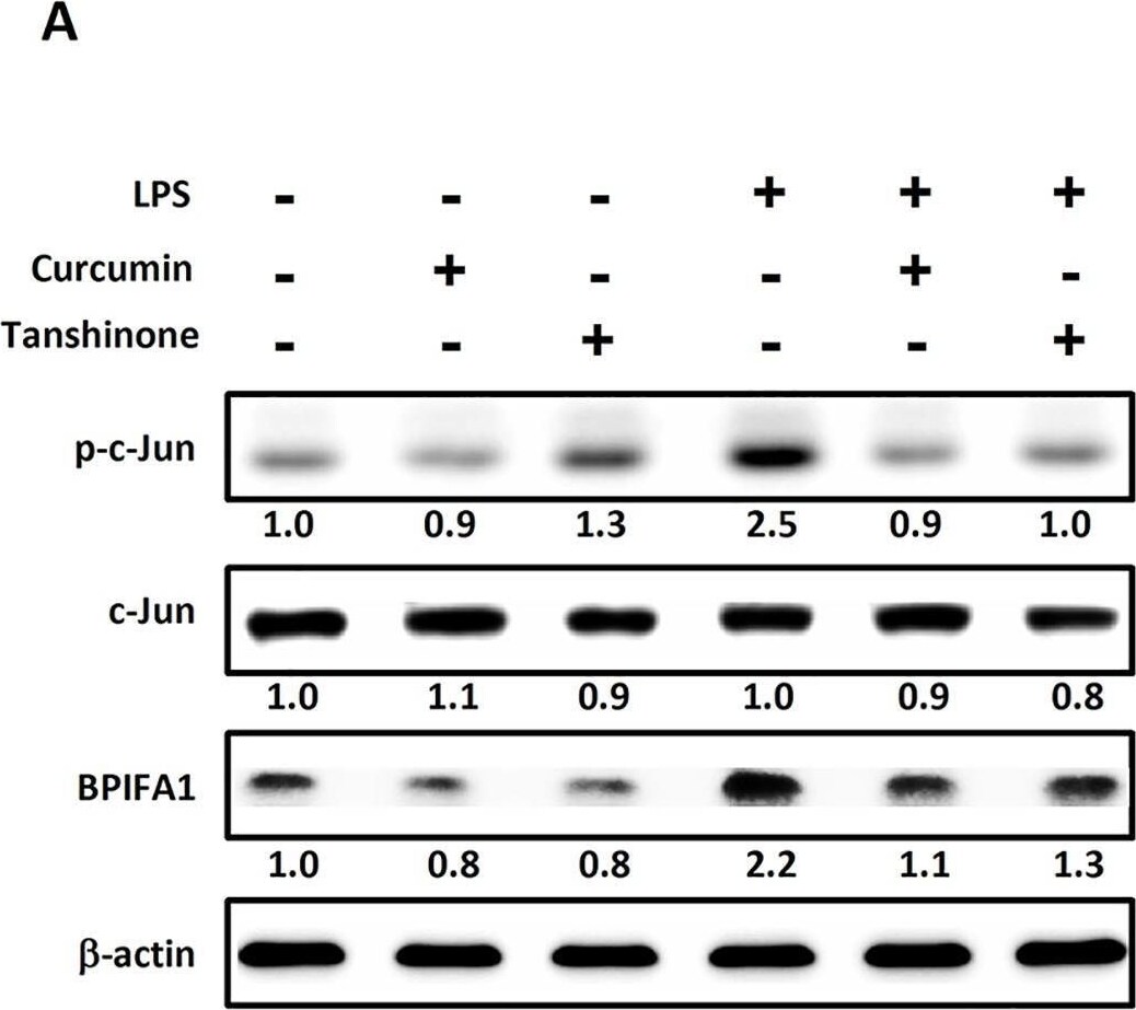
Detection of PLUNC by Western Blot
AP-1 is involved in LPS-induced BPIFA1 expression. Cells were pretreated with 10 μM curcumin or tanshinone (inhibitors of c-Jun) for 30 min, followed by incubation with LPS (10 μg/ml) for 2 h. Protein expression levels were determined using western blot and normalized to those of beta -actin. The expression level of each protein is quantified by signal intensity and is indicated at the bottom of each lane. The quantitative analysis of western blot for three independent experiments was shown in S3 Fig. ANOVA with Tukey’s test was used to compare the overall difference between the groups. P < 0.05 was considered statistically significant. (B) Cells were transfected with AP-1-Luc reporter and incubated with LPS (10 μg/ml) for another 2 h. Cell lysates were subjected to luciferase activity assays to determine AP-1 luciferase activity. The results represented mean and standard deviation values from three independent experiments. *, P < 0.05 compared between two groups. Image collected and cropped by CiteAb from the following open publication (https://pubmed.ncbi.nlm.nih.gov/26646664), licensed under a CC-BY license. Not internally tested by R&D Systems.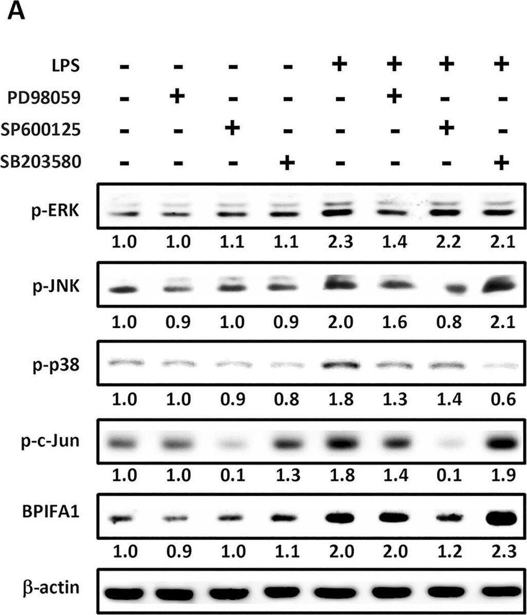
Detection of PLUNC by Western Blot
JNK/c-Jun pathway is involved in LPS-mediated up-regulation of BPIFA1 expression in nasal epithelial cells.(A) Cells were pretreated for 1 h with 20 μM PD98059 (ERK inhibitor), 10 μM SP600125 (JNK inhibitor), or 20 μM SB203580 (p38 inhibitor) and incubated with 10 μg/ml LPS for 2 h. (B) Cells were transfected with a JNK-dominant negative (DN-JNK) mutant for 24 h or pretreated with SP600125 for 30 min prior to incubation with LPS for 1 h. The protein expression levels were determined using western blot. beta -actin was used as the loading control. The western blots were carried out independently in triplicate and results were representative of one of three independent experiments. The expression level of each protein was quantified by signal intensity and was indicated at the bottom of each lane. The quantitative analysis of western blot for three independent experiments was shown in S2 Fig. ANOVA with Tukey’s test was used to compare the overall difference between the groups. P < 0.05 was considered statistically significant. Image collected and cropped by CiteAb from the following open publication (https://pubmed.ncbi.nlm.nih.gov/26646664), licensed under a CC-BY license. Not internally tested by R&D Systems.Applications for Human PLUNC Antibody (251512)
Western Blot
Sample: Recombinant Human PLUNC
Reviewed Applications
Read 1 review rated 5 using MAB1897 in the following applications:
Formulation, Preparation, and Storage
Purification
Reconstitution
Reconstitute at 0.5 mg/mL in sterile PBS. For liquid material, refer to CoA for concentration.
Formulation
Shipping
Stability & Storage
- 12 months from date of receipt, -20 to -70 °C as supplied.
- 1 month, 2 to 8 °C under sterile conditions after reconstitution.
- 6 months, -20 to -70 °C under sterile conditions after reconstitution.
Calculators
Background: PLUNC
Long Name
Alternate Names
Entrez Gene IDs
Gene Symbol
UniProt
Additional PLUNC Products
Product Documents for Human PLUNC Antibody (251512)
Certificate of Analysis
To download a Certificate of Analysis, please enter a lot or batch number in the search box below.
Note: Certificate of Analysis not available for kit components.
Product Specific Notices for Human PLUNC Antibody (251512)
For research use only
Related Research Areas
Citations for Human PLUNC Antibody (251512)
 Powered by Bioz
Customer Reviews for Human PLUNC Antibody (251512) (1)
Have you used Human PLUNC Antibody (251512)?
Submit a review and receive an Amazon gift card!
$25/€18/£15/$25CAN/¥2500 for a review with an image
$10/€7/£6/$10CAN/¥1110 for a review without an image
Submit a review
Customer Images
-
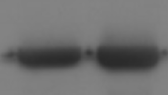
Application: Western BlotSample Tested: Salivary samplesSpecies: HumanVerified Customer | Posted 08/06/2021

There are no reviews that match your criteria.
Protocols
Find general support by application which include: protocols, troubleshooting, illustrated assays, videos and webinars.
- Cellular Response to Hypoxia Protocols
- R&D Systems Quality Control Western Blot Protocol
- Troubleshooting Guide: Western Blot Figures
- Western Blot Conditions
- Western Blot Protocol
- Western Blot Protocol for Cell Lysates
- Western Blot Troubleshooting
- Western Blot Troubleshooting Guide
- View all Protocols, Troubleshooting, Illustrated assays and Webinars