P4HB (Prolyl 4-hydroxylase beta chain; also PDI) is a 60 kDa member of the protein disulfide isomerase family. As an intracellular homodimer, it forms a tetrameric complex with P4H alpha chains to form an active prolyl 4 hydroxylase. This catalyses the hydroxylation of proline in collagen. On the cell surface, it reduces disulfide bonds in HIV that allow the virus to fuse with CXCR4 and enter susceptible cells. Mature human P4HB is 491 amino acids (aa) in length. It contains two TRX domains (aa 25 134 and 368 475) plus an ER retention sequence (aa 505 508). There is one potential isoform that shows an 11 aa substitution for the first 162 amino acids. Over aa 18 505, human P4HB shares 94% aa identity with mouse P4HB.
Human Protein Disulfide Isomerase/P4HB Antibody
R&D Systems | Catalog # AF4236

Key Product Details
Species Reactivity
Human
Applications
Western Blot, Immunocytochemistry, Simple Western, Immunoprecipitation
Label
Unconjugated
Antibody Source
Polyclonal Goat IgG
Loading...
Product Specifications
Immunogen
Chinese hamster ovary cell line CHO-derived recombinant human P4HB
Asp18-Lys505
Accession # P07237
Asp18-Lys505
Accession # P07237
Specificity
Detects human P4HB in direct ELISAs and Western blots.
Clonality
Polyclonal
Host
Goat
Isotype
IgG
Scientific Data Images for Human Protein Disulfide Isomerase/P4HB Antibody
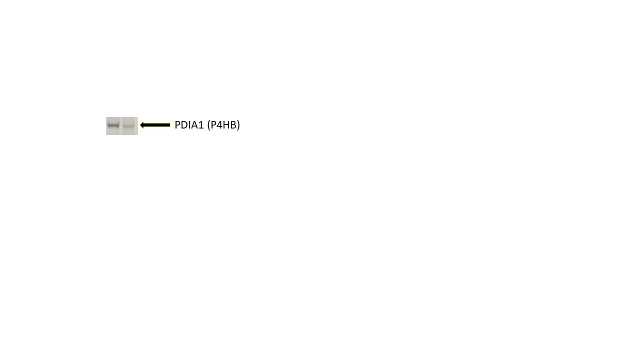
Detection of Human Protein Disulfide Isomerase/P4HB by Western Blot.
Western blot shows lysates of PANC-1 human pancreatic carcinoma cell line and MCF-7 human breast cancer cell line. PVDF membrane was probed with 1 µg/mL of Goat Anti-Human Protein Disulfide Isomerase/ P4HB Antigen Affinity-purified Polyclonal Antibody (Catalog # AF4236) followed by HRP-conjugated Anti-Goat IgG Secondary Antibody (Catalog # HAF017). A specific band was detected for Protein Disulfide Isomerase/P4HB at approximately 57 kDa (as indicated). This experiment was conducted under reducing conditions and using Immunoblot Buffer Group 1.
Protein Disulfide Isomerase/P4HB in HeLa Human Cell Line.
Protein Disulfide Isomerase/P4HB was detected in immersion fixed HeLa human cervical epithelial carcinoma cell line using Goat Anti-Human Protein Disulfide Isomerase/P4HB Antigen Affinity-purified Polyclonal Antibody (Catalog # AF4236) at 1.7 µg/mL for 3 hours at room temperature. Cells were stained using the NorthernLights™ 557-conjugated Anti-Goat IgG Secondary Antibody (red; Catalog # NL001) and counterstained with DAPI (blue). Specific staining was localized to cytoplasm. View our protocol for Fluorescent ICC Staining of Cells on Coverslips.
Detection of Human Protein Disulfide Isomerase/P4HB by Simple WesternTM.
Simple Western lane view shows lysates of PANC-1 human pancreatic carcinoma cell line and MCF-7 human breast cancer cell line, loaded at 0.2 mg/mL. A specific band was detected for Protein Disulfide Isomerase/P4HB at approximately 59 kDa (as indicated) using 10 µg/mL of Goat Anti-Human Protein Disulfide Isomerase/P4HB Antigen Affinity-purified Polyclonal Antibody (Catalog # AF4236) followed by 1:50 dilution of HRP-conjugated Anti-Goat IgG Secondary Antibody (Catalog # HAF109). This experiment was conducted under reducing conditions and using the 12-230 kDa separation system.Applications for Human Protein Disulfide Isomerase/P4HB Antibody
Application
Recommended Usage
Immunocytochemistry
1-15 µg/mL
Sample: Immersion fixed HeLa human cervical epithelial carcinoma cell line
Sample: Immersion fixed HeLa human cervical epithelial carcinoma cell line
Immunoprecipitation
25 µg/mL
Sample: Conditioned cell culture medium spiked with Recombinant Human Protein Disulfide Isomerase/P4HB (Catalog # 4236-DI), see our available Western blot detection antibodies
Sample: Conditioned cell culture medium spiked with Recombinant Human Protein Disulfide Isomerase/P4HB (Catalog # 4236-DI), see our available Western blot detection antibodies
Simple Western
10 µg/mL
Sample: PANC‑1 human pancreatic carcinoma cell line and MCF‑7 human breast cancer cell line
Sample: PANC‑1 human pancreatic carcinoma cell line and MCF‑7 human breast cancer cell line
Western Blot
1 µg/mL
Sample: PANC‑1 human pancreatic carcinoma cell line and MCF‑7 human breast cancer cell line
Sample: PANC‑1 human pancreatic carcinoma cell line and MCF‑7 human breast cancer cell line
Reviewed Applications
Read 1 review rated 5 using AF4236 in the following applications:
Formulation, Preparation, and Storage
Purification
Antigen Affinity-purified
Reconstitution
Reconstitute at 0.2 mg/mL in sterile PBS. For liquid material, refer to CoA for concentration.
Loading...
Formulation
Lyophilized from a 0.2 μm filtered solution in PBS with Trehalose. *Small pack size (SP) is supplied either lyophilized or as a 0.2 µm filtered solution in PBS.
Shipping
Lyophilized product is shipped at ambient temperature. Liquid small pack size (-SP) is shipped with polar packs. Upon receipt, store immediately at the temperature recommended below.
Stability & Storage
Use a manual defrost freezer and avoid repeated freeze-thaw cycles.
- 12 months from date of receipt, -20 to -70 °C as supplied.
- 1 month, 2 to 8 °C under sterile conditions after reconstitution.
- 6 months, -20 to -70 °C under sterile conditions after reconstitution.
Calculators
Background: Protein Disulfide Isomerase/P4HB
Long Name
Proline 4-Hydroxylase, Beta Polypeptide
Alternate Names
DSI, ERBA2L, GIT, PDI, PDIA1, PO4DB, PO4HB, PROHB, Thbp
Gene Symbol
P4HB
UniProt
Additional Protein Disulfide Isomerase/P4HB Products
Product Documents for Human Protein Disulfide Isomerase/P4HB Antibody
Certificate of Analysis
To download a Certificate of Analysis, please enter a lot or batch number in the search box below.
Note: Certificate of Analysis not available for kit components.
Product Specific Notices for Human Protein Disulfide Isomerase/P4HB Antibody
For research use only
Related Research Areas
Citations for Human Protein Disulfide Isomerase/P4HB Antibody
 Powered by Bioz
Customer Reviews for Human Protein Disulfide Isomerase/P4HB Antibody (1)
5 out of 5
1 Customer Rating
Have you used Human Protein Disulfide Isomerase/P4HB Antibody?
Submit a review and receive an Amazon gift card!
$25/€18/£15/$25CAN/¥2500 Yen for a review with an image
$10/€7/£6/$10CAN/¥1110 Yen for a review without an image
Submit a review
Customer Images
Showing
1
-
1 of
1 review
Showing All
Filter By:
-
Application: Western BlotSample Tested: RPMI 8226 human multiple myeloma cell lineSpecies: HumanVerified Customer | Posted 01/10/2018

There are no reviews that match your criteria.
Protocols
Find general support by application which include: protocols, troubleshooting, illustrated assays, videos and webinars.
- Appropriate Fixation of IHC/ICC Samples
- Cellular Response to Hypoxia Protocols
- ClariTSA™ Fluorophore Kits
- Detection & Visualization of Antibody Binding
- ICC Cell Smear Protocol for Suspension Cells
- ICC Immunocytochemistry Protocol Videos
- ICC for Adherent Cells
- Immunocytochemistry (ICC) Protocol
- Immunocytochemistry Troubleshooting
- Immunofluorescence of Organoids Embedded in Cultrex Basement Membrane Extract
- Immunohistochemistry (IHC) and Immunocytochemistry (ICC) Protocols
- Immunoprecipitation Protocol
- Preparing Samples for IHC/ICC Experiments
- Preventing Non-Specific Staining (Non-Specific Binding)
- Primary Antibody Selection & Optimization
- Protocol for VisUCyte™ HRP Polymer Detection Reagent
- Protocol for the Fluorescent ICC Staining of Cell Smears - Graphic
- Protocol for the Fluorescent ICC Staining of Cultured Cells on Coverslips - Graphic
- Protocol for the Preparation and Fluorescent ICC Staining of Cells on Coverslips
- Protocol for the Preparation and Fluorescent ICC Staining of Non-adherent Cells
- Protocol for the Preparation and Fluorescent ICC Staining of Stem Cells on Coverslips
- Protocol for the Preparation of a Cell Smear for Non-adherent Cell ICC - Graphic
- R&D Systems Quality Control Western Blot Protocol
- TUNEL and Active Caspase-3 Detection by IHC/ICC Protocol
- The Importance of IHC/ICC Controls
- Troubleshooting Guide: Western Blot Figures
- Western Blot Conditions
- Western Blot Protocol
- Western Blot Protocol for Cell Lysates
- Western Blot Troubleshooting
- Western Blot Troubleshooting Guide
- View all Protocols, Troubleshooting, Illustrated assays and Webinars
Loading...