Human Rotatin, or RTTN, is a 2,226 amino acids (aa) protein discovered in a large-scale gene trap screen for factors that govern left-right specification. RTTN plays a role in the maintenance of a normal ciliary structure and is required for correct asymmetric expression of NODAL, LEFTY and PITX2. In humans, RTTN mutations link aberrant ciliary function to abnormal development and organization of the cortex and might be a cause of Primary Microcephaly, Primordial Dwarfism and other brain malformations.
Human RTTN Antibody (2311A)
R&D Systems | Catalog # MAB9966
Recombinant Monoclonal Antibody.

Key Product Details
Species Reactivity
Human
Applications
Immunohistochemistry, Western Blot, ELISA, Immunocytochemistry
Label
Unconjugated
Antibody Source
Recombinant Monoclonal Rabbit IgG Clone # 2311A
Loading...
Product Specifications
Immunogen
Synthetic peptide containing human RTTN
Accession # Q86VV8
Accession # Q86VV8
Specificity
Detects human RTTN in direct ELISAs.
Clonality
Monoclonal
Host
Rabbit
Isotype
IgG
Scientific Data Images for Human RTTN Antibody (2311A)
Detection of Human RTTN by Western Blot.
Western blot shows lysates of HeLa human cervical epithelial carcinoma cell line and Jurkat human acute T cell leukemia cell line. PVDF membrane was probed with 0.5 µg/mL of Rabbit Anti-Human RTTN Monoclonal Antibody (Catalog # MAB9966) followed by HRP-conjugated Anti-Rabbit IgG Secondary Antibody (Catalog # HAF008). A specific band was detected for RTTN at approximately 248 kDa (as indicated). This experiment was conducted under reducing conditions and using Immunoblot Buffer Group 1.
RTTN in HDLM‑2 Human Cell Line.
RTTN was detected in immersion fixed HDLM-2 human Hodgkin's lymphoma cell line using Rabbit Anti-Human RTTN Monoclonal Antibody (Catalog # MAB9966) at 8 µg/mL for 3 hours at room temperature. Cells were stained using the NorthernLights™ 557-conjugated Anti-Rabbit IgG Secondary Antibody (red; Catalog # NL004) and counterstained with DAPI (blue). Specific staining was localized to cytoplasm. View our protocol for Fluorescent ICC Staining of Non-adherent Cells.
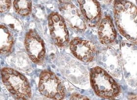
RTTN in Human Stomach.
RTTN was detected in immersion fixed paraffin-embedded sections of human stomach using Rabbit Anti-Human RTTN Monoclonal Antibody (Catalog # MAB9966) at 1 µg/mL for 1 hour at room temperature followed by incubation with the Anti-Rabbit IgG VisUCyte™ HRP Polymer Antibody (Catalog # VC003). Tissue was stained using DAB (brown) and counterstained with hematoxylin (blue). Specific staining was localized to cytoplasm in gastric glands. View our protocol for IHC Staining with VisUCyte HRP Polymer Detection Reagents.Applications for Human RTTN Antibody (2311A)
Application
Recommended Usage
ELISA
This antibody functions as an ELISA detection antibody when paired with Mouse Anti-Human RTTN Monoclonal Antibody(Catalog # MAB99661).
This product is intended for assay development on various assay platforms requiring antibody pairs.
Immunocytochemistry
8-25 µg/mL
Sample: Immersion fixed HDLM-2 human Hodgkin's lymphoma cell line
Sample: Immersion fixed HDLM-2 human Hodgkin's lymphoma cell line
Immunohistochemistry
1-25 µg/mL
Sample: Immersion fixed paraffin-embedded sections of human stomach
Sample: Immersion fixed paraffin-embedded sections of human stomach
Western Blot
0.5 µg/mL
Sample: HeLa human cervical epithelial carcinoma cell line and Jurkat human acute T cell leukemia cell line
Sample: HeLa human cervical epithelial carcinoma cell line and Jurkat human acute T cell leukemia cell line
Reviewed Applications
Read 1 review rated 5 using MAB9966 in the following applications:
Formulation, Preparation, and Storage
Purification
Protein A or G purified from cell culture supernatant
Reconstitution
Reconstitute at 0.5 mg/mL in sterile PBS. For liquid material, refer to CoA for concentration.
Loading...
Formulation
Lyophilized from a 0.2 μm filtered solution in PBS with Trehalose. *Small pack size (SP) is supplied either lyophilized or as a 0.2 µm filtered solution in PBS.
Shipping
Lyophilized product is shipped at ambient temperature. Liquid small pack size (-SP) is shipped with polar packs. Upon receipt, store immediately at the temperature recommended below.
Stability & Storage
Use a manual defrost freezer and avoid repeated freeze-thaw cycles.
- 12 months from date of receipt, -20 to -70 °C, as supplied.
- 1 month, 2 to 8 °C under sterile conditions after opening.
- 6 months, -20 to -70 °C under sterile conditions after opening.
Calculators
Background: RTTN
Long Name
Rotatin
Alternate Names
MSSP, PMGYS, Rotatin
Gene Symbol
RTTN
UniProt
Additional RTTN Products
Product Documents for Human RTTN Antibody (2311A)
Certificate of Analysis
To download a Certificate of Analysis, please enter a lot or batch number in the search box below.
Note: Certificate of Analysis not available for kit components.
Product Specific Notices for Human RTTN Antibody (2311A)
For research use only
Customer Reviews for Human RTTN Antibody (2311A) (1)
5 out of 5
1 Customer Rating
Have you used Human RTTN Antibody (2311A)?
Submit a review and receive an Amazon gift card!
$25/€18/£15/$25CAN/¥2500 Yen for a review with an image
$10/€7/£6/$10CAN/¥1110 Yen for a review without an image
Submit a review
Customer Images
Showing
1
-
1 of
1 review
Showing All
Filter By:
-
Application: ImmunohistochemistrySample Tested: Stomach tissueSpecies: HumanVerified Customer | Posted 07/04/2022

There are no reviews that match your criteria.
Protocols
Find general support by application which include: protocols, troubleshooting, illustrated assays, videos and webinars.
- Antigen Retrieval Protocol (PIER)
- Antigen Retrieval for Frozen Sections Protocol
- Appropriate Fixation of IHC/ICC Samples
- Cellular Response to Hypoxia Protocols
- Chromogenic IHC Staining of Formalin-Fixed Paraffin-Embedded (FFPE) Tissue Protocol
- Chromogenic Immunohistochemistry Staining of Frozen Tissue
- ClariTSA™ Fluorophore Kits
- Detection & Visualization of Antibody Binding
- ELISA Sample Preparation & Collection Guide
- ELISA Troubleshooting Guide
- Fluorescent IHC Staining of Frozen Tissue Protocol
- Graphic Protocol for Heat-induced Epitope Retrieval
- Graphic Protocol for the Preparation and Fluorescent IHC Staining of Frozen Tissue Sections
- Graphic Protocol for the Preparation and Fluorescent IHC Staining of Paraffin-embedded Tissue Sections
- Graphic Protocol for the Preparation of Gelatin-coated Slides for Histological Tissue Sections
- How to Run an R&D Systems DuoSet ELISA
- How to Run an R&D Systems Quantikine ELISA
- How to Run an R&D Systems Quantikine™ QuicKit™ ELISA
- ICC Cell Smear Protocol for Suspension Cells
- ICC Immunocytochemistry Protocol Videos
- ICC for Adherent Cells
- IHC Sample Preparation (Frozen sections vs Paraffin)
- Immunocytochemistry (ICC) Protocol
- Immunocytochemistry Troubleshooting
- Immunofluorescence of Organoids Embedded in Cultrex Basement Membrane Extract
- Immunofluorescent IHC Staining of Formalin-Fixed Paraffin-Embedded (FFPE) Tissue Protocol
- Immunohistochemistry (IHC) and Immunocytochemistry (ICC) Protocols
- Immunohistochemistry Frozen Troubleshooting
- Immunohistochemistry Paraffin Troubleshooting
- Preparing Samples for IHC/ICC Experiments
- Preventing Non-Specific Staining (Non-Specific Binding)
- Primary Antibody Selection & Optimization
- Protocol for Heat-Induced Epitope Retrieval (HIER)
- Protocol for Making a 4% Formaldehyde Solution in PBS
- Protocol for VisUCyte™ HRP Polymer Detection Reagent
- Protocol for the Fluorescent ICC Staining of Cell Smears - Graphic
- Protocol for the Fluorescent ICC Staining of Cultured Cells on Coverslips - Graphic
- Protocol for the Preparation & Fixation of Cells on Coverslips
- Protocol for the Preparation and Chromogenic IHC Staining of Frozen Tissue Sections
- Protocol for the Preparation and Chromogenic IHC Staining of Frozen Tissue Sections - Graphic
- Protocol for the Preparation and Chromogenic IHC Staining of Paraffin-embedded Tissue Sections
- Protocol for the Preparation and Chromogenic IHC Staining of Paraffin-embedded Tissue Sections - Graphic
- Protocol for the Preparation and Fluorescent ICC Staining of Cells on Coverslips
- Protocol for the Preparation and Fluorescent ICC Staining of Non-adherent Cells
- Protocol for the Preparation and Fluorescent ICC Staining of Stem Cells on Coverslips
- Protocol for the Preparation and Fluorescent IHC Staining of Frozen Tissue Sections
- Protocol for the Preparation and Fluorescent IHC Staining of Paraffin-embedded Tissue Sections
- Protocol for the Preparation of Gelatin-coated Slides for Histological Tissue Sections
- Protocol for the Preparation of a Cell Smear for Non-adherent Cell ICC - Graphic
- Quantikine HS ELISA Kit Assay Principle, Alkaline Phosphatase
- Quantikine HS ELISA Kit Principle, Streptavidin-HRP Polymer
- R&D Systems Quality Control Western Blot Protocol
- Sandwich ELISA (Colorimetric) – Biotin/Streptavidin Detection Protocol
- Sandwich ELISA (Colorimetric) – Direct Detection Protocol
- TUNEL and Active Caspase-3 Detection by IHC/ICC Protocol
- The Importance of IHC/ICC Controls
- Troubleshooting Guide: ELISA
- Troubleshooting Guide: Immunohistochemistry
- Troubleshooting Guide: Western Blot Figures
- Western Blot Conditions
- Western Blot Protocol
- Western Blot Protocol for Cell Lysates
- Western Blot Troubleshooting
- Western Blot Troubleshooting Guide
- View all Protocols, Troubleshooting, Illustrated assays and Webinars
Loading...