S100A4, also known as Metastasin, Mtsl and Calvasculin, is an 11 kDa member of the S100 (soluble in 100% saturated ammonium sulfate) family of proteins (1-5). The S100 family is further classified as a member of the EF-hand superfamily of Ca++-binding proteins. These participate in both calcium-dependent and
calcium-independent protein-protein interactions. The hallmark of this superfamily is the EF-hand motif that consists of a Ca++-binding site flanked by two alpha -helices (helix E and helix F) that were originally identified in a right-handed model of carp muscle calcium-binding protein (6). Human S100A4 is 101 amino acids (aa) in length (1, 2). It contains two EF hand domains, one between aa 12-47, and a second between aa 50-85. The first domain has a 14 aa cation-binding motif and binds Ca++ with low affinity. The second Ca++-binding motif is 12 aa in length and binds Ca++ with high affinity. S100A4 has no classical signal sequence but is secreted from cells (3, 7). Human S100A4 shares 93% and 95% aa identity with mouse and canine S100A4, respectively. S100A4 reportedly exists as a dimer (8, 9, 10). Extracellular S100A4 is reported to induce MMP production, activate MMPs, promote neurite outgrowth and stimulate cardiomyocyte proliferation (4, 10, 11, 12, 13). Within the cell, dimers are likely the functional unit. Here, they are constitutive homo- or heterodimers (with S100A1) that interact with Ca++, undergo a conformational change, and subsequently bind to cytoplasmic targets. Known targets include p53, myosin heavy chain II, F-actin and liprin beta 1 (4, 14). In general, it can be said that S100A4 blocks target phosphorylation and multimerization (4, 7, 14). S100A4 activity has been associated with cell transformation. It seems likely this is either coincidental, or a consequence, rather than a cause of transformation (3).

Key Product Details
Validated by
Knockout/Knockdown
Species Reactivity
Validated:
Human
Cited:
Human
Applications
Validated:
Knockout Validated, Immunohistochemistry, Western Blot, Immunocytochemistry
Cited:
Bioassay
Label
Unconjugated
Antibody Source
Monoclonal Mouse IgG1 Clone # 922813
Loading...
Product Specifications
Immunogen
E. coli-derived recombinant human S100A4
Met1-Ser101
Accession # P26447
Met1-Ser101
Accession # P26447
Specificity
Detects human S100A4 in direct ELISAs.
Clonality
Monoclonal
Host
Mouse
Isotype
IgG1
Scientific Data Images for Human S100A4 Antibody (922813)
Detection of Human and Mouse S100A4 by Western Blot.
Western blot shows lysates of HeLa human cervical epithelial carcinoma cell line and NIH-3T3 mouse embryonic fibroblast cell line. PVDF membrane was probed with 2 µg/mL of Mouse Anti-Human S100A4 Monoclonal Antibody (Catalog # MAB4137) followed by HRP-conjugated Anti-Mouse IgG Secondary Antibody (Catalog # HAF018). A specific band was detected for S100A4 at approximately 11 kDa (as indicated). This experiment was conducted under reducing conditions and using Immunoblot Buffer Group 1.
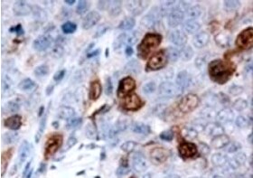
S100A4 in Human Cervical Cancer Tissue.
S100A4 was detected in immersion fixed paraffin-embedded sections of human cervical cancer tissue using Mouse Anti-Human S100A4 Monoclonal Antibody (Catalog # MAB4137) at 15 µg/mL overnight at 4 °C. Tissue was stained using the Anti-Mouse HRP-DAB Cell & Tissue Staining Kit (brown; Catalog # CTS002) and counterstained with hematoxylin (blue). Specific staining was localized to cytoplasm in cancer cells. View our protocol for Chromogenic IHC Staining of Paraffin-embedded Tissue Sections.
S100A4 in A549 Human Cell Line.
S100A4 was detected in immersion fixed A549 human lung carcinoma cells unstimulated (lower panel) and stimulated with the EMT Inducing Media Supplement (upper panel; Catalog # CCM017) using Mouse Anti-Human S100A4 Monoclonal Antibody (Catalog # MAB4137) at 1 µg/mL for 3 hours at room temperature. Cells were stained using the NorthernLights™ 557-conjugated Anti-Mouse IgG Secondary Antibody (red; Catalog # NL007) and counterstained with DAPI (blue). Specific staining was localized to cytoplasm. View our protocol for Fluorescent ICC Staining of Cells on Coverslips.
Western Blot Shows Human S100A4 Specificity by Using Knockout Cell Line.
Western blot shows lysates of HeLa human cervical epithelial carcinoma parental cell line and S100A4 knockout HeLa cell line (KO). PVDF membrane was probed with 2 µg/mL of Mouse Anti-Human S100A4 Monoclonal Antibody (Catalog # MAB4137) followed by HRP-conjugated Anti-Mouse IgG Secondary Antibody (Catalog # HAF018). A specific band was detected for S100A4 at approximately 12 kDa (as indicated) in the parental HeLa cell line, but is not detectable in knockout HeLa cell line. GAPDH (Catalog # MAB5718) is shown as a loading control. This experiment was conducted under reducing conditions and using Immunoblot Buffer Group 1.Applications for Human S100A4 Antibody (922813)
Application
Recommended Usage
Immunocytochemistry
8-25 µg/mL
Sample: Immersion fixed A549 human lung carcinoma cells unstimulated and stimulated with the EMT Inducing Media Supplement (Catalog # CCM017)
Sample: Immersion fixed A549 human lung carcinoma cells unstimulated and stimulated with the EMT Inducing Media Supplement (Catalog # CCM017)
Immunohistochemistry
8-25 µg/mL
Sample: Immersion fixed paraffin-embedded sections of human cervical cancer tissue
Sample: Immersion fixed paraffin-embedded sections of human cervical cancer tissue
Knockout Validated
S100A4
is specifically detected in HeLa human cervical epithelial carcinoma parental cell line but is not detectable in
S100A4 knockout HeLa cell line.
Western Blot
2 µg/mL
Sample: HeLa human cervical epithelial carcinoma cell line and NIH‑3T3 mouse embryonic fibroblast cell line
Sample: HeLa human cervical epithelial carcinoma cell line and NIH‑3T3 mouse embryonic fibroblast cell line
Reviewed Applications
Read 4 reviews rated 4.5 using MAB4137 in the following applications:
Formulation, Preparation, and Storage
Purification
Protein A or G purified from hybridoma culture supernatant
Reconstitution
Reconstitute at 0.5 mg/mL in sterile PBS. For liquid material, refer to CoA for concentration.
Loading...
Formulation
Lyophilized from a 0.2 μm filtered solution in PBS with Trehalose. *Small pack size (SP) is supplied either lyophilized or as a 0.2 µm filtered solution in PBS.
Shipping
Lyophilized product is shipped at ambient temperature. Liquid small pack size (-SP) is shipped with polar packs. Upon receipt, store immediately at the temperature recommended below.
Stability & Storage
Use a manual defrost freezer and avoid repeated freeze-thaw cycles.
- 12 months from date of receipt, -20 to -70 °C as supplied.
- 1 month, 2 to 8 °C under sterile conditions after reconstitution.
- 6 months, -20 to -70 °C under sterile conditions after reconstitution.
Calculators
Background: S100A4
References
- Engelkamp, D. et al. (1992) Biochemistry 31:10258.
- Tomida, Y. et al. (1992) Biochem. Biophys. Res. Commun. 189:1310.
- Garrett, S.C. et al. (2006) J. Biol. Chem. 281:677.
- Santamaria-Kisiel, L. et al. (2006) Biochem. J. 396:201.
- Donato, R. (2001) Int. J. Biochem. Mol. Biol. 33:637.
- Kretsinger, R.H. and C.E. Nockolds (1973) J. Biol. Chem. 248:3313.
- Helfman, D.M. et al. (2005) Br. J. Cancer 92:1955.
- Burkitt, W.I. et al. (2003) Biochem. Soc. Trans. 31:985.
- Vallaly, K.M. et al. (2002) Biochemistry 41:12670.
- Novitskaya, V. et al. (2000) J. Biol. Chem. 275:41278.
- Stary, M. et al. (2006) Biochem. Biophys. Res. Commun. 343:555.
- Semov, A. et al. (2005) J. Biol. Chem. 280:20833.
- Saleem, M. et al. (2006) Proc. Natl. Acad. Sci. USA 103:14825.
- Kriajevska, M. et al. (2002) J. Biol. Chem. 277:5229.
Long Name
S100 Calcium Binding Protein A4
Alternate Names
18A2, 42A, CAPL, MTS1, P9KA, PEL98
Gene Symbol
S100A4
UniProt
Additional S100A4 Products
Product Documents for Human S100A4 Antibody (922813)
Certificate of Analysis
To download a Certificate of Analysis, please enter a lot or batch number in the search box below.
Note: Certificate of Analysis not available for kit components.
Product Specific Notices for Human S100A4 Antibody (922813)
For research use only
Related Research Areas
Citations for Human S100A4 Antibody (922813)
 Powered by Bioz
Customer Reviews for Human S100A4 Antibody (922813) (4)
4.5 out of 5
4 Customer Ratings
Have you used Human S100A4 Antibody (922813)?
Submit a review and receive an Amazon gift card!
$25/€18/£15/$25CAN/¥2500 Yen for a review with an image
$10/€7/£6/$10CAN/¥1110 Yen for a review without an image
Submit a review
Customer Images
Showing
1
-
4 of
4 reviews
Showing All
Filter By:
-
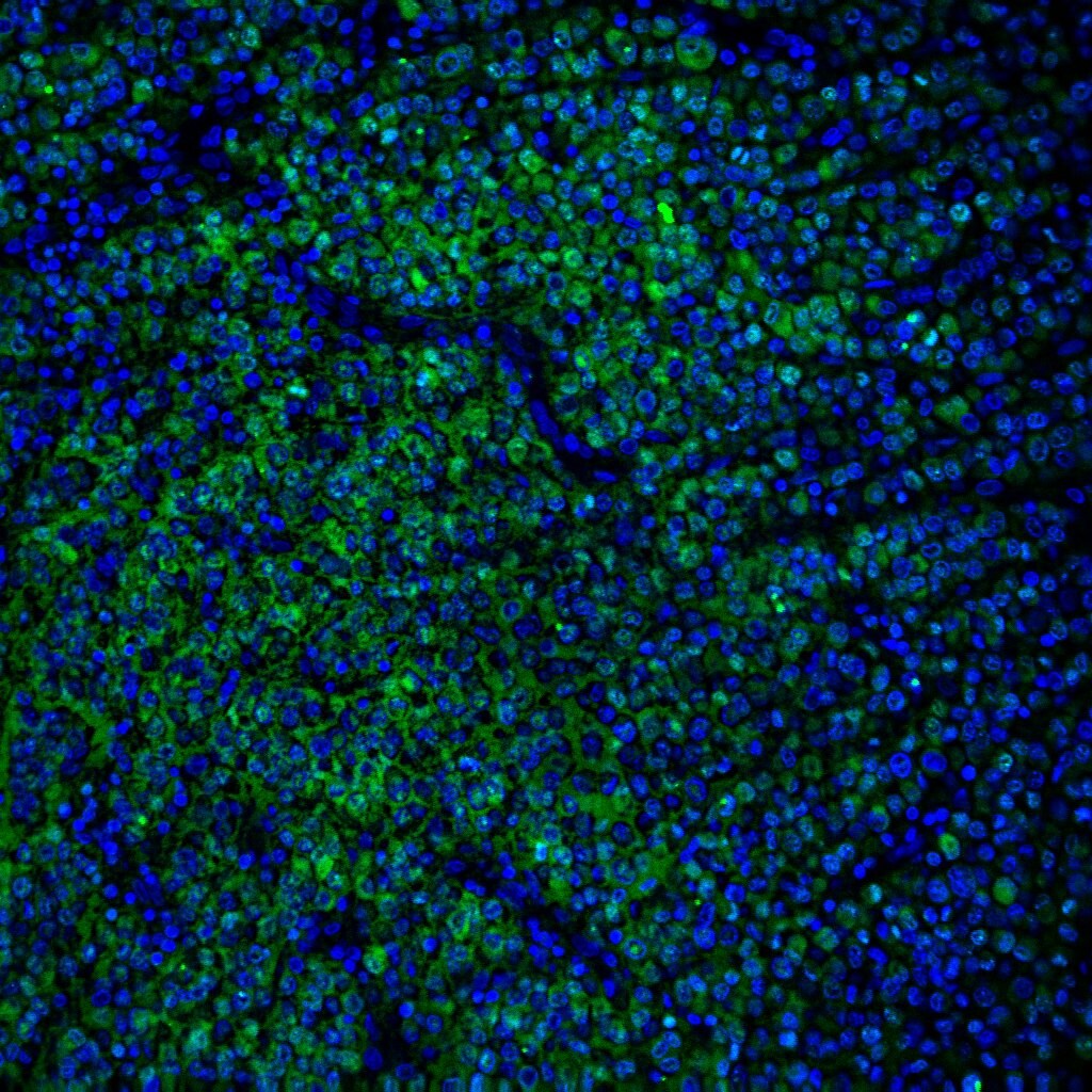
Application: ImmunohistochemistrySample Tested: Cervical cancer tissueSpecies: HumanVerified Customer | Posted 07/13/2022

-
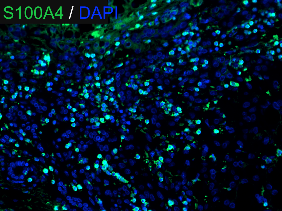
Application: Immunocytochemistry/ImmunofluorescenceSample Tested: Melanoma tissueSpecies: HumanVerified Customer | Posted 10/01/2021
-
Application: Immunocytochemistry/ImmunofluorescenceSample Tested: Adult brainSpecies: MouseVerified Customer | Posted 08/30/2020Dilution 1:200
-
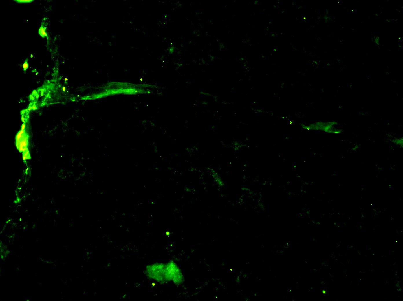
Application: Immunocytochemistry/ImmunofluorescenceSample Tested: fibrotic liverSpecies: HumanVerified Customer | Posted 03/12/2018
There are no reviews that match your criteria.
Protocols
Find general support by application which include: protocols, troubleshooting, illustrated assays, videos and webinars.
- Antigen Retrieval Protocol (PIER)
- Antigen Retrieval for Frozen Sections Protocol
- Appropriate Fixation of IHC/ICC Samples
- Cellular Response to Hypoxia Protocols
- Chromogenic IHC Staining of Formalin-Fixed Paraffin-Embedded (FFPE) Tissue Protocol
- Chromogenic Immunohistochemistry Staining of Frozen Tissue
- ClariTSA™ Fluorophore Kits
- Detection & Visualization of Antibody Binding
- Fluorescent IHC Staining of Frozen Tissue Protocol
- Graphic Protocol for Heat-induced Epitope Retrieval
- Graphic Protocol for the Preparation and Fluorescent IHC Staining of Frozen Tissue Sections
- Graphic Protocol for the Preparation and Fluorescent IHC Staining of Paraffin-embedded Tissue Sections
- Graphic Protocol for the Preparation of Gelatin-coated Slides for Histological Tissue Sections
- ICC Cell Smear Protocol for Suspension Cells
- ICC Immunocytochemistry Protocol Videos
- ICC for Adherent Cells
- IHC Sample Preparation (Frozen sections vs Paraffin)
- Immunocytochemistry (ICC) Protocol
- Immunocytochemistry Troubleshooting
- Immunofluorescence of Organoids Embedded in Cultrex Basement Membrane Extract
- Immunofluorescent IHC Staining of Formalin-Fixed Paraffin-Embedded (FFPE) Tissue Protocol
- Immunohistochemistry (IHC) and Immunocytochemistry (ICC) Protocols
- Immunohistochemistry Frozen Troubleshooting
- Immunohistochemistry Paraffin Troubleshooting
- Preparing Samples for IHC/ICC Experiments
- Preventing Non-Specific Staining (Non-Specific Binding)
- Primary Antibody Selection & Optimization
- Protocol for Heat-Induced Epitope Retrieval (HIER)
- Protocol for Making a 4% Formaldehyde Solution in PBS
- Protocol for VisUCyte™ HRP Polymer Detection Reagent
- Protocol for the Fluorescent ICC Staining of Cell Smears - Graphic
- Protocol for the Fluorescent ICC Staining of Cultured Cells on Coverslips - Graphic
- Protocol for the Preparation & Fixation of Cells on Coverslips
- Protocol for the Preparation and Chromogenic IHC Staining of Frozen Tissue Sections
- Protocol for the Preparation and Chromogenic IHC Staining of Frozen Tissue Sections - Graphic
- Protocol for the Preparation and Chromogenic IHC Staining of Paraffin-embedded Tissue Sections
- Protocol for the Preparation and Chromogenic IHC Staining of Paraffin-embedded Tissue Sections - Graphic
- Protocol for the Preparation and Fluorescent ICC Staining of Cells on Coverslips
- Protocol for the Preparation and Fluorescent ICC Staining of Non-adherent Cells
- Protocol for the Preparation and Fluorescent ICC Staining of Stem Cells on Coverslips
- Protocol for the Preparation and Fluorescent IHC Staining of Frozen Tissue Sections
- Protocol for the Preparation and Fluorescent IHC Staining of Paraffin-embedded Tissue Sections
- Protocol for the Preparation of Gelatin-coated Slides for Histological Tissue Sections
- Protocol for the Preparation of a Cell Smear for Non-adherent Cell ICC - Graphic
- R&D Systems Quality Control Western Blot Protocol
- TUNEL and Active Caspase-3 Detection by IHC/ICC Protocol
- The Importance of IHC/ICC Controls
- Troubleshooting Guide: Immunohistochemistry
- Troubleshooting Guide: Western Blot Figures
- Western Blot Conditions
- Western Blot Protocol
- Western Blot Protocol for Cell Lysates
- Western Blot Troubleshooting
- Western Blot Troubleshooting Guide
- View all Protocols, Troubleshooting, Illustrated assays and Webinars
Loading...