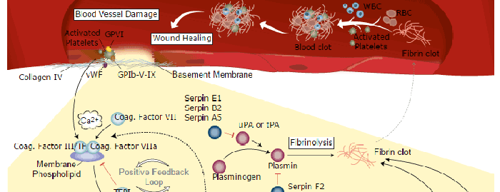
Serpin F2 is a member of the Serpin superfamily and the primary physiological inhibitor of the serine protease plasmin, which is responsible for the dissolution of fibrin clots (1, 2). In addition to plasmin, Serpin F2 is also an efficient inhibitor of trypsin and chymotrypsin (3). Liver and kidney are major sites of Serpin F2 production and other tissues such as muscle, intestine, central nervous system, and placenta also express its mRNA at a moderate level. The tissue expression pattern of Serpin F2 indicates that it is a key regulator of plasmin-mediated proteolysis in these tissues (4). Human Serpin F2 is synthesized as a 491 amino acid precursor with a 27 amino acid signal peptide. The secreted protein has a short propeptide (residues 28‑39) and a mature chain (residues 40‑491). The presence of the propeptide did not affect its ability to inhibit plasmin but reduced its cross-linking ability to fibrin (5).
Human Serpin F2/alpha 2-Antiplasmin Antibody (236122)
R&D Systems | Catalog # MAB1470

Scientific images may be resized, cropped, or adjusted for brightness or contrast for presentation purposes. These updates are limited to presentation and do not affect the underlying scientific interpretation of the data. Where available, additional source data may be provided upon request.
Key Product Details
Validated by
Species Reactivity
Validated:
Cited:
Applications
Validated:
Cited:
Label
Antibody Source
Product Specifications
Immunogen
Met28-Lys491
Accession # P08697
Specificity
Clonality
Host
Isotype
Scientific Data Images for Human Serpin F2/alpha 2-Antiplasmin Antibody (236122)
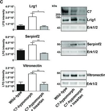
Detection of Mouse Serpin F2/alpha 2-Antiplasmin by Western Blot
Proteomics analysis reveals losartan-mediated reversion of early fibrotic changes in RDEBHeat map of cluster analysis of protein abundances determined by label-free quantification mass spectrometry. Extracted ion currents were used to determine protein abundances, and respective intensities were log2-transformed and normalized (z-score). Samples were clustered hierarchically, and protein abundances were clustered by k-means. Cluster sizes are indicated by color code on the left. Clusters 3, 4, 5, 8, and 9 highlight losartan-induced changes. The general patterns of protein abundance in clusters 5, 8, and 9 are similar in wild-type and losartan-treated C7-hypomorphic mice and thus contain downstream targets of losartan involved in RDEB disease progression. WT, wild-type; H, hypomorphic; H + Los, losartan-treated C7-hypomorphic skin.Proteins from cluster 5 carrying GO terms related to inflammation were short-listed and analyzed on potential interactions using default settings in STRING DB (confidence score 0.4) (Szklarczyk et al, 2015).Bar graphs show abundance of selected representative proteins in cluster 5 that were normalized by losartan treatment. Shown to the left are the mean ± S.E.M. of the normalized protein abundance (LFQ intensity) of groups of individual mice corresponding to wild-type, untreated, and losartan-treated C7-hypomorphic mice as indicated in the figure. Unpaired t-test was used to calculate significance. Abundance of Lrg1: *P-value C7-hypomorphic vs. losartan-treated C7-hypomorphic mice = 0.022; abundance of serpin f2: **P-value untreated C7-hypomorphic vs. losartan-treated C7-hypomorphic mice = 0.0084; abundance of vitronectin: **P-value untreated C7-hypomorphic vs. losartan-treated C7-hypomorphic mice = 0.0094. Right, validation of proteomics analysis by Western blotting of independent biological replicates. Representative Western blots of skin lysates from wild-type, untreated C7-hypomorphic, and 7-week losartan-treated C7-hypomorphic mice noApplications for Human Serpin F2/alpha 2-Antiplasmin Antibody (236122)
Immunoprecipitation
Sample: Conditioned cell culture medium spiked with Recombinant Human Serpin F2/ alpha 2‑Antiplasmin (Catalog # 1470-PI), see our available Western blot detection antibodies
Western Blot
Sample: Recombinant Human Serpin F2/ alpha 2‑Antiplasmin (Catalog # 1470-PI)
Formulation, Preparation, and Storage
Purification
Reconstitution
Reconstitute at 0.5 mg/mL in sterile PBS. For liquid material, refer to CoA for concentration.
Formulation
Shipping
Stability & Storage
- 12 months from date of receipt, -20 to -70 °C as supplied.
- 1 month, 2 to 8 °C under sterile conditions after reconstitution.
- 6 months, -20 to -70 °C under sterile conditions after reconstitution.
Calculators
Background: Serpin F2/alpha 2-Antiplasmin
References
- Tone, M. et al. (1987) J. Biochem. 102:1033.
- Silverman, G.A. et al. (2001) J. Biol. Chem. 276:33293.
- Potempa, J. et al. (1988) Science 241:699.
- Menoud, P.-A. et al. (1996) J. Clin. Invest. 97:2478.
- Sumi, Y. et al. (1989) J. Biochem. 106:703.
Alternate Names
Gene Symbol
UniProt
Additional Serpin F2/alpha 2-Antiplasmin Products
Product Documents for Human Serpin F2/alpha 2-Antiplasmin Antibody (236122)
Certificate of Analysis
To download a Certificate of Analysis, please enter a lot or batch number in the search box below.
Note: Certificate of Analysis not available for kit components.
Product Specific Notices for Human Serpin F2/alpha 2-Antiplasmin Antibody (236122)
For research use only
Citations for Human Serpin F2/alpha 2-Antiplasmin Antibody (236122)
 Powered by Bioz
Customer Reviews for Human Serpin F2/alpha 2-Antiplasmin Antibody (236122)
There are currently no reviews for this product. Be the first to review Human Serpin F2/alpha 2-Antiplasmin Antibody (236122) and earn rewards!
Have you used Human Serpin F2/alpha 2-Antiplasmin Antibody (236122)?
Submit a review and receive an Amazon gift card!
$25/€18/£15/$25CAN/¥2500 for a review with an image
$10/€7/£6/$10CAN/¥1110 for a review without an image
Submit a review
Protocols
Find general support by application which include: protocols, troubleshooting, illustrated assays, videos and webinars.
- Cellular Response to Hypoxia Protocols
- Immunoprecipitation Protocol
- R&D Systems Quality Control Western Blot Protocol
- Troubleshooting Guide: Western Blot Figures
- Western Blot Conditions
- Western Blot Protocol
- Western Blot Protocol for Cell Lysates
- Western Blot Troubleshooting
- Western Blot Troubleshooting Guide
- View all Protocols, Troubleshooting, Illustrated assays and Webinars
Associated Pathways