Key Product Details

Species Reactivity

Validated:

Human

Cited:

Human, Mouse

Applications

Validated:

Immunohistochemistry, Western Blot, Immunocytochemistry

Cited:

Immunohistochemistry, Immunohistochemistry-Paraffin, Immunohistochemistry-Frozen, Western Blot, Immunoprecipitation, ELISA Development

Label

Unconjugated

Antibody Source

Polyclonal Goat IgG
Loading...

Product Specifications

Immunogen

Mouse myeloma cell line NS0-derived recombinant human sFRP-1 (R&D Systems, Catalog # 1384-SF) 
Ser32-Lys314
Accession # AAB70793

Specificity

Detects human sFRP-1 in direct ELISAs and Western blots. In direct ELISAs, less than 2% cross-reactivity with recombinant human (rh) sFRP-2, and rhsFRP-5 is observed.

Clonality

Polyclonal

Host

Goat

Isotype

IgG

Scientific Data Images for Human sFRP‑1 Antibody

sFRP-1 antibody in Human Kidney by Immunohistochemistry (IHC-P).

sFRP‑1 in Human Kidney.

sFRP-1 was detected in immersion fixed paraffin-embedded sections of human kidney using Goat Anti-Human sFRP-1 Antigen Affinity-purified Polyclonal Antibody (Catalog # AF1384) at 1 µg/mL overnight at 4 °C. Tissue was stained using the Anti-Goat HRP-DAB Cell & Tissue Staining Kit (brown; Catalog # CTS008) and counterstained with hematoxylin (blue). Specific staining was localized to epithelial cell cytoplasm in convoluted tubules. View our protocol for Chromogenic IHC Staining of Paraffin-embedded Tissue Sections.

sFRP-1 antibody in MBA-MB-468 Human Cell Line by Immunocytochemistry (ICC).

sFRP‑1 in MBA‑MB‑468 Human Cell Line.

sFRP-1 was detected in immersion fixed MBA-MB-468 human breast cancer cell line using Goat Anti-Human sFRP-1 Antigen Affinity-purified Polyclonal Antibody (Catalog # AF1384) at 15 µg/mL for 3 hours at room temperature. Cells were stained using the NorthernLights™ 557-conjugated Anti-Goat IgG Secondary Antibody (red; Catalog # NL001) and counterstained with DAPI (blue). Specific staining was localized to cytoplasm. View our protocol for Fluorescent ICC Staining of Cells on Coverslips.

Applications for Human sFRP‑1 Antibody

Application
Recommended Usage

Immunocytochemistry

5-15 µg/mL
Sample: Immersion fixed MBA-MB-468 human breast cancer cell line

Immunohistochemistry

1-15 µg/mL
Sample: Immersion fixed paraffin-embedded sections of human kidney

Western Blot

0.1 µg/mL
Sample: Recombinant Human sFRP‑1 (Histidine-tagged) (Catalog # 1384-SF)

Reviewed Applications

Read 3 reviews rated 3 using AF1384 in the following applications:

Formulation, Preparation, and Storage

Purification

Antigen Affinity-purified

Reconstitution

Reconstitute at 0.2 mg/mL in sterile PBS. For liquid material, refer to CoA for concentration.


Loading...

Formulation

Lyophilized from a 0.2 μm filtered solution in PBS with Trehalose. *Small pack size (SP) is supplied either lyophilized or as a 0.2 µm filtered solution in PBS.

Shipping

Lyophilized product is shipped at ambient temperature. Liquid small pack size (-SP) is shipped with polar packs. Upon receipt, store immediately at the temperature recommended below.

Stability & Storage

Use a manual defrost freezer and avoid repeated freeze-thaw cycles.
  • 12 months from date of receipt, -20 to -70 °C as supplied.
  • 1 month, 2 to 8 °C under sterile conditions after reconstitution.
  • 6 months, -20 to -70 °C under sterile conditions after reconstitution.

Calculators

The reconstitution calculator allows you to quickly calculate the volume of a reagent to reconstitute your vial. Simply enter the mass of reagent and the target concentration and the calculator will determine the rest.

=
÷

Background: sFRP-1

Secreted Frizzled Related Proteins (sFRPs) are a family of secreted, soluble vertebrate glycoproteins which contain homology to the Wnt-binding domain of the Frizzled (Fz) family of transmembrane receptors. sFRPs are approximately 30-35 kDa in size and are comprised of 3 domains: a signal sequence; an N-terminal Fz cysteine-rich domain (CRD) with 10 conserved cysteines; and a C-terminal heparin-binding region with weak homology to Netrin. The Fz CRD mediates Wnt-binding and is present in all Fz and sFRP family members (1).

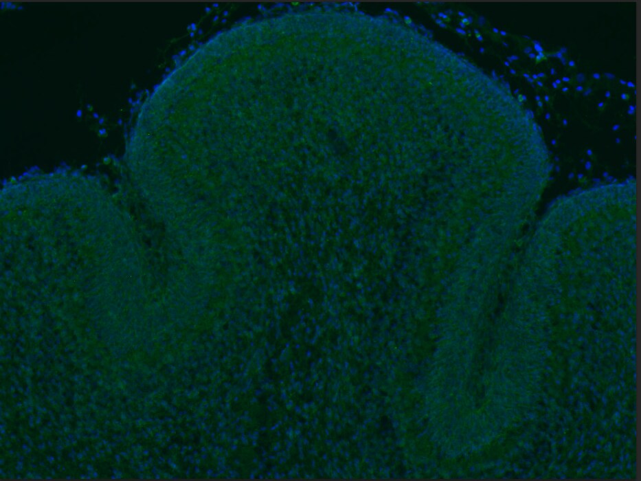
sFRP-1, also known as secreted apoptosis-related protein 2 (SARP-2), FRP and FrzA, is expressed in the embryonic kidney, eye, brain, teeth, salivary gland and small intestine, most often at sites of epithelial-mesenchyme interaction (5). Expression in the adult animal is strong in the eye, kidney, and heart and also prevalent in the brain and lung (2, 5). sFRP-1 was first characterized as a protein that enhances the sensitivity of cells to apoptotic stimuli (3) and as an antagonist of Wnt signaling in Xenopus embryos (4). It is also characterized as a tumor suppressor in breast (6) and cervical carcinomas (7). In contrast, sFRP-1 is expressed by the majority of malignant gliomas (8) and contributes to the development of uterine leiomyomas (9), suggesting that the role of sFRP-1 is dependent on cell context. sFRP-1 has diverse activities, from inducing angiogenesis (10) in a variety of in vivo models to helping regulate Wnt-4 signaling (with sFRP-2) in renal organogenesis (11). Mouse and human sFRP-1 proteins share 94% amino acid identity (1).

References

  1. Jones, S. et al. (2002) Bioessays 24:811.
  2. Rattner, A. et al. (1997) Proc. Natl. Acad. Sci. USA 94:2859.
  3. Melkonyan, H. et al. (1997) Proc. Natl. Acad. Sci. USA 94:13636.
  4. Finch, P. et al. (1997) Proc. Natl. Acad. Sci. USA 94:6770.
  5. Leimeister, C. et al. (1998) Mech. Dev. 75:29.
  6. Ugolini, F. et al. (2001) Oncogene 20:5810.
  7. Ko, J. et al. (2002) Exp. Cell. Res. 280:280.
  8. Roth, W. et al. (2000) Oncogene 19:4210.
  9. Fukuhara, K, et al. (2002) J. Clin. Endocr. Metab. 87:1729.
  10. Dufourcq, P. et al. (2002) Circulation 106: 3097.
  11. Yoshino, K. et al. (2001) Mech. Dev. 102:45.

Long Name

Secreted Frizzled Related Protein 1

Alternate Names

FrzA, SARP-2, sFRP1

Entrez Gene IDs

6422 (Human); 20377 (Mouse); 84402 (Rat)

Gene Symbol

SFRP1

UniProt

Additional sFRP-1 Products

Product Documents for Human sFRP‑1 Antibody

Certificate of Analysis

To download a Certificate of Analysis, please enter a lot or batch number in the search box below.

Note: Certificate of Analysis not available for kit components.

Product Specific Notices for Human sFRP‑1 Antibody

For research use only

Related Research Areas

Citations for Human sFRP‑1 Antibody

Customer Reviews for Human sFRP‑1 Antibody (3)

3 out of 5
3 Customer Ratings
5 Stars
0%
4 Stars
67%
3 Stars
0%
2 Stars
0%
1 Stars
33%

Have you used Human sFRP‑1 Antibody?

Submit a review and receive an Amazon gift card!

$25/€18/£15/$25CAN/¥2500 Yen for a review with an image

$10/€7/£6/$10CAN/¥1110 Yen for a review without an image

Submit a review
Amazon Gift Card

Customer Images


Showing  1 - 3 of 3 reviews Showing All
Filter By:
  • Human sFRP-1 Antibody
    Name: Cathy Guo
    Application: Immunofluorescence
    Sample Tested: Frozen section from 4% PFA fixed mouse brain.
    Species: Mouse
    Verified Customer | Posted 10/22/2020
    Sfrp14 (Green) _DAPI (blue& ) on P4 mouse cerebellum.
    Human sFRP‑1 Antibody AF1384
    Bio-Techne Response
    This review was submitted through the legacy Novus Innovators Program, reflecting a new species or application tested on a primary antibody.
  • Human sFRP-1 Antibody
    Name: Anonymous
    Application: Microarray
    Sample Tested: EDTA Plasma
    Species: Human
    Verified Customer | Posted 01/03/2020
    Human sFRP‑1 Antibody AF1384
  • Human sFRP-1 Antibody
    Name: Anonymous
    Application: Microarray
    Sample Tested: EDTA Plasma
    Species: Human
    Verified Customer | Posted 10/31/2018

There are no reviews that match your criteria.

Showing  1 - 3 of 3 reviews Showing All

Protocols

Find general support by application which include: protocols, troubleshooting, illustrated assays, videos and webinars.

FAQs

No product specific FAQs exist for this product.

View all FAQs for Antibodies