SREBP2 (Sterol Regulatory Element-Binding Protein 2; also bHLHD2 and SREBF2) is a 120-125 kDa member of the SREBP family of proteins. It is ubiquitously expressed and found in the intracellular membrane fraction of cells. SREBP2 is a transcriptional factor initially embedded in the ER as an inactive precursor associated with SCAP. When necessary, SCAP mediates SREBP2 transfer to the Golgi, where two resident proteases remove the N-terminus from SREBP2, and the N-terminus is transported into the nucleus. Here, SREBP2 acts as a transcription factor, activating the LDLR and cholesterol synthesis genes. The human SREBP2 precursor is an 1141 amino acid (aa) two transmembrane protein whose N- and C-termini are cytoplasmic. The two cytoplasmic domains span aa 1-479 and 555-1141, respectively. Proteolytic cleavage between Leu484-Cys485 generates the 64-66 kDa SREBP2 transcription factor. This fragment contains a bHLH DNA binding domain (aa 330-380) and one Leu zipper region (aa 381-401). Homodimerization of SREBP2 is necessary for nuclear translocation. There is one potential isoform that shows a deletion of aa 274-276 coupled to a 96 aa substitution for aa 580-1141. A second isoform (known in rodent) shows a premature truncation after Val463 and runs at 55 kDa in SDS-PAGE. Over aa 242-450, human SREBP2 shares 97% aa identity with mouse SREBP2.

Key Product Details
Species Reactivity
Validated:
Human
Cited:
Human, Mouse
Applications
Validated:
Immunohistochemistry, Immunocytochemistry
Cited:
Immunohistochemistry, Western Blot
Label
Unconjugated
Antibody Source
Monoclonal Mouse IgG1 Clone # 751512
Loading...
Product Specifications
Immunogen
E. coli-derived recombinant human SREBP2
Leu242-Asp450
Accession # Q12772
Leu242-Asp450
Accession # Q12772
Specificity
Detects human SREBP2 in ELISA.
Clonality
Monoclonal
Host
Mouse
Isotype
IgG1
Scientific Data Images for Human SREBP2 Antibody (751512)
SREBP2 in HepG2 Human Cell Line.
SREBP2 was detected in immersion fixed HepG2 human hepatocellular carcinoma cell line using Mouse Anti-Human SREBP2 Monoclonal Antibody (Catalog # MAB7119) at 10 µg/mL for 3 hours at room temperature. Cells were stained using the NorthernLights™ 557-conjugated Anti-Mouse IgG Secondary Antibody (red; Catalog # NL007) and counterstained with DAPI (blue). Specific staining was localized to cell surfaces and cytoplasm. View our protocol for Fluorescent ICC Staining of Cells on Coverslips.
SREBP2 in Human Kidney.
SREBP2 was detected in immersion fixed paraffin-embedded sections of human kidney using Mouse Anti-Human SREBP2 Monoclonal Antibody (Catalog # MAB7119) at 15 µg/mL overnight at 4 °C. Tissue was stained using the Anti-Mouse HRP-DAB Cell & Tissue Staining Kit (brown; Catalog # CTS002) and counterstained with hematoxylin (blue). Specific staining was localized to epithelial cells in convoluted tubules. View our protocol for Chromogenic IHC Staining of Paraffin-embedded Tissue Sections.Applications for Human SREBP2 Antibody (751512)
Application
Recommended Usage
Immunocytochemistry
8-25 µg/mL
Sample: Immersion fixed HepG2 human hepatocellular carcinoma cell line
Sample: Immersion fixed HepG2 human hepatocellular carcinoma cell line
Immunohistochemistry
8-25 µg/mL
Sample: Immersion fixed paraffin-embedded sections of human kidney
Sample: Immersion fixed paraffin-embedded sections of human kidney
Reviewed Applications
Read 1 review rated 5 using MAB7119 in the following applications:
Formulation, Preparation, and Storage
Purification
Protein A or G purified from hybridoma culture supernatant
Reconstitution
Reconstitute at 0.5 mg/mL in sterile PBS. For liquid material, refer to CoA for concentration.
Loading...
Formulation
Lyophilized from a 0.2 μm filtered solution in PBS with Trehalose. See Certificate of Analysis for details.
*Small pack size (-SP) is supplied either lyophilized or as a 0.2 µm filtered solution in PBS.
*Small pack size (-SP) is supplied either lyophilized or as a 0.2 µm filtered solution in PBS.
Shipping
Lyophilized product is shipped at ambient temperature. Liquid small pack size (-SP) is shipped with polar packs. Upon receipt, store immediately at the temperature recommended below.
Stability & Storage
Use a manual defrost freezer and avoid repeated freeze-thaw cycles.
- 12 months from date of receipt, -20 to -70 °C as supplied.
- 1 month, 2 to 8 °C under sterile conditions after reconstitution.
- 6 months, -20 to -70 °C under sterile conditions after reconstitution.
Calculators
Background: SREBP2
Long Name
Sterol Regulatory Element Binding Transcription Factor 2
Alternate Names
bHLhd2, SREBF2
Gene Symbol
SREBF2
UniProt
Additional SREBP2 Products
Product Documents for Human SREBP2 Antibody (751512)
Certificate of Analysis
To download a Certificate of Analysis, please enter a lot or batch number in the search box below.
Note: Certificate of Analysis not available for kit components.
Product Specific Notices for Human SREBP2 Antibody (751512)
For research use only
Citations for Human SREBP2 Antibody (751512)
 Powered by Bioz
Customer Reviews for Human SREBP2 Antibody (751512) (1)
5 out of 5
1 Customer Rating
Have you used Human SREBP2 Antibody (751512)?
Submit a review and receive an Amazon gift card!
$25/€18/£15/$25CAN/¥2500 Yen for a review with an image
$10/€7/£6/$10CAN/¥1110 Yen for a review without an image
Submit a review
Customer Images
Showing
1
-
1 of
1 review
Showing All
Filter By:
-
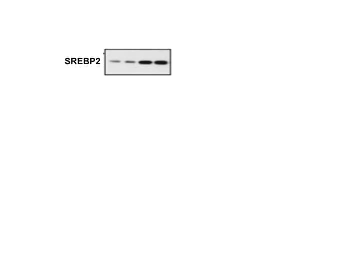
Application: Western BlotSample Tested: iPS2 human induced pluripotent stem cellsSpecies: HumanVerified Customer | Posted 01/17/2018

There are no reviews that match your criteria.
Protocols
Find general support by application which include: protocols, troubleshooting, illustrated assays, videos and webinars.
- Antigen Retrieval Protocol (PIER)
- Antigen Retrieval for Frozen Sections Protocol
- Appropriate Fixation of IHC/ICC Samples
- Cellular Response to Hypoxia Protocols
- Chromogenic IHC Staining of Formalin-Fixed Paraffin-Embedded (FFPE) Tissue Protocol
- Chromogenic Immunohistochemistry Staining of Frozen Tissue
- ClariTSA™ Fluorophore Kits
- Detection & Visualization of Antibody Binding
- Fluorescent IHC Staining of Frozen Tissue Protocol
- Graphic Protocol for Heat-induced Epitope Retrieval
- Graphic Protocol for the Preparation and Fluorescent IHC Staining of Frozen Tissue Sections
- Graphic Protocol for the Preparation and Fluorescent IHC Staining of Paraffin-embedded Tissue Sections
- Graphic Protocol for the Preparation of Gelatin-coated Slides for Histological Tissue Sections
- ICC Cell Smear Protocol for Suspension Cells
- ICC Immunocytochemistry Protocol Videos
- ICC for Adherent Cells
- IHC Sample Preparation (Frozen sections vs Paraffin)
- Immunocytochemistry (ICC) Protocol
- Immunocytochemistry Troubleshooting
- Immunofluorescence of Organoids Embedded in Cultrex Basement Membrane Extract
- Immunofluorescent IHC Staining of Formalin-Fixed Paraffin-Embedded (FFPE) Tissue Protocol
- Immunohistochemistry (IHC) and Immunocytochemistry (ICC) Protocols
- Immunohistochemistry Frozen Troubleshooting
- Immunohistochemistry Paraffin Troubleshooting
- Preparing Samples for IHC/ICC Experiments
- Preventing Non-Specific Staining (Non-Specific Binding)
- Primary Antibody Selection & Optimization
- Protocol for Heat-Induced Epitope Retrieval (HIER)
- Protocol for Making a 4% Formaldehyde Solution in PBS
- Protocol for VisUCyte™ HRP Polymer Detection Reagent
- Protocol for the Fluorescent ICC Staining of Cell Smears - Graphic
- Protocol for the Fluorescent ICC Staining of Cultured Cells on Coverslips - Graphic
- Protocol for the Preparation & Fixation of Cells on Coverslips
- Protocol for the Preparation and Chromogenic IHC Staining of Frozen Tissue Sections
- Protocol for the Preparation and Chromogenic IHC Staining of Frozen Tissue Sections - Graphic
- Protocol for the Preparation and Chromogenic IHC Staining of Paraffin-embedded Tissue Sections
- Protocol for the Preparation and Chromogenic IHC Staining of Paraffin-embedded Tissue Sections - Graphic
- Protocol for the Preparation and Fluorescent ICC Staining of Cells on Coverslips
- Protocol for the Preparation and Fluorescent ICC Staining of Non-adherent Cells
- Protocol for the Preparation and Fluorescent ICC Staining of Stem Cells on Coverslips
- Protocol for the Preparation and Fluorescent IHC Staining of Frozen Tissue Sections
- Protocol for the Preparation and Fluorescent IHC Staining of Paraffin-embedded Tissue Sections
- Protocol for the Preparation of Gelatin-coated Slides for Histological Tissue Sections
- Protocol for the Preparation of a Cell Smear for Non-adherent Cell ICC - Graphic
- TUNEL and Active Caspase-3 Detection by IHC/ICC Protocol
- The Importance of IHC/ICC Controls
- Troubleshooting Guide: Immunohistochemistry
- View all Protocols, Troubleshooting, Illustrated assays and Webinars
Loading...
Associated Pathways
