TACE is a member of the ADAM family that contains A Disintegrin And Metalloprotease-like domain. Like other membrane-anchored ADAMs, TACE consists of a pro domain with a cysteine switch and furin cleavage sequence, a catalytic domain with the zinc-binding site and Met-turn expected for reprolysins, a disintegrin-like domain, a cysteine-rich domain, an EGF-like domain, a transmembrane domain, and the cytoplasmic domain. In addition to its ability to release the 17 kDa extracellular form of tumor necrosis factor-alpha (TNF-alpha ) from the 26 kDa membrane-anchored TNF-alpha, TACE also plays an essential role in shedding ectodomains from a variety of proteins such as L-Selectin, Transforming Growth Factor-alpha, Amyloid Protein Precursor, and Notch-1 receptor. TACE mRNA is present in virtually every tissue and TACE protein resides both on the cell surface and in the cell.

Key Product Details
Species Reactivity
Validated:
Human
Cited:
Human
Applications
Validated:
Immunohistochemistry, Western Blot
Cited:
Immunohistochemistry-Paraffin, Western Blot
Label
Unconjugated
Antibody Source
Polyclonal Goat IgG
Loading...
Product Specifications
Immunogen
S. frugiperda insect ovarian cell line Sf 21-derived recombinant human TACE/ADAM17
Arg215-Asn671
Accession # P78536
Arg215-Asn671
Accession # P78536
Specificity
Detects human TACE/ADAM17 in direct ELISAs and Western blots.
Clonality
Polyclonal
Host
Goat
Isotype
IgG
Scientific Data Images for Human TACE/ADAM17 Antibody
Detection of Human TACE/ADAM17 by Western Blot.
Western blot shows lysates of human heart tissue. PVDF membrane was probed with 1 µg/mL of Goat Anti-Human TACE/ADAM17 Antigen Affinity-purified Polyclonal Antibody (Catalog # AF9301) followed by HRP-conjugated Anti-Goat IgG Secondary Antibody (Catalog # HAF019). A specific band was detected for TACE/ADAM17 at approximately 100 kDa (as indicated). This experiment was conducted under reducing conditions and using Immunoblot Buffer Group 1.
TACE/ADAM17 in Human Colon.
TACE/ADAM17 was detected in immersion fixed paraffin-embedded sections of human colon using Goat Anti-Human TACE/ADAM17 Antigen Affinity-purified Polyclonal Antibody (Catalog # AF9301) at 3 µg/mL overnight at 4 °C. Before incubation with the primary antibody, tissue was subjected to heat-induced epitope retrieval using Antigen Retrieval Reagent-Basic (Catalog # CTS013). Tissue was stained using the Anti-Goat HRP-DAB Cell & Tissue Staining Kit (brown; Catalog # CTS008) and counterstained with hematoxylin (blue). Specific staining was localized to epithelial cells. View our protocol for Chromogenic IHC Staining of Paraffin-embedded Tissue Sections.Applications for Human TACE/ADAM17 Antibody
Application
Recommended Usage
Immunohistochemistry
5-15 µg/mL
Sample: Immersion fixed paraffin-embedded sections of human colon
Sample: Immersion fixed paraffin-embedded sections of human colon
Western Blot
1 µg/mL
Sample: Human heart tissue
Sample: Human heart tissue
Reviewed Applications
Read 1 review rated 5 using AF9301 in the following applications:
Formulation, Preparation, and Storage
Purification
Antigen Affinity-purified
Reconstitution
Sterile PBS to a final concentration of 0.2 mg/mL. For liquid material, refer to CoA for concentration.
Loading...
Formulation
Lyophilized from a 0.2 μm filtered solution in PBS with Trehalose. *Small pack size (SP) is supplied either lyophilized or as a 0.2 µm filtered solution in PBS.
Shipping
Lyophilized product is shipped at ambient temperature. Liquid small pack size (-SP) is shipped with polar packs. Upon receipt, store immediately at the temperature recommended below.
Stability & Storage
Use a manual defrost freezer and avoid repeated freeze-thaw cycles.
- 12 months from date of receipt, -20 to -70 °C as supplied.
- 1 month, 2 to 8 °C under sterile conditions after reconstitution.
- 6 months, -20 to -70 °C under sterile conditions after reconstitution.
Calculators
Background: TACE/ADAM17
References
- Black, R.A. and J.D. Becherer (1998) in Tumor Necrosis Factor alpha -Converting Enzyme. Barrett, A.J. et al. (eds): Handbook of Proteolytic Enzymes, San Diego: Academic Press, p. 1315.
- Primakoff, P. and D.G. Myles (2000) Trends in Genetics 16:83.
Long Name
TNF-alpha Converting Enzyme
Alternate Names
ADAM17, CD156b
Gene Symbol
ADAM17
UniProt
Additional TACE/ADAM17 Products
Product Documents for Human TACE/ADAM17 Antibody
Certificate of Analysis
To download a Certificate of Analysis, please enter a lot or batch number in the search box below.
Note: Certificate of Analysis not available for kit components.
Product Specific Notices for Human TACE/ADAM17 Antibody
For research use only
Related Research Areas
Citations for Human TACE/ADAM17 Antibody
 Powered by Bioz
Customer Reviews for Human TACE/ADAM17 Antibody (1)
5 out of 5
1 Customer Rating
Have you used Human TACE/ADAM17 Antibody?
Submit a review and receive an Amazon gift card!
$25/€18/£15/$25CAN/¥2500 Yen for a review with an image
$10/€7/£6/$10CAN/¥1110 Yen for a review without an image
Submit a review
Customer Images
Showing
1
-
1 of
1 review
Showing All
Filter By:
-
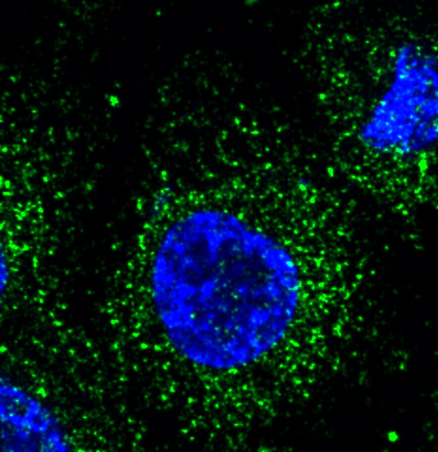
Application: Immunocytochemistry/ImmunofluorescenceSample Tested: THP-1 human acute monocytic leukemia cell lineSpecies: HumanVerified Customer | Posted 11/17/2018

There are no reviews that match your criteria.
Protocols
Find general support by application which include: protocols, troubleshooting, illustrated assays, videos and webinars.
- Antigen Retrieval Protocol (PIER)
- Antigen Retrieval for Frozen Sections Protocol
- Appropriate Fixation of IHC/ICC Samples
- Cellular Response to Hypoxia Protocols
- Chromogenic IHC Staining of Formalin-Fixed Paraffin-Embedded (FFPE) Tissue Protocol
- Chromogenic Immunohistochemistry Staining of Frozen Tissue
- ClariTSA™ Fluorophore Kits
- Detection & Visualization of Antibody Binding
- Fluorescent IHC Staining of Frozen Tissue Protocol
- Graphic Protocol for Heat-induced Epitope Retrieval
- Graphic Protocol for the Preparation and Fluorescent IHC Staining of Frozen Tissue Sections
- Graphic Protocol for the Preparation and Fluorescent IHC Staining of Paraffin-embedded Tissue Sections
- Graphic Protocol for the Preparation of Gelatin-coated Slides for Histological Tissue Sections
- IHC Sample Preparation (Frozen sections vs Paraffin)
- Immunofluorescent IHC Staining of Formalin-Fixed Paraffin-Embedded (FFPE) Tissue Protocol
- Immunohistochemistry (IHC) and Immunocytochemistry (ICC) Protocols
- Immunohistochemistry Frozen Troubleshooting
- Immunohistochemistry Paraffin Troubleshooting
- Preparing Samples for IHC/ICC Experiments
- Preventing Non-Specific Staining (Non-Specific Binding)
- Primary Antibody Selection & Optimization
- Protocol for Heat-Induced Epitope Retrieval (HIER)
- Protocol for Making a 4% Formaldehyde Solution in PBS
- Protocol for VisUCyte™ HRP Polymer Detection Reagent
- Protocol for the Preparation & Fixation of Cells on Coverslips
- Protocol for the Preparation and Chromogenic IHC Staining of Frozen Tissue Sections
- Protocol for the Preparation and Chromogenic IHC Staining of Frozen Tissue Sections - Graphic
- Protocol for the Preparation and Chromogenic IHC Staining of Paraffin-embedded Tissue Sections
- Protocol for the Preparation and Chromogenic IHC Staining of Paraffin-embedded Tissue Sections - Graphic
- Protocol for the Preparation and Fluorescent IHC Staining of Frozen Tissue Sections
- Protocol for the Preparation and Fluorescent IHC Staining of Paraffin-embedded Tissue Sections
- Protocol for the Preparation of Gelatin-coated Slides for Histological Tissue Sections
- R&D Systems Quality Control Western Blot Protocol
- TUNEL and Active Caspase-3 Detection by IHC/ICC Protocol
- The Importance of IHC/ICC Controls
- Troubleshooting Guide: Immunohistochemistry
- Troubleshooting Guide: Western Blot Figures
- Western Blot Conditions
- Western Blot Protocol
- Western Blot Protocol for Cell Lysates
- Western Blot Troubleshooting
- Western Blot Troubleshooting Guide
- View all Protocols, Troubleshooting, Illustrated assays and Webinars
Loading...
Associated Pathways

