The Transferrin Receptor (TfR or TfR-1, designated CD71) is a type 2 transmembrane glycoprotein expressed on erythroid progenitors, muscle cells and proliferating cells as a 188 kDa disulfide-linked homodimer of 95 kDa monomers (1-4). As the major mediator of cellular iron uptake, it binds and internalizes diferric transferrin, allowing iron release at the low pH of the endosome (2, 5). The human TfR cDNA encodes 760 amino acids (aa) including a 67 aa N-terminal intracellular domain, a 21 aa transmembrane domain, and a 672 aa extracellular domain (ECD) with helical, peptidase (nonfunctional), and ligand binding domains, including an RGD potential integrin binding site (5). Human TfR ECD shares 75 - 80% aa identity with mouse, rat, feline, canine, equine, porcine and bovine TfR. A 679 aa alternately spliced form begins at aa 82 and is presumably secreted, while in an 804 aa form, 44 aa are inserted at aa 518 within the peptidase region (6). Most soluble TfR (sTfR) arises from trypsin proteolysis at aa 100, producing the circulating form of TfR (3). sTfR concentration in plasma or serum is proportional to total TfR and can be increased by iron deficiency (3). Erythroid progenitors, which use iron for hemoglobin synthesis, normally account for the bulk of total body TfR production (3). Since rapidly growing cells require iron to replicate DNA, cancer cells can express up to 5-fold more TfR than quiescent cells in the surrounding tissue (2, 4). Antibody targeting of TfR can inhibit tumor cell proliferation and induce apoptosis (2, 4). The hereditary hemochromatosis protein HFE competes with diferric transferrin for binding to TfR, and targets TfR for degradation rather than recycling (2, 5). TfR has been reported to have ferritin-independent functions in T cell development, immunological synapse formation and galectin-3-mediated cell death, and to be a cell entry receptor for New World hemorrhagic fever arenaviruses (2, 4, 7).
Human TfR (Transferrin R) Antibody
R&D Systems | Catalog # AF2474
 by Western Blot.")
Key Product Details
Species Reactivity
Validated:
Human
Cited:
Human, Mouse, Transgenic Mouse
Applications
Validated:
Immunohistochemistry, Western Blot, Blockade of Receptor-ligand Interaction
Cited:
Immunohistochemistry-Frozen, Western Blot, Neutralization, Surface Plasmon Resonance
Label
Unconjugated
Antibody Source
Polyclonal Goat IgG
Loading...
Product Specifications
Immunogen
Human placenta-derived TfR (Transferrin R)
Specificity
Detects human TfR (Transferrin R) in direct ELISAs and Western blots.
Clonality
Polyclonal
Host
Goat
Isotype
IgG
Endotoxin Level
<0.10 EU per 1 μg of the antibody by the LAL method.
Scientific Data Images for Human TfR (Transferrin R) Antibody
Detection of Human TfR (Transferrin R) by Western Blot.
Western blot shows lysates of ZR-75 human breast cancer cell line, U937 human histiocytic lymphoma cell line, and K562 human chronic myelogenous leukemia cell line. PVDF membrane was probed with 0.25 µg/mL of Goat Anti-Human TfR (Transferrin R) Antigen Affinity-purified Polyclonal Antibody (Catalog # AF2474) followed by HRP-conjugated Anti-Goat IgG Secondary Antibody (Catalog # HAF017). A specific band was detected for TfR (Transferrin R) at approximately 95 kDa (as indicated). This experiment was conducted under reducing conditions and using Immunoblot Buffer Group 1. in Human Liver.")
TfR (Transferrin R) in Human Liver.
TfR (Transferrin R) was detected in immersion fixed paraffin-embedded sections of human liver using Goat Anti-Human TfR (Transferrin R) Antigen Affinity-purified Polyclonal Antibody (Catalog # AF2474) at 5 µg/mL overnight at 4 °C. Tissue was stained using the Anti-Goat HRP-DAB Cell & Tissue Staining Kit (brown; Catalog # CTS008) and counterstained with hematoxylin (blue). Specific labeling was localized to the plasma membrane of hepatocytes. View our protocol for Chromogenic IHC Staining of Paraffin-embedded Tissue Sections. by Western Blot")
Detection of Mouse TfR (Transferrin R) by Western Blot
Effect of Pcsk9 deletion on LDLR and CD81 levels in age- and sex-matched Pcsk9-/- mice (n = 5) and their wild type littermates (n = 4).Shown are (A) mean ± SE serum LDL-C levels and (B) Western blot analysis of CD81 and LDLR levels in liver extracts with TfR as loading control, in animals sacrificed after a 4-hour fast. Lanes in panel B represent samples from individual mice. LDL-C, low-density lipoprotein cholesterol; LDLR, low-density lipoprotein receptor; PCSK9, proprotein convertase subtilisin/kexin type 9; SE, standard error; TfR, transferrin receptor; wt, wild type. Image collected and cropped by CiteAb from the following publication (https://dx.plos.org/10.1371/journal.pone.0154498), licensed under a CC-BY license. Not internally tested by R&D Systems. by Western Blot")
Detection of TfR (Transferrin R) by Western Blot
shFXN have altered iron metabolism proteins. RNA was reverse transcribed and amplified for (A) Transferrin receptor (TfR), (B) Ferroportin (Fpn), and Beta-2-microglobulin (B2M) as a housekeeping control, transcript abundance was quantified using the delta delta Ct method normalized to the empty vector endothelial cells (EVEC). Total protein lysates were run on a 12% bis-tris SDS-PAGE, transferred to nitrocellulose, and probed for (C) TfR and (D) Fpn protein expression against Tata-Binding Protein (TBP) as a housekeeping gene. Each protein is normalized to that of TBP as quantified by densitometry, normalized to pooled EVEC values. (E) Representative blot is shown. TfR is visualized with Alexa-647 (Red), and Fpn with Alexa-488 (Green). Student’s t-test alpha = 0.05. *p < 0.05, **p < 0.01. (A) EVEC; n = 2 shFXN; n = 3. (B) EVEC; n = 1, and shFXN2; n = 2. (C–E) EVEC; n = 6 and shFXN; n = 8. Image collected and cropped by CiteAb from the following open publication (https://www.frontiersin.org/articles/10.3389/fmolb.2023.1299201/full), licensed under a CC-BY license. Not internally tested by R&D Systems.Applications for Human TfR (Transferrin R) Antibody
Application
Recommended Usage
Blockade of Receptor-ligand Interaction
In a functional ELISA, 0.1-0.5 µg/mL of this antibody will block 50% of the binding of 25 ng/mL of Recombinant Human TfR (Transferrin R) (Catalog # 2474-TR) to immobilized human Holo-Transferrin (Catalog # 2914-HT) coated at 500 ng/mL (100 µL/well). At 10 μg/mL, this antibody will block >90% of the binding.
Immunohistochemistry
5-15 µg/mL
Sample: Immersion fixed paraffin-embedded sections of human bladder, breast, and liver
Sample: Immersion fixed paraffin-embedded sections of human bladder, breast, and liver
Western Blot
0.25 µg/mL
Sample: ZR‑75 human breast cancer cell line, U937 human histiocytic lymphoma cell line, and K562 human chronic myelogenous leukemia cell line
Sample: ZR‑75 human breast cancer cell line, U937 human histiocytic lymphoma cell line, and K562 human chronic myelogenous leukemia cell line
Reviewed Applications
Read 4 reviews rated 4.5 using AF2474 in the following applications:
Formulation, Preparation, and Storage
Purification
Antigen Affinity-purified
Reconstitution
Reconstitute at 0.2 mg/mL in sterile PBS. For liquid material, refer to CoA for concentration.
Loading...
Formulation
Lyophilized from a 0.2 μm filtered solution in PBS with Trehalose. *Small pack size (SP) is supplied either lyophilized or as a 0.2 µm filtered solution in PBS.
Shipping
Lyophilized product is shipped at ambient temperature. Liquid small pack size (-SP) is shipped with polar packs. Upon receipt, store immediately at the temperature recommended below.
Stability & Storage
Use a manual defrost freezer and avoid repeated freeze-thaw cycles.
- 12 months from date of receipt, -20 to -70 °C as supplied.
- 1 month, 2 to 8 °C under sterile conditions after reconstitution.
- 6 months, -20 to -70 °C under sterile conditions after reconstitution.
Calculators
Background: TfR (Transferrin R)
References
- Schneider, C. et al. (1984) Nature 311:675.
- Daniels, T.R. et al. (2006) Clin. Immunol. 121:144.
- Skikne, B.S. (2008) Am. J. Hematol. 83:872.
- Macedo, M.F. and M. deSousa (2008) Inflamm. Allergy Drug Targets 7:41.
- Aisen, P. (2004) Int. J. Biochem. Cell Biol. 36:2137.
- Entrez protein Accession # EAW53671, EAW53672.
- Radoshitzky, S.R. et al. (2007) Nature 446:92.
Long Name
Transferrin Receptor
Alternate Names
CD71, TfR (TransferrinR), TFR1, TFRC, TRFR
Gene Symbol
TFRC
Additional TfR (Transferrin R) Products
Product Documents for Human TfR (Transferrin R) Antibody
Certificate of Analysis
To download a Certificate of Analysis, please enter a lot or batch number in the search box below.
Note: Certificate of Analysis not available for kit components.
Product Specific Notices for Human TfR (Transferrin R) Antibody
For research use only
Citations for Human TfR (Transferrin R) Antibody
 Powered by Bioz
Customer Reviews for Human TfR (Transferrin R) Antibody (4)
4.5 out of 5
4 Customer Ratings
Have you used Human TfR (Transferrin R) Antibody?
Submit a review and receive an Amazon gift card!
$25/€18/£15/$25CAN/¥2500 Yen for a review with an image
$10/€7/£6/$10CAN/¥1110 Yen for a review without an image
Submit a review
Customer Images
Showing
1
-
4 of
4 reviews
Showing All
Filter By:
-
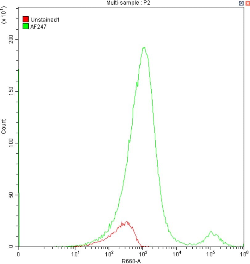
Application: Flow CytometrySample Tested: K562 human chronic myelogenous leukemia cell lineSpecies: cellVerified Customer | Posted 12/11/20251:100 dilution

-
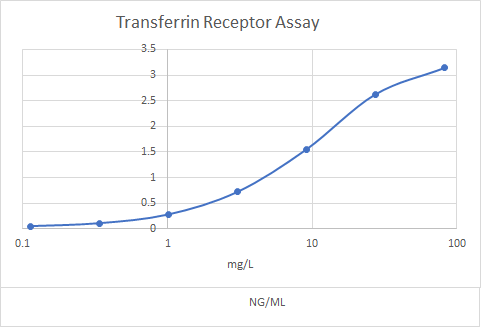
Application: ELISASample Tested: Heparin PlasmaSpecies: HumanVerified Customer | Posted 05/12/2021Coated wells with this antibody to measure transferrin receptor in an immunoassay.
-
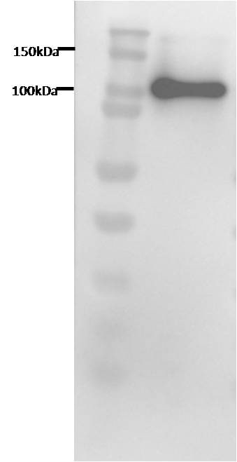
Application: Western BlotSample Tested: Brain Vascular Smooth Muscle CellsSpecies: HumanVerified Customer | Posted 02/08/2019
-
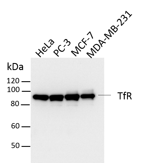
Application: Western BlotSample Tested: HeLa cells, PC-3 human prostate cancer cell line, MCF-7 human breast cancer cell line and MDA-MB-231 human breast cancer cell lineSpecies: HumanVerified Customer | Posted 07/23/2018Total cell lysates from HeLa, PC-3, MCF-7 and MDA-MB-231 were subjected to western blot. PVDF membrane were probed with 0.5 um/ml Human TfR Antibody (AF2474). A specific band was detected for TfR at approximately 90 kDa. This experiment was conducted under reducing conditions
There are no reviews that match your criteria.
Protocols
Find general support by application which include: protocols, troubleshooting, illustrated assays, videos and webinars.
- Antigen Retrieval Protocol (PIER)
- Antigen Retrieval for Frozen Sections Protocol
- Appropriate Fixation of IHC/ICC Samples
- Cellular Response to Hypoxia Protocols
- Chromogenic IHC Staining of Formalin-Fixed Paraffin-Embedded (FFPE) Tissue Protocol
- Chromogenic Immunohistochemistry Staining of Frozen Tissue
- ClariTSA™ Fluorophore Kits
- Detection & Visualization of Antibody Binding
- Fluorescent IHC Staining of Frozen Tissue Protocol
- Graphic Protocol for Heat-induced Epitope Retrieval
- Graphic Protocol for the Preparation and Fluorescent IHC Staining of Frozen Tissue Sections
- Graphic Protocol for the Preparation and Fluorescent IHC Staining of Paraffin-embedded Tissue Sections
- Graphic Protocol for the Preparation of Gelatin-coated Slides for Histological Tissue Sections
- IHC Sample Preparation (Frozen sections vs Paraffin)
- Immunofluorescent IHC Staining of Formalin-Fixed Paraffin-Embedded (FFPE) Tissue Protocol
- Immunohistochemistry (IHC) and Immunocytochemistry (ICC) Protocols
- Immunohistochemistry Frozen Troubleshooting
- Immunohistochemistry Paraffin Troubleshooting
- Preparing Samples for IHC/ICC Experiments
- Preventing Non-Specific Staining (Non-Specific Binding)
- Primary Antibody Selection & Optimization
- Protocol for Heat-Induced Epitope Retrieval (HIER)
- Protocol for Making a 4% Formaldehyde Solution in PBS
- Protocol for VisUCyte™ HRP Polymer Detection Reagent
- Protocol for the Preparation & Fixation of Cells on Coverslips
- Protocol for the Preparation and Chromogenic IHC Staining of Frozen Tissue Sections
- Protocol for the Preparation and Chromogenic IHC Staining of Frozen Tissue Sections - Graphic
- Protocol for the Preparation and Chromogenic IHC Staining of Paraffin-embedded Tissue Sections
- Protocol for the Preparation and Chromogenic IHC Staining of Paraffin-embedded Tissue Sections - Graphic
- Protocol for the Preparation and Fluorescent IHC Staining of Frozen Tissue Sections
- Protocol for the Preparation and Fluorescent IHC Staining of Paraffin-embedded Tissue Sections
- Protocol for the Preparation of Gelatin-coated Slides for Histological Tissue Sections
- R&D Systems Quality Control Western Blot Protocol
- TUNEL and Active Caspase-3 Detection by IHC/ICC Protocol
- The Importance of IHC/ICC Controls
- Troubleshooting Guide: Immunohistochemistry
- Troubleshooting Guide: Western Blot Figures
- Western Blot Conditions
- Western Blot Protocol
- Western Blot Protocol for Cell Lysates
- Western Blot Troubleshooting
- Western Blot Troubleshooting Guide
- View all Protocols, Troubleshooting, Illustrated assays and Webinars
Loading...