TL1A is a type II transmembrane protein belonging to the TNF superfamily and has been designated TNF superfamily member 15 (TNFSF15). Human TL1A is a 251 aa protein consisting of a 35 aa cytoplasmic domain, a 24 aa transmembrane region and a 192 aa C-terminal extracellular domain. It is a longer variant of the previously cloned TL1 (also known as VEGI) that is possibly a cloning artifact. TL1A is predominantly expressed in endothelial cells and its expression is inducible by TNF-alpha and IL-1 alpha. TL1A binds with high affinity to death receptor 3 (DR3), which is now designated TNF receptor superfamily member 25 (TNFRSF25). DR3 was formerly designated TNFRSF12 when it was thought to be the receptor for TWEAK/TNFSF12. DR3 is expressed primarily on activated T cells. Depending on the cell context, ligation of DR3 by TL1A can trigger one of two signaling pathways, activation of the transcription factor NF-kappa-B or activation of caspases and apoptosis. On primary T cells, TL1A induces NF-kappa-B activation and a costimulatory signal to increase IL-2 responsiveness and the secretion of proinflammatory cytokines. However, in a tumor cell line, TF-1, TL1A has been shown to induce caspase activity and apoptosis. These effects of TL1A are blocked by the secreted, soluble decoy receptor 3 (DcR3), also known as TR6 and TNFRSF6B, which compete with DR3 for binding to TL1A. Consistent with the observed in vitro activities, TL1A promotes ex vivo splenocyte expansion and enhances in vivo graft-versus-host-response.
Human TL1A/TNFSF15 Antibody (2116D)
R&D Systems | Catalog # MAB74421
Recombinant Monoclonal Antibody.

Key Product Details
Species Reactivity
Human
Applications
Immunohistochemistry, Western Blot, Neutralization
Label
Unconjugated
Antibody Source
Recombinant Monoclonal Rabbit IgG Clone # 2116D
Loading...
Product Specifications
Immunogen
E. coli-derived recombinant human TL1A/TNFSF15
Leu72-Leu251
Accession # O95150
Leu72-Leu251
Accession # O95150
Specificity
Detects human TL1A/TNFSF15 in direct ELISAs and Western blots.
Clonality
Monoclonal
Host
Rabbit
Isotype
IgG
Endotoxin Level
<0.10 EU per 1 μg of the antibody by the LAL method.
Scientific Data Images for Human TL1A/TNFSF15 Antibody (2116D)
Detection of Human TL1A/TNFSF15 by Western Blot.
Western blot shows lysates of human prostate tissue, human lung tissue, and HUVEC human umbilical vein endothelial cells. PVDF membrane was probed with 1 µg/mL of Rabbit Anti-Human TL1A/ TNFSF15 Monoclonal Antibody (Catalog # MAB74421) followed by HRP-conjugated Anti-Rabbit IgG Secondary Antibody (Catalog # HAF008). A specific band was detected for TL1A/TNFSF15 at approximately 22 kDa (as indicated). This experiment was conducted under reducing conditions and using Immunoblot Buffer Group 1.
TL1A/TNFSF15 in Human Prostate Cancer Tissue.
TL1A/TNFSF15 was detected in immersion fixed paraffin-embedded sections of human prostate cancer tissue using Rabbit Anti-Human TL1A/TNFSF15 Monoclonal Antibody (Catalog # MAB74421) at 0.3 µg/mL for 1 hour at room temperature followed by incubation with the Anti-Mouse IgG VisUCyte™ HRP Polymer Antibody (Catalog # VC001). Tissue was stained using DAB (brown) and counterstained with hematoxylin (blue). Specific staining was localized to cytoplasm of epithelial cells. View our protocol for IHC Staining with VisUCyte HRP Polymer Detection Reagents.
Apoptosis Induced by TL1A/TNFSF15 and Neutralization by Human TL1A/TNFSF15 Antibody.
Recombinant Human TL1A/TNFSF15 (Catalog # 1319-TL) induces apoptosis in the TF-1 human erythroleukemic cell line in a dose-dependent manner (orange line), as measured by Resazurin (Catalog # AR002). Apoptosis elicited by Recombinant Human TL1A/TNFSF15 (80 ng/mL) is neutralized (green line) by increasing concentrations of Rabbit Anti-Human TL1A/ TNFSF15 Monoclonal Antibody (Catalog # MAB74421). The ND50 is typically 0.04-0.2 ug/mL.Applications for Human TL1A/TNFSF15 Antibody (2116D)
Application
Recommended Usage
Immunohistochemistry
0.3-25 µg/mL
Sample: Immersion fixed paraffin-embedded sections of human prostate cancer tissue
Sample: Immersion fixed paraffin-embedded sections of human prostate cancer tissue
Western Blot
1 µg/mL
Sample: Human prostate tissue, human lung tissue, and HUVEC human umbilical vein endothelial cells
Sample: Human prostate tissue, human lung tissue, and HUVEC human umbilical vein endothelial cells
Neutralization
Measured
by its ability to neutralize TL1A/TNFSF15-induced apoptosis in the
TF‑1 human erythroleukemic cell line. The Neutralization Dose
(ND50) is typically 0.04-0.2 ug/mL in the
presence of 80 ng/mL Recombinant Human TL1A/TNFSF15.
Reviewed Applications
Read 1 review rated 5 using MAB74421 in the following applications:
Formulation, Preparation, and Storage
Purification
Protein A or G purified from cell culture supernatant
Reconstitution
Reconstitute at 0.5 mg/mL in sterile PBS. For liquid material, refer to CoA for concentration.
Loading...
Formulation
Lyophilized from a 0.2 μm filtered solution in PBS with Trehalose. *Small pack size (SP) is supplied either lyophilized or as a 0.2 µm filtered solution in PBS.
Shipping
Lyophilized product is shipped at ambient temperature. Liquid small pack size (-SP) is shipped with polar packs. Upon receipt, store immediately at the temperature recommended below.
Stability & Storage
Use a manual defrost freezer and avoid repeated freeze-thaw cycles.
- 12 months from date of receipt, -20 to -70 °C as supplied.
- 1 month, 2 to 8 °C under sterile conditions after reconstitution.
- 6 months, -20 to -70 °C under sterile conditions after reconstitution.
Calculators
Background: TL1A/TNFSF15
References
- Migone, T.S. et al. (2002) Immunity 16:479.
Long Name
TNF-like 1
Alternate Names
TNFSF15, VEGI
Gene Symbol
TNFSF15
UniProt
Additional TL1A/TNFSF15 Products
Product Documents for Human TL1A/TNFSF15 Antibody (2116D)
Certificate of Analysis
To download a Certificate of Analysis, please enter a lot or batch number in the search box below.
Note: Certificate of Analysis not available for kit components.
Product Specific Notices for Human TL1A/TNFSF15 Antibody (2116D)
For research use only
Customer Reviews for Human TL1A/TNFSF15 Antibody (2116D) (1)
5 out of 5
1 Customer Rating
Have you used Human TL1A/TNFSF15 Antibody (2116D)?
Submit a review and receive an Amazon gift card!
$25/€18/£15/$25CAN/¥2500 Yen for a review with an image
$10/€7/£6/$10CAN/¥1110 Yen for a review without an image
Submit a review
Customer Images
Showing
1
-
1 of
1 review
Showing All
Filter By:
-
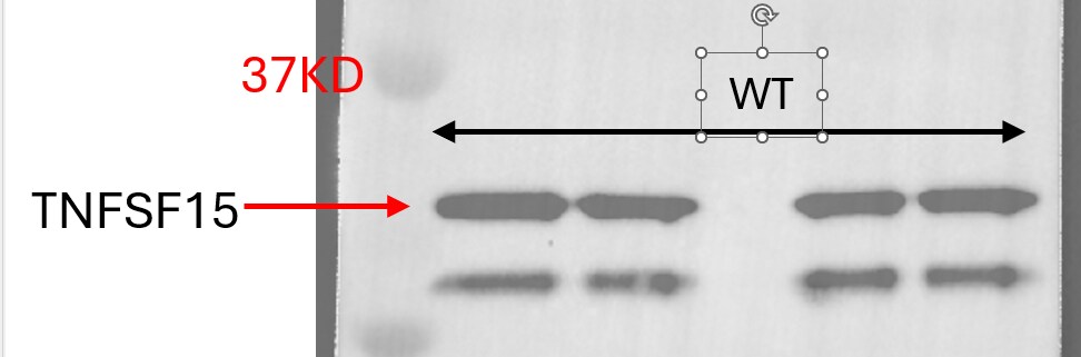
Application: Western BlotSample Tested: MonocytesVerified Customer | Posted 01/13/2026Western blot for TNFSF-15 DectectionI used this antibody to detect TNFSF 15 in endothelial cell with western blot in 1:1000 dilution and it worked very well.

There are no reviews that match your criteria.
Protocols
Find general support by application which include: protocols, troubleshooting, illustrated assays, videos and webinars.
- Antigen Retrieval Protocol (PIER)
- Antigen Retrieval for Frozen Sections Protocol
- Appropriate Fixation of IHC/ICC Samples
- Cellular Response to Hypoxia Protocols
- Chromogenic IHC Staining of Formalin-Fixed Paraffin-Embedded (FFPE) Tissue Protocol
- Chromogenic Immunohistochemistry Staining of Frozen Tissue
- ClariTSA™ Fluorophore Kits
- Detection & Visualization of Antibody Binding
- Fluorescent IHC Staining of Frozen Tissue Protocol
- Graphic Protocol for Heat-induced Epitope Retrieval
- Graphic Protocol for the Preparation and Fluorescent IHC Staining of Frozen Tissue Sections
- Graphic Protocol for the Preparation and Fluorescent IHC Staining of Paraffin-embedded Tissue Sections
- Graphic Protocol for the Preparation of Gelatin-coated Slides for Histological Tissue Sections
- IHC Sample Preparation (Frozen sections vs Paraffin)
- Immunofluorescent IHC Staining of Formalin-Fixed Paraffin-Embedded (FFPE) Tissue Protocol
- Immunohistochemistry (IHC) and Immunocytochemistry (ICC) Protocols
- Immunohistochemistry Frozen Troubleshooting
- Immunohistochemistry Paraffin Troubleshooting
- Preparing Samples for IHC/ICC Experiments
- Preventing Non-Specific Staining (Non-Specific Binding)
- Primary Antibody Selection & Optimization
- Protocol for Heat-Induced Epitope Retrieval (HIER)
- Protocol for Making a 4% Formaldehyde Solution in PBS
- Protocol for VisUCyte™ HRP Polymer Detection Reagent
- Protocol for the Preparation & Fixation of Cells on Coverslips
- Protocol for the Preparation and Chromogenic IHC Staining of Frozen Tissue Sections
- Protocol for the Preparation and Chromogenic IHC Staining of Frozen Tissue Sections - Graphic
- Protocol for the Preparation and Chromogenic IHC Staining of Paraffin-embedded Tissue Sections
- Protocol for the Preparation and Chromogenic IHC Staining of Paraffin-embedded Tissue Sections - Graphic
- Protocol for the Preparation and Fluorescent IHC Staining of Frozen Tissue Sections
- Protocol for the Preparation and Fluorescent IHC Staining of Paraffin-embedded Tissue Sections
- Protocol for the Preparation of Gelatin-coated Slides for Histological Tissue Sections
- R&D Systems Quality Control Western Blot Protocol
- TUNEL and Active Caspase-3 Detection by IHC/ICC Protocol
- The Importance of IHC/ICC Controls
- Troubleshooting Guide: Immunohistochemistry
- Troubleshooting Guide: Western Blot Figures
- Western Blot Conditions
- Western Blot Protocol
- Western Blot Protocol for Cell Lysates
- Western Blot Troubleshooting
- Western Blot Troubleshooting Guide
- View all Protocols, Troubleshooting, Illustrated assays and Webinars
Loading...
Associated Pathways

