TMEM119 (Transmembrane Protein 119, also known as Osteoblast Induction Factor or OBIF), is an approximately 38-kDa type 1 transmembrane protein that is predominantly expressed in osteoblasts and is upregulated during osteoblastic differentiation (1, 2). TMEM119 is also expressed in a cell line of microglia, and TMEM119 immunoreactivity is observed in a specific subset of microglia in brains of neurodegenerative diseases, such as Alzheimers disease (3). Mature human TMEM119 consists of a 71 amino acid (aa) extracellular domain (ECD), a 21 aa transmembrane segment, and a 166 aa cytoplasmic domain. Within the ECD, human TMEM119 shares 78% and 75% aa sequence identity with mouse and rat TMEM119, respectively. TMEM-119 is involved in the osteoblast differentiation and bone development by acting as a ligand and has been reported to contribute to the proliferation, migration, and invasion of osteosarcoma cells, as well as functioning as an oncogene in osteosarcoma (3, 4).
Human TMEM119 Antibody (1023426)
R&D Systems | Catalog # MAB10313

Key Product Details
Species Reactivity
Validated:
Human
Cited:
Human
Applications
Validated:
Immunohistochemistry, Flow Cytometry, CyTOF-ready
Cited:
Immunohistochemistry
Label
Unconjugated
Antibody Source
Monoclonal Mouse IgG1 Clone # 1023426
Loading...
Product Specifications
Immunogen
Chinese Hamster Ovary cell line, CHO derived human TREM119
Arg26-Met96
Accession # Q4V9L6.1
Arg26-Met96
Accession # Q4V9L6.1
Specificity
Detects human TREM119 in direct ELISAs.
Clonality
Monoclonal
Host
Mouse
Isotype
IgG1
Scientific Data Images for Human TMEM119 Antibody (1023426)
Detection of TMEM119 in HEK293 Human Cell Line transfected with Human TMEM119 and eGFP by Flow Cytometry
HEK293 human embryonic kidney cell line transfected with either (A) human TMEM119 or (B) irrelevant protein, and eGFP, was stained with Mouse anti-human TMEM119 monoclonal antibody (Catalog # MAB10313) followed by Allophycocyanin-conjugated anti-Mouse IgG Secondary Antibody (F0101B). Quadrant markers were set based on control antibody staining (MAB002, data not shown). Staining was performed using our Staining Membrane-Associated Proteins protocol.
TMEM119 in Human Brain Cortex Tissue.
TMEM119 was detected in immersion fixed paraffin-embedded sections of human brain cortex tissue using Mouse Anti-Human TMEM119 Monoclonal Antibody (Catalog # MAB10313) at 5 µg/mL for 1 hour at room temperature followed by incubation with the Anti-Mouse IgG VisUCyte™ HRP Polymer Antibody (VC001). Before incubation with the primary antibody, tissue was subjected to heat-induced epitope retrieval using Antigen Retrieval Reagent-Basic (CTS013). Tissue was stained using DAB (brown) and counterstained with hematoxylin (blue). Specific staining was localized to gilal cells. Staining was performed using our IHC Staining with VisUCyte HRP Polymer Detection Reagents protocol.Applications for Human TMEM119 Antibody (1023426)
Application
Recommended Usage
CyTOF-ready
Ready to be labeled using established conjugation methods. No BSA or other carrier proteins that could interfere with conjugation.
Flow Cytometry
0.25 µg/106 cells
Sample: HEK293 Human Cell Line transfected with Human TMEM119 and eGFP
Sample: HEK293 Human Cell Line transfected with Human TMEM119 and eGFP
Immunohistochemistry
5-25 µg/mL
Sample: Immersion fixed paraffin-embedded sections of human brain cortex tissue
Sample: Immersion fixed paraffin-embedded sections of human brain cortex tissue
Reviewed Applications
Read 1 review rated 5 using MAB10313 in the following applications:
Flow Cytometry Panel Builder
Bio-Techne Knows Flow Cytometry
Save time and reduce costly mistakes by quickly finding compatible reagents using the Panel Builder Tool.
Advanced Features
- Spectra Viewer - Custom analysis of spectra from multiple fluorochromes
- Spillover Popups - Visualize the spectra of individual fluorochromes
- Antigen Density Selector - Match fluorochrome brightness with antigen density

Formulation, Preparation, and Storage
Purification
Protein A or G purified from cell culture supernatant
Reconstitution
Reconstitute at 0.5 mg/mL in sterile PBS. For liquid material, refer to CoA for concentration.
Loading...
Formulation
Lyophilized from a 0.2 μm filtered solution in PBS with Trehalose. See Certificate of Analysis for details.
*Small pack size (-SP) is supplied either lyophilized or as a 0.2 µm filtered solution in PBS.
*Small pack size (-SP) is supplied either lyophilized or as a 0.2 µm filtered solution in PBS.
Shipping
Lyophilized product is shipped at ambient temperature. Liquid small pack size (-SP) is shipped with polar packs. Upon receipt, store immediately at the temperature recommended below.
Stability & Storage
Use a manual defrost freezer and avoid repeated freeze-thaw cycles.
- 12 months from date of receipt, -20 to -70 °C as supplied.
- 1 month, 2 to 8 °C under sterile conditions after reconstitution.
- 6 months, -20 to -70 °C under sterile conditions after reconstitution.
Calculators
Background: TMEM119
References
- Jiang, Z,H. et al. (2017) Expt & Mol Med. 49:e329.
- Mizuhashi, K. et al. (2012) Dev. Growth Differ. 54:474.
- Satoh, J. et al. (2016) Neuropathol. 36:39.
- Kanamoto, T. et al. (2009) BMC Develop. Biol. 9:70.
Long Name
Transmembrane Protein 119
Alternate Names
OBIF
Gene Symbol
TMEM119
UniProt
Additional TMEM119 Products
Product Documents for Human TMEM119 Antibody (1023426)
Certificate of Analysis
To download a Certificate of Analysis, please enter a lot or batch number in the search box below.
Note: Certificate of Analysis not available for kit components.
Product Specific Notices for Human TMEM119 Antibody (1023426)
For research use only
Related Research Areas
Citations for Human TMEM119 Antibody (1023426)
 Powered by Bioz
Customer Reviews for Human TMEM119 Antibody (1023426) (1)
5 out of 5
1 Customer Rating
Have you used Human TMEM119 Antibody (1023426)?
Submit a review and receive an Amazon gift card!
$25/€18/£15/$25CAN/¥2500 Yen for a review with an image
$10/€7/£6/$10CAN/¥1110 Yen for a review without an image
Submit a review
Customer Images
Showing
1
-
1 of
1 review
Showing All
Filter By:
-
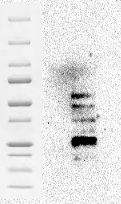
Application: Western BlotSample Tested: Colon tissueSpecies: MouseVerified Customer | Posted 09/18/2024

There are no reviews that match your criteria.
Protocols
Find general support by application which include: protocols, troubleshooting, illustrated assays, videos and webinars.
- 7-Amino Actinomycin D (7-AAD) Cell Viability Flow Cytometry Protocol
- Antigen Retrieval Protocol (PIER)
- Antigen Retrieval for Frozen Sections Protocol
- Appropriate Fixation of IHC/ICC Samples
- Cellular Response to Hypoxia Protocols
- Chromogenic IHC Staining of Formalin-Fixed Paraffin-Embedded (FFPE) Tissue Protocol
- Chromogenic Immunohistochemistry Staining of Frozen Tissue
- ClariTSA™ Fluorophore Kits
- Detection & Visualization of Antibody Binding
- Extracellular Membrane Flow Cytometry Protocol
- Flow Cytometry Protocol for Cell Surface Markers
- Flow Cytometry Protocol for Staining Membrane Associated Proteins
- Flow Cytometry Staining Protocols
- Flow Cytometry Troubleshooting Guide
- Fluorescent IHC Staining of Frozen Tissue Protocol
- Graphic Protocol for Heat-induced Epitope Retrieval
- Graphic Protocol for the Preparation and Fluorescent IHC Staining of Frozen Tissue Sections
- Graphic Protocol for the Preparation and Fluorescent IHC Staining of Paraffin-embedded Tissue Sections
- Graphic Protocol for the Preparation of Gelatin-coated Slides for Histological Tissue Sections
- IHC Sample Preparation (Frozen sections vs Paraffin)
- Immunofluorescent IHC Staining of Formalin-Fixed Paraffin-Embedded (FFPE) Tissue Protocol
- Immunohistochemistry (IHC) and Immunocytochemistry (ICC) Protocols
- Immunohistochemistry Frozen Troubleshooting
- Immunohistochemistry Paraffin Troubleshooting
- Intracellular Flow Cytometry Protocol Using Alcohol (Methanol)
- Intracellular Flow Cytometry Protocol Using Detergents
- Intracellular Nuclear Staining Flow Cytometry Protocol Using Detergents
- Intracellular Staining Flow Cytometry Protocol Using Alcohol Permeabilization
- Intracellular Staining Flow Cytometry Protocol Using Detergents to Permeabilize Cells
- Preparing Samples for IHC/ICC Experiments
- Preventing Non-Specific Staining (Non-Specific Binding)
- Primary Antibody Selection & Optimization
- Propidium Iodide Cell Viability Flow Cytometry Protocol
- Protocol for Heat-Induced Epitope Retrieval (HIER)
- Protocol for Liperfluo
- Protocol for Making a 4% Formaldehyde Solution in PBS
- Protocol for VisUCyte™ HRP Polymer Detection Reagent
- Protocol for the Characterization of Human Th22 Cells
- Protocol for the Characterization of Human Th9 Cells
- Protocol for the Preparation & Fixation of Cells on Coverslips
- Protocol for the Preparation and Chromogenic IHC Staining of Frozen Tissue Sections
- Protocol for the Preparation and Chromogenic IHC Staining of Frozen Tissue Sections - Graphic
- Protocol for the Preparation and Chromogenic IHC Staining of Paraffin-embedded Tissue Sections
- Protocol for the Preparation and Chromogenic IHC Staining of Paraffin-embedded Tissue Sections - Graphic
- Protocol for the Preparation and Fluorescent IHC Staining of Frozen Tissue Sections
- Protocol for the Preparation and Fluorescent IHC Staining of Paraffin-embedded Tissue Sections
- Protocol for the Preparation of Gelatin-coated Slides for Histological Tissue Sections
- Protocol: Annexin V and PI Staining by Flow Cytometry
- Protocol: Annexin V and PI Staining for Apoptosis by Flow Cytometry
- TUNEL and Active Caspase-3 Detection by IHC/ICC Protocol
- The Importance of IHC/ICC Controls
- Troubleshooting Guide: Fluorokine Flow Cytometry Kits
- Troubleshooting Guide: Immunohistochemistry
- View all Protocols, Troubleshooting, Illustrated assays and Webinars
Loading...