Vesicle-associated membrane protein (VAMP)-associated protein B (VAP-B; also VAMP-B) is an ~30 kDa ubiquitously expressed type IV transmembrane protein belonging to the VAP family (1, 2). It is found in endoplasmic reticulum (ER), Golgi and other membranes as a homodimer or a heterodimer with VAP-A, probably associating through a GxxxG motif in the transmembrane regions (1, 2). Human VAP-B cDNA encodes 243 amino acids (aa) that include a 222 aa cytoplasmic domain and a 21 aa C-terminal membrane anchor. The cytoplasmic domain contains a mobile sperm protein (MSP) domain (aa 7‑124) and a coiled-coil region (aa 159‑196). Human VAP-B shares 90%, 89%, 96%, 96% and 94% aa identity with mouse, rat, canine, bovine and porcine VAP-B, respectively. VAP-A and VAP-B MSP domains recruit FFAT (two phenylalanines in an acidic tract)-motif-containing proteins to the cytosolic surface of ER membranes (2‑4). FFAT proteins mediate many of the effects of VAPs on regulation of membrane transport, phospholipid biosynthesis, microtubule organization, and the unfolded protein response (2, 3). VAPs also interact with some SNARE and viral proteins (2). A human polymorphism of VAP-B, P56S, is found in three familial motor neuron diseases, notably the amylotrophic lateral sclerosis variant ALS8 (2). It produces a non-functional protein that can dimerize with and inhibit function of normal VAP-B, leading formation of intracellular aggregates and increased ER-stress-induced death of motor neurons (5‑7). It can also promote cleavage and secretion of soluble VAP-B, which can then function as a ligand for EPH receptors (8). A naturally occurring 99 aa isoform of VAP-B that diverges at aa 71 within the MSP domain is termed VAP-C (1, 9). It also appears to be a negative regulator of VAP-A and VAP-B (9). While VAP-B is used by hepatitis C virus (HCV) for its propagation, VAP-C inhibits HCV propagation (9).

Key Product Details
Species Reactivity
Validated:
Human
Cited:
Human
Applications
Validated:
Immunohistochemistry, Western Blot, Intracellular Staining by Flow Cytometry, CyTOF-ready
Cited:
Western Blot, Immunocytochemistry, Proximity Ligation Assay
Label
Unconjugated
Antibody Source
Monoclonal Mouse IgG1 Clone # 736904
Loading...
Product Specifications
Immunogen
E. coli-derived recombinant human VAP-B
Ala2-Pro132
Accession # O95292
Ala2-Pro132
Accession # O95292
Specificity
Detects human VAP-B in direct ELISAs and Western blots. In direct ELISAs, approximately 25% cross-reactivity
with recombinant human VAP-A is observed.
Clonality
Monoclonal
Host
Mouse
Isotype
IgG1
Scientific Data Images for Human VAP-B Antibody (736904)
Detection of Human VAP‑B by Western Blot.
Western blot shows lysates of HepG2 human hepatocellular carcinoma cell line, SH-SY5Y human neuroblastoma cell line, and human heart tissue. PVDF membrane was probed with 0.1 µg/mL of Mouse Anti-Human VAP-B Monoclonal Antibody (Catalog # MAB58551) followed by HRP-conjugated Anti-Mouse IgG Secondary Antibody (Catalog # HAF007). A specific band was detected for VAP-B at approximately 32-33 kDa (as indicated). This experiment was conducted under reducing conditions and using Immunoblot Buffer Group 1.
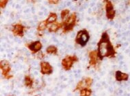
VAP‑B in Human Brain.
VAP-B was detected in immersion fixed paraffin-embedded sections of human brain (medulla) using Mouse Anti-Human VAP-B Monoclonal Antibody (Catalog # MAB58551) at 15 µg/mL overnight at 4 °C. Before incubation with the primary antibody, tissue was subjected to heat-induced epitope retrieval using Antigen Retrieval Reagent-Basic (Catalog # CTS013). Tissue was stained using the Anti-Mouse HRP-DAB Cell & Tissue Staining Kit (brown; Catalog # CTS002) and counterstained with hematoxylin (blue). Specific staining was localized to the cytoplasm of neurons. View our protocol for Chromogenic IHC Staining of Paraffin-embedded Tissue Sections.
Detection of VAPB in T98G Human Cell Line by Flow Cytometry.
T98G human glioblastoma cell line was stained with Mouse Anti-Human VAP-B Monoclonal Antibody (Catalog # MAB58551, filled histogram) or isotype control antibody (Catalog # MAB002, open histogram), followed by Allophycocyanin-conjugated Anti-Mouse IgG Secondary Antibody (Catalog # F0101B). To facilitate intracellular staining, cells were fixed with paraformaldehye and permeabilized with saponin.Applications for Human VAP-B Antibody (736904)
Application
Recommended Usage
CyTOF-ready
Ready to be labeled using established conjugation methods. No BSA or other carrier proteins that could interfere with conjugation.
Immunohistochemistry
8-25 µg/mL
Sample: Immersion fixed paraffin-embedded sections of human brain (medulla)
Sample: Immersion fixed paraffin-embedded sections of human brain (medulla)
Intracellular Staining by Flow Cytometry
2.5 µg/106 cells
Sample: T98G human glioblastoma cell line fixed with paraformaldehye and permeabilized with saponin.
Sample: T98G human glioblastoma cell line fixed with paraformaldehye and permeabilized with saponin.
Western Blot
0.1 µg/mL
Sample: HepG2 human hepatocellular carcinoma cell line, SH‑SY5Y human neuroblastoma cell line, and human heart tissue
Sample: HepG2 human hepatocellular carcinoma cell line, SH‑SY5Y human neuroblastoma cell line, and human heart tissue
Reviewed Applications
Read 1 review rated 5 using MAB58551 in the following applications:
Flow Cytometry Panel Builder
Bio-Techne Knows Flow Cytometry
Save time and reduce costly mistakes by quickly finding compatible reagents using the Panel Builder Tool.
Advanced Features
- Spectra Viewer - Custom analysis of spectra from multiple fluorochromes
- Spillover Popups - Visualize the spectra of individual fluorochromes
- Antigen Density Selector - Match fluorochrome brightness with antigen density

Formulation, Preparation, and Storage
Purification
Protein A or G purified from hybridoma culture supernatant
Reconstitution
Sterile PBS to a final concentration of 0.5 mg/mL. For liquid material, refer to CoA for concentration.
Loading...
Formulation
Lyophilized from a 0.2 μm filtered solution in PBS with Trehalose. *Small pack size (SP) is supplied either lyophilized or as a 0.2 µm filtered solution in PBS.
Shipping
Lyophilized product is shipped at ambient temperature. Liquid small pack size (-SP) is shipped with polar packs. Upon receipt, store immediately at the temperature recommended below.
Stability & Storage
Use a manual defrost freezer and avoid repeated freeze-thaw cycles.
- 12 months from date of receipt, -20 to -70 °C as supplied.
- 1 month, 2 to 8 °C under sterile conditions after reconstitution.
- 6 months, -20 to -70 °C under sterile conditions after reconstitution.
Calculators
Background: VAP-B
References
- Nishimura, Y. et al. (1999) Biochem. Biophys. Res. Commun. 254:21.
- Lev, S. et al. (2008) Trends Cell Biol. 18:282.
- Peretti, D. et al., 2008, Mol. Biol. Cell 19:3871.
- Kaiser, S.E. et al., 2005, Structure 13:1035.
- Prosser, D.C. et al. (2008) J. Cell Sci. 121:3052.
- Gkogkas, C. et al. (2008) Hum. Mol. Genet. 17:1517.
- Suzuki, H. et al. (2009) J. Neurochem. 108:973.
- Tsuda, H. et al. (2008) Cell 133:963.
- Kukihara, H. et al. (2009) J. Virol. 83:7959.
Long Name
VAMP [Vesicle-associated Membrane Protein]-associated Protein B and C
Alternate Names
ALS8, VAMP-B, VAMP-C, VAP-C, VAPB
Gene Symbol
VAPB
UniProt
Additional VAP-B Products
Product Documents for Human VAP-B Antibody (736904)
Certificate of Analysis
To download a Certificate of Analysis, please enter a lot or batch number in the search box below.
Note: Certificate of Analysis not available for kit components.
Product Specific Notices for Human VAP-B Antibody (736904)
For research use only
Citations for Human VAP-B Antibody (736904)
 Powered by Bioz
Customer Reviews for Human VAP-B Antibody (736904) (1)
5 out of 5
1 Customer Rating
Have you used Human VAP-B Antibody (736904)?
Submit a review and receive an Amazon gift card!
$25/€18/£15/$25CAN/¥2500 Yen for a review with an image
$10/€7/£6/$10CAN/¥1110 Yen for a review without an image
Submit a review
Customer Images
Showing
1
-
1 of
1 review
Showing All
Filter By:
-
Application: ImmunohistochemistrySample Tested: Brain tissueSpecies: HumanVerified Customer | Posted 02/09/2022Paraffin-embedded sections of human brain were incubated at 15 ug/mL overnight at 4 C.

There are no reviews that match your criteria.
Protocols
Find general support by application which include: protocols, troubleshooting, illustrated assays, videos and webinars.
- 7-Amino Actinomycin D (7-AAD) Cell Viability Flow Cytometry Protocol
- Antigen Retrieval Protocol (PIER)
- Antigen Retrieval for Frozen Sections Protocol
- Appropriate Fixation of IHC/ICC Samples
- Cellular Response to Hypoxia Protocols
- Chromogenic IHC Staining of Formalin-Fixed Paraffin-Embedded (FFPE) Tissue Protocol
- Chromogenic Immunohistochemistry Staining of Frozen Tissue
- ClariTSA™ Fluorophore Kits
- Detection & Visualization of Antibody Binding
- Extracellular Membrane Flow Cytometry Protocol
- Flow Cytometry Protocol for Cell Surface Markers
- Flow Cytometry Protocol for Staining Membrane Associated Proteins
- Flow Cytometry Staining Protocols
- Flow Cytometry Troubleshooting Guide
- Fluorescent IHC Staining of Frozen Tissue Protocol
- Graphic Protocol for Heat-induced Epitope Retrieval
- Graphic Protocol for the Preparation and Fluorescent IHC Staining of Frozen Tissue Sections
- Graphic Protocol for the Preparation and Fluorescent IHC Staining of Paraffin-embedded Tissue Sections
- Graphic Protocol for the Preparation of Gelatin-coated Slides for Histological Tissue Sections
- IHC Sample Preparation (Frozen sections vs Paraffin)
- Immunofluorescent IHC Staining of Formalin-Fixed Paraffin-Embedded (FFPE) Tissue Protocol
- Immunohistochemistry (IHC) and Immunocytochemistry (ICC) Protocols
- Immunohistochemistry Frozen Troubleshooting
- Immunohistochemistry Paraffin Troubleshooting
- Intracellular Flow Cytometry Protocol Using Alcohol (Methanol)
- Intracellular Flow Cytometry Protocol Using Detergents
- Intracellular Nuclear Staining Flow Cytometry Protocol Using Detergents
- Intracellular Staining Flow Cytometry Protocol Using Alcohol Permeabilization
- Intracellular Staining Flow Cytometry Protocol Using Detergents to Permeabilize Cells
- Preparing Samples for IHC/ICC Experiments
- Preventing Non-Specific Staining (Non-Specific Binding)
- Primary Antibody Selection & Optimization
- Propidium Iodide Cell Viability Flow Cytometry Protocol
- Protocol for Heat-Induced Epitope Retrieval (HIER)
- Protocol for Liperfluo
- Protocol for Making a 4% Formaldehyde Solution in PBS
- Protocol for VisUCyte™ HRP Polymer Detection Reagent
- Protocol for the Characterization of Human Th22 Cells
- Protocol for the Characterization of Human Th9 Cells
- Protocol for the Preparation & Fixation of Cells on Coverslips
- Protocol for the Preparation and Chromogenic IHC Staining of Frozen Tissue Sections
- Protocol for the Preparation and Chromogenic IHC Staining of Frozen Tissue Sections - Graphic
- Protocol for the Preparation and Chromogenic IHC Staining of Paraffin-embedded Tissue Sections
- Protocol for the Preparation and Chromogenic IHC Staining of Paraffin-embedded Tissue Sections - Graphic
- Protocol for the Preparation and Fluorescent IHC Staining of Frozen Tissue Sections
- Protocol for the Preparation and Fluorescent IHC Staining of Paraffin-embedded Tissue Sections
- Protocol for the Preparation of Gelatin-coated Slides for Histological Tissue Sections
- Protocol: Annexin V and PI Staining by Flow Cytometry
- Protocol: Annexin V and PI Staining for Apoptosis by Flow Cytometry
- R&D Systems Quality Control Western Blot Protocol
- TUNEL and Active Caspase-3 Detection by IHC/ICC Protocol
- The Importance of IHC/ICC Controls
- Troubleshooting Guide: Fluorokine Flow Cytometry Kits
- Troubleshooting Guide: Immunohistochemistry
- Troubleshooting Guide: Western Blot Figures
- Western Blot Conditions
- Western Blot Protocol
- Western Blot Protocol for Cell Lysates
- Western Blot Troubleshooting
- Western Blot Troubleshooting Guide
- View all Protocols, Troubleshooting, Illustrated assays and Webinars
Loading...