Mouse Integrin alpha E/CD103 Antibody
R&D Systems | Catalog # AF1990

Key Product Details
Species Reactivity
Validated:
Mouse
Cited:
Mouse, Transgenic Mouse
Applications
Validated:
Western Blot, Flow Cytometry, CyTOF-ready
Cited:
Immunohistochemistry, Immunohistochemistry-Frozen, Flow Cytometry, CyTOF-reported
Label
Unconjugated
Antibody Source
Polyclonal Goat IgG
Loading...
Product Specifications
Immunogen
Mouse myeloma cell line NS0-derived recombinant mouse Integrin alpha E/CD103
Phe20-Arg1113
Accession # Q60677
Phe20-Arg1113
Accession # Q60677
Specificity
Detects mouse Integrin alpha E/CD103 in direct ELISAs and Western blots.
Clonality
Polyclonal
Host
Goat
Isotype
IgG
Scientific Data Images for Mouse Integrin alpha E/CD103 Antibody
Detection of Integrin alpha E/CD103 in Mouse splenocytes by Flow Cytometry
Mouse splenocytes were stained with Rat Anti-Mouse CD3 APC‑conjugated Monoclonal Antibody (Catalog # FAB4841A) and either (A) Goat Anti-Mouse Integrin alpha E/CD103 Antigen Affinity-purified Polyclonal Antibody (Catalog # AF1990) or (B) isotype control antibody (Catalog # AB-108-C) followed by Phycoerythrin-conjugated Anti-Goat IgG Secondary Antibody (Catalog # F0107). View our protocol for Staining Membrane-associated Proteins.
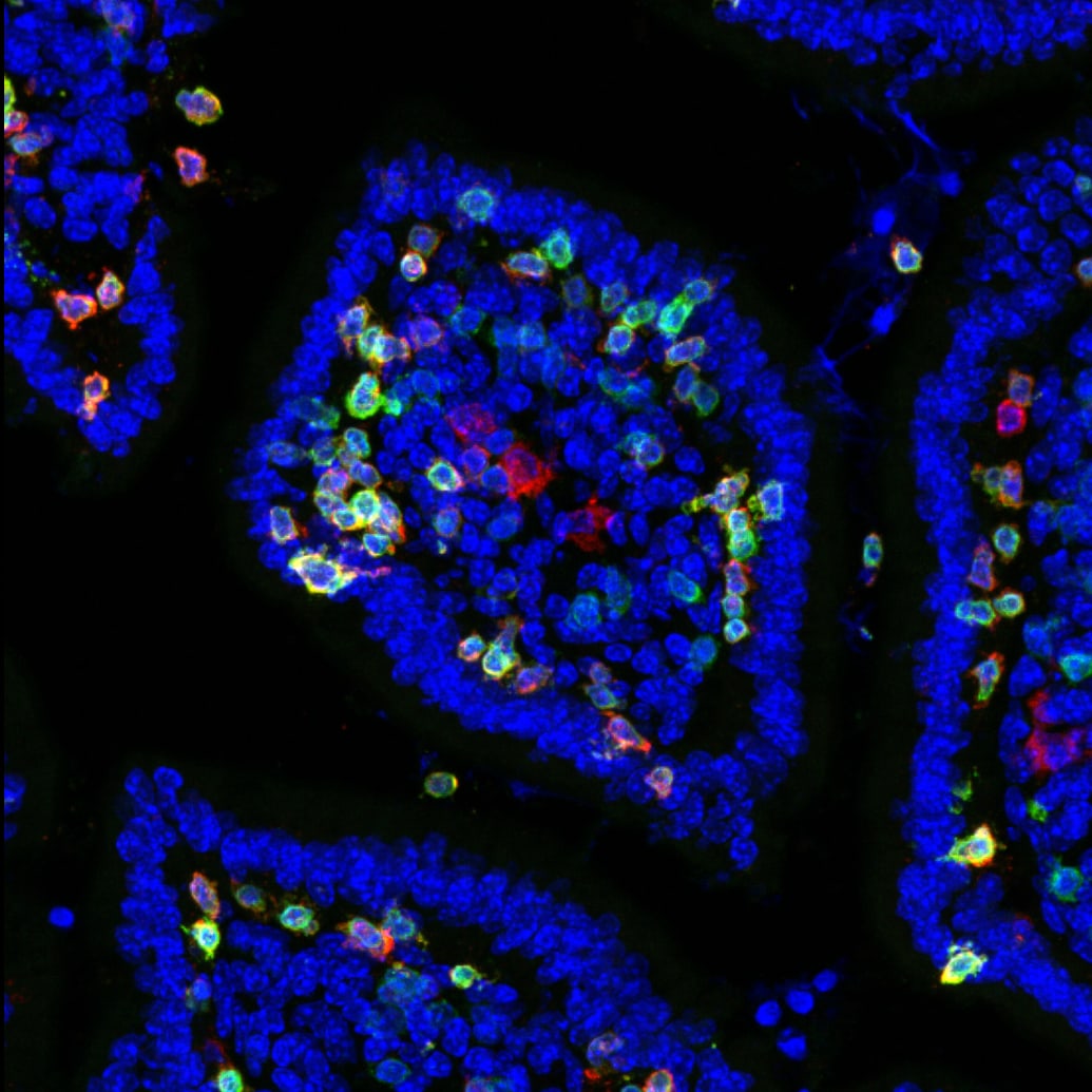
Detection of Mouse Integrin alpha E/CD103 by Immunohistochemistry
AXL upregulation in TKI-resistant HCC inversely correlates with tumor infiltration of immune cells and may predict treatment response. A Lists of upregulated druggable targets in sorafenib-resistant HepG2 cells and lenvatinib-resistant HCC patient-derived xenograft (PDX). The numbers and the color scale bar indicate the expression fold-change of resistant samples versus sensitive samples. B Bubble plots showing the correlation analysis of AXL with immune gene signatures in sorafenib-resistant samples from the datasets GSE176151 (left) and GSE151412 (right). The color scale bar and the size of the dots indicate the Pearson correlation coefficient. C Representative multiplex immunofluorescence images (top) of staining of CD103, CD8 alpha, AXL, and DAPI in residual xenografts after treatment with DMOS, Sora, or Lenva. Scale bar = 50 µm. D Immunotherapy response prediction (top) of TCGA-LIHC patients using Tumor immune dysfunction and exclusion (TIDE) analysis. Heatmap (bottom) of TIDE scores and the corresponding AXL expression (normalized count) of patients from TCGA-LIHC dataset. E Violin plot of AXL expression in immunotherapy responders and non-responders as predicted by TIDE. F Heatmap showing AXL expression, T-cell dysfunction score, and T-cell exclusion score of patients from TCGA-LIHC dataset. G Violin plots of T-cell dysfunction score (left) and T-cell exclusion score (right) in AXL-high and AXL-low patients. *p < 0.05; **p < 0.01; ***p < 0.001 on a two-tailed unpaired Student’s t test. Image collected and cropped by CiteAb from the following open publication (https://www.nature.com/articles/s41419-024-06493-0), licensed under a CC-BY license. Not internally tested by R&D Systems.Applications for Mouse Integrin alpha E/CD103 Antibody
Application
Recommended Usage
CyTOF-ready
Ready to be labeled using established conjugation methods. No BSA or other carrier proteins that could interfere with conjugation.
Flow Cytometry
2.5 µg/106 cells
Sample: See below
Sample: See below
Western Blot
0.1 µg/mL
Sample: Recombinant Mouse Integrin alpha E/CD103
Sample: Recombinant Mouse Integrin alpha E/CD103
Reviewed Applications
Read 1 review rated 5 using AF1990 in the following applications:
Flow Cytometry Panel Builder
Bio-Techne Knows Flow Cytometry
Save time and reduce costly mistakes by quickly finding compatible reagents using the Panel Builder Tool.
Advanced Features
- Spectra Viewer - Custom analysis of spectra from multiple fluorochromes
- Spillover Popups - Visualize the spectra of individual fluorochromes
- Antigen Density Selector - Match fluorochrome brightness with antigen density

Formulation, Preparation, and Storage
Purification
Antigen Affinity-purified
Reconstitution
Reconstitute at 0.2 mg/mL in sterile PBS. For liquid material, refer to CoA for concentration.
Loading...
Formulation
Lyophilized from a 0.2 μm filtered solution in PBS with Trehalose. See Certificate of Analysis for details.
*Small pack size (-SP) is supplied either lyophilized or as a 0.2 µm filtered solution in PBS.
*Small pack size (-SP) is supplied either lyophilized or as a 0.2 µm filtered solution in PBS.
Shipping
Lyophilized product is shipped at ambient temperature. Liquid small pack size (-SP) is shipped with polar packs. Upon receipt, store immediately at the temperature recommended below.
Stability & Storage
Use a manual defrost freezer and avoid repeated freeze-thaw cycles.
- 12 months from date of receipt, -20 to -70 °C as supplied.
- 1 month, 2 to 8 °C under sterile conditions after reconstitution.
- 6 months, -20 to -70 °C under sterile conditions after reconstitution.
Calculators
Background: Integrin alpha E/CD103
Alternate Names
CD103, HUMINAE, ITGAE
Gene Symbol
ITGAE
UniProt
Additional Integrin alpha E/CD103 Products
Product Documents for Mouse Integrin alpha E/CD103 Antibody
Certificate of Analysis
To download a Certificate of Analysis, please enter a lot or batch number in the search box below.
Note: Certificate of Analysis not available for kit components.
Product Specific Notices for Mouse Integrin alpha E/CD103 Antibody
For research use only
Citations for Mouse Integrin alpha E/CD103 Antibody
 Powered by Bioz
Customer Reviews for Mouse Integrin alpha E/CD103 Antibody (1)
5 out of 5
1 Customer Rating
Have you used Mouse Integrin alpha E/CD103 Antibody?
Submit a review and receive an Amazon gift card!
$25/€18/£15/$25CAN/¥2500 Yen for a review with an image
$10/€7/£6/$10CAN/¥1110 Yen for a review without an image
Submit a review
Customer Images
Showing
1
-
1 of
1 review
Showing All
Filter By:
-
Application: Immunocytochemistry/ImmunofluorescenceSample Tested: Adult small intestineSpecies: MouseVerified Customer | Posted 07/09/2019Cryosections from PLP immersion fixed tissue were stained for Itgae (1:100) for 4h @ RT. After washing, tissue was stained with Donkey anti-Goat alexa546 2ndary antibody for 1h at RT. CD3 (green), Itgae (red), Hoechst (blue).

There are no reviews that match your criteria.
Protocols
Find general support by application which include: protocols, troubleshooting, illustrated assays, videos and webinars.
- 7-Amino Actinomycin D (7-AAD) Cell Viability Flow Cytometry Protocol
- Cellular Response to Hypoxia Protocols
- Extracellular Membrane Flow Cytometry Protocol
- Flow Cytometry Protocol for Cell Surface Markers
- Flow Cytometry Protocol for Staining Membrane Associated Proteins
- Flow Cytometry Staining Protocols
- Flow Cytometry Troubleshooting Guide
- Intracellular Flow Cytometry Protocol Using Alcohol (Methanol)
- Intracellular Flow Cytometry Protocol Using Detergents
- Intracellular Nuclear Staining Flow Cytometry Protocol Using Detergents
- Intracellular Staining Flow Cytometry Protocol Using Alcohol Permeabilization
- Intracellular Staining Flow Cytometry Protocol Using Detergents to Permeabilize Cells
- Propidium Iodide Cell Viability Flow Cytometry Protocol
- Protocol for Liperfluo
- Protocol for the Characterization of Human Th22 Cells
- Protocol for the Characterization of Human Th9 Cells
- Protocol: Annexin V and PI Staining by Flow Cytometry
- Protocol: Annexin V and PI Staining for Apoptosis by Flow Cytometry
- R&D Systems Quality Control Western Blot Protocol
- Troubleshooting Guide: Fluorokine Flow Cytometry Kits
- Troubleshooting Guide: Western Blot Figures
- Western Blot Conditions
- Western Blot Protocol
- Western Blot Protocol for Cell Lysates
- Western Blot Troubleshooting
- Western Blot Troubleshooting Guide
- View all Protocols, Troubleshooting, Illustrated assays and Webinars
Loading...
Associated Pathways

