LAMP-1 (lysosome-associated membrane glycoprotein 1; also P2B and CD107a) is a 120 kDa member of the LAMP family of proteins. It is a highly glycosylated type I transmembrane protein whose N-terminus projects either extracellularly or into the lumen of lysosomes. LAMP-1 may protect lysosomal membranes from enzymatic attack or mediate fusion of phagosomes with lysosomes. Mature mouse LAMP1 is 382 amino acids (aa) in length. It contains a 346 aa lumenal domain (aa 25‑370) and a 12 aa cytoplasmic region. Over aa 25‑370, mouse LAMP1 shares 82% and 62% aa identity with rat and human LAMP1, respectively.
Mouse LAMP-1/CD107a Lumenal Domain Antibody
R&D Systems | Catalog # AF4320

Key Product Details
Species Reactivity
Validated:
Mouse
Cited:
Human, Mouse, Transgenic Mouse
Applications
Validated:
Western Blot, Intracellular Staining by Flow Cytometry, Immunocytochemistry, CyTOF-ready
Cited:
Immunohistochemistry, Immunohistochemistry-Paraffin, Western Blot, Immunocytochemistry
Label
Unconjugated
Antibody Source
Polyclonal Goat IgG
Loading...
Product Specifications
Immunogen
Mouse myeloma cell line NS0-derived recombinant mouse LAMP1/CD107a
Leu25-Asn370
Accession # P11438
Leu25-Asn370
Accession # P11438
Specificity
Detects mouse LAMP1/CD107a Lumenal Domain in direct ELISAs and Western blots. In direct ELISAs, less than 10% cross‑reactivity with recombinant human LAMP1 is observed.
Clonality
Polyclonal
Host
Goat
Isotype
IgG
Scientific Data Images for Mouse LAMP-1/CD107a Lumenal Domain Antibody
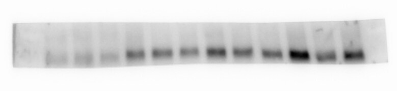
Detection of Mouse LAMP‑1/CD107a by Western Blot.
Western blot shows lysates of mouse small intestine tissue and NIH-3T3 mouse embryonic fibroblast cell line. PVDF membrane was probed with 0.2 µg/mL of Goat Anti-Mouse LAMP-1/CD107a Lumenal Domain Antigen Affinity-purified Polyclonal Antibody (Catalog # AF4320) followed by HRP-conjugated Anti-Goat IgG Secondary Antibody (Catalog # HAF017). A specific band was detected for LAMP-1/CD107a at approximately 100-120 kDa (as indicated). This experiment was conducted under reducing conditions and using Immunoblot Buffer Group 1.
Detection of LAMP-1 in RAW 264.7 Mouse Cell Line by Flow Cytometry.
RAW 264.7 mouse monocyte/macrophage cell line was stained with Goat Anti-Mouse LAMP1/CD107a Lumenal Domain Antigen Affinity-purified Polyclonal Antibody (Catalog # AF4320, filled histogram) or control antibody (Catalog # AB-108-C, open histogram), followed by Allophycocyanin-conjugated Anti-Goat IgG Secondary Antibody (Catalog # F0108). To facilitate intracellular staining, cells were fixed with Flow Cytometry Fixation Buffer (Catalog # FC004) and permeabilized with Flow Cytometry Permeabilization/Wash Buffer I (Catalog # FC005). View our protocol for Staining Intracellular Molecules.
LAMP1/CD107a in RAW 264.7 Mouse Cell Line.
LAMP1/CD107a was detected in immersion fixed RAW 264.7 mouse monocyte/macrophage cell line using Goat Anti-Mouse LAMP1/CD107a Lumenal Domain Antigen Affinity-purified Polyclonal Antibody (Catalog # AF4320) at 10 µg/mL for 3 hours at room temperature. Cells were stained using the NorthernLights™ 557-conjugated Anti-Goat IgG Secondary Antibody (red; Catalog # NL001) and counterstained with DAPI (blue). Specific staining was localized to cytoplasm. View our protocol for Fluorescent ICC Staining of Cells on Coverslips.Applications for Mouse LAMP-1/CD107a Lumenal Domain Antibody
Application
Recommended Usage
CyTOF-ready
Ready to be labeled using established conjugation methods. No BSA or other carrier proteins that could interfere with conjugation.
Immunocytochemistry
5-15 µg/mL
Sample: Immersion fixed RAW 264.7 mouse monocyte/macrophage cell line
Sample: Immersion fixed RAW 264.7 mouse monocyte/macrophage cell line
Intracellular Staining by Flow Cytometry
0.25 µg/106 cells
Sample: RAW 264.7 mouse monocyte/macrophage cell line fixed with Flow Cytometry Fixation Buffer (Catalog # FC004) and permeabilized with Flow Cytometry Permeabilization/Wash Buffer I (Catalog # FC005)
Sample: RAW 264.7 mouse monocyte/macrophage cell line fixed with Flow Cytometry Fixation Buffer (Catalog # FC004) and permeabilized with Flow Cytometry Permeabilization/Wash Buffer I (Catalog # FC005)
Western Blot
0.2 µg/mL
Sample: Mouse small intestine tissue and NIH‑3T3 mouse embryonic fibroblast cell line
Sample: Mouse small intestine tissue and NIH‑3T3 mouse embryonic fibroblast cell line
Reviewed Applications
Read 2 reviews rated 5 using AF4320 in the following applications:
Flow Cytometry Panel Builder
Bio-Techne Knows Flow Cytometry
Save time and reduce costly mistakes by quickly finding compatible reagents using the Panel Builder Tool.
Advanced Features
- Spectra Viewer - Custom analysis of spectra from multiple fluorochromes
- Spillover Popups - Visualize the spectra of individual fluorochromes
- Antigen Density Selector - Match fluorochrome brightness with antigen density

Formulation, Preparation, and Storage
Purification
Antigen Affinity-purified
Reconstitution
Reconstitute at 0.2 mg/mL in sterile PBS. For liquid material, refer to CoA for concentration.
Loading...
Formulation
Lyophilized from a 0.2 μm filtered solution in PBS with Trehalose. *Small pack size (SP) is supplied either lyophilized or as a 0.2 µm filtered solution in PBS.
Shipping
Lyophilized product is shipped at ambient temperature. Liquid small pack size (-SP) is shipped with polar packs. Upon receipt, store immediately at the temperature recommended below.
Stability & Storage
Use a manual defrost freezer and avoid repeated freeze-thaw cycles.
- 12 months from date of receipt, -20 to -70 °C as supplied.
- 1 month, 2 to 8 °C under sterile conditions after reconstitution.
- 6 months, -20 to -70 °C under sterile conditions after reconstitution.
Calculators
Background: LAMP-1/CD107a
Long Name
Lysosome-associated Membrane Glycoprotein 1
Alternate Names
CD107a, LAMP1
Gene Symbol
LAMP1
UniProt
Additional LAMP-1/CD107a Products
Product Documents for Mouse LAMP-1/CD107a Lumenal Domain Antibody
Certificate of Analysis
To download a Certificate of Analysis, please enter a lot or batch number in the search box below.
Note: Certificate of Analysis not available for kit components.
Product Specific Notices for Mouse LAMP-1/CD107a Lumenal Domain Antibody
For research use only
Citations for Mouse LAMP-1/CD107a Lumenal Domain Antibody
 Powered by Bioz
Customer Reviews for Mouse LAMP-1/CD107a Lumenal Domain Antibody (2)
5 out of 5
2 Customer Ratings
Have you used Mouse LAMP-1/CD107a Lumenal Domain Antibody?
Submit a review and receive an Amazon gift card!
$25/€18/£15/$25CAN/¥2500 Yen for a review with an image
$10/€7/£6/$10CAN/¥1110 Yen for a review without an image
Submit a review
Customer Images
Showing
1
-
2 of
2 reviews
Showing All
Filter By:
-
Application: Western BlotSample Tested: Primary sorted Oligodendrocytes and Primary oligodendrocyte culturesSpecies: MouseVerified Customer | Posted 11/21/2019Clean band at predicted molecular weight, 10 ug of protein loaded per lane.Dilution of 0.2 mg/mL stock: 1:1000 Great antibody for each application tested so far on mouse tissue and cells: wb, IHC-Fr, ICC

-
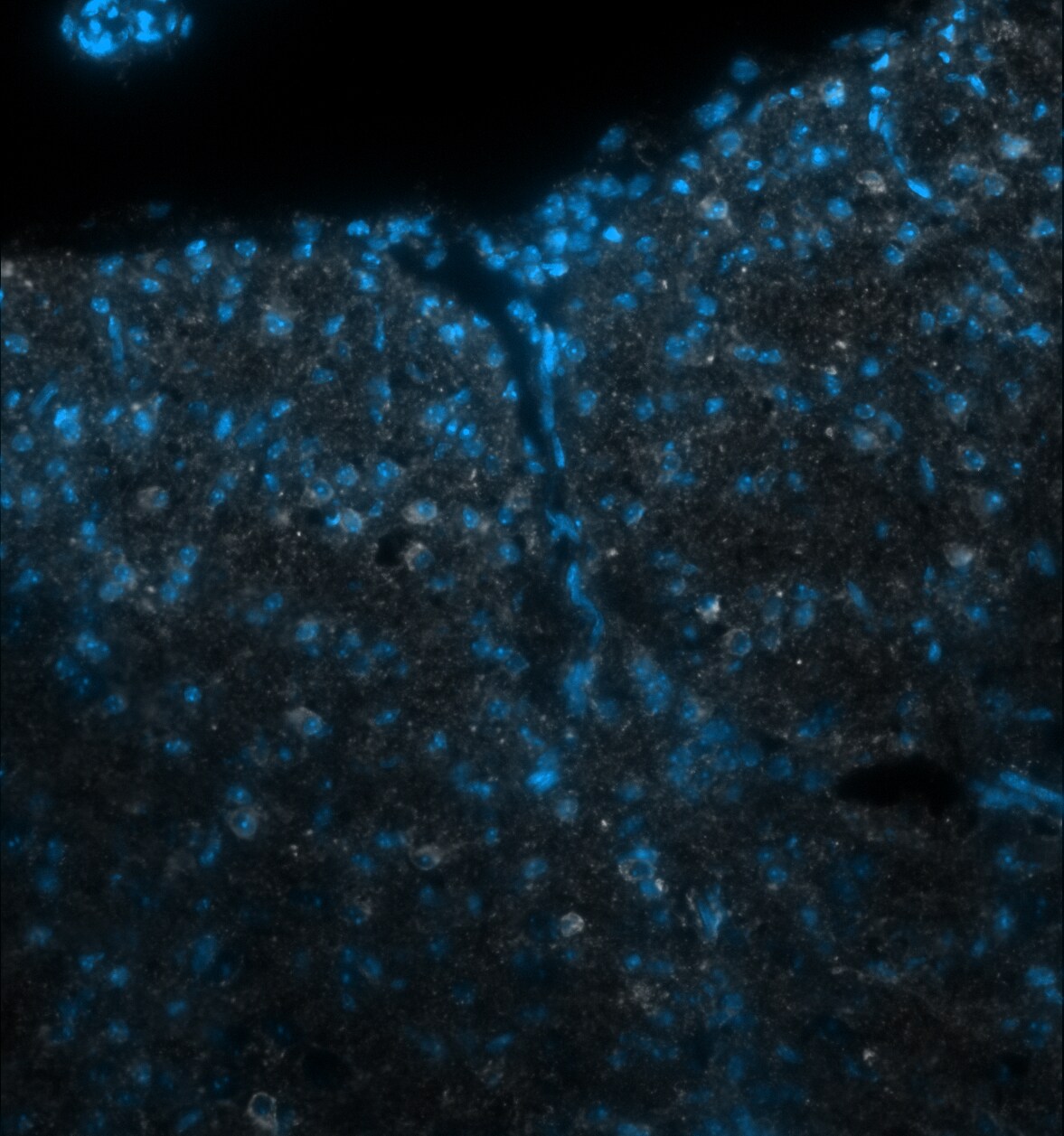
Application: Immunohistochemistry-FrozenSample Tested: Adult spinal cord and Primary culture of oligodendrocytesSpecies: MouseVerified Customer | Posted 11/21/2019P10 mouse ventral funiculus thoracic spinal cord stained with Lamp1 antibody (white) and Dapi.Dilution of 0.2 mg/mL stock: 1:40
There are no reviews that match your criteria.
Protocols
Find general support by application which include: protocols, troubleshooting, illustrated assays, videos and webinars.
- 7-Amino Actinomycin D (7-AAD) Cell Viability Flow Cytometry Protocol
- Appropriate Fixation of IHC/ICC Samples
- Cellular Response to Hypoxia Protocols
- ClariTSA™ Fluorophore Kits
- Detection & Visualization of Antibody Binding
- Extracellular Membrane Flow Cytometry Protocol
- Flow Cytometry Protocol for Cell Surface Markers
- Flow Cytometry Protocol for Staining Membrane Associated Proteins
- Flow Cytometry Staining Protocols
- Flow Cytometry Troubleshooting Guide
- ICC Cell Smear Protocol for Suspension Cells
- ICC Immunocytochemistry Protocol Videos
- ICC for Adherent Cells
- Immunocytochemistry (ICC) Protocol
- Immunocytochemistry Troubleshooting
- Immunofluorescence of Organoids Embedded in Cultrex Basement Membrane Extract
- Immunohistochemistry (IHC) and Immunocytochemistry (ICC) Protocols
- Intracellular Flow Cytometry Protocol Using Alcohol (Methanol)
- Intracellular Flow Cytometry Protocol Using Detergents
- Intracellular Nuclear Staining Flow Cytometry Protocol Using Detergents
- Intracellular Staining Flow Cytometry Protocol Using Alcohol Permeabilization
- Intracellular Staining Flow Cytometry Protocol Using Detergents to Permeabilize Cells
- Preparing Samples for IHC/ICC Experiments
- Preventing Non-Specific Staining (Non-Specific Binding)
- Primary Antibody Selection & Optimization
- Propidium Iodide Cell Viability Flow Cytometry Protocol
- Protocol for Liperfluo
- Protocol for VisUCyte™ HRP Polymer Detection Reagent
- Protocol for the Characterization of Human Th22 Cells
- Protocol for the Characterization of Human Th9 Cells
- Protocol for the Fluorescent ICC Staining of Cell Smears - Graphic
- Protocol for the Fluorescent ICC Staining of Cultured Cells on Coverslips - Graphic
- Protocol for the Preparation and Fluorescent ICC Staining of Cells on Coverslips
- Protocol for the Preparation and Fluorescent ICC Staining of Non-adherent Cells
- Protocol for the Preparation and Fluorescent ICC Staining of Stem Cells on Coverslips
- Protocol for the Preparation of a Cell Smear for Non-adherent Cell ICC - Graphic
- Protocol: Annexin V and PI Staining by Flow Cytometry
- Protocol: Annexin V and PI Staining for Apoptosis by Flow Cytometry
- R&D Systems Quality Control Western Blot Protocol
- TUNEL and Active Caspase-3 Detection by IHC/ICC Protocol
- The Importance of IHC/ICC Controls
- Troubleshooting Guide: Fluorokine Flow Cytometry Kits
- Troubleshooting Guide: Western Blot Figures
- Western Blot Conditions
- Western Blot Protocol
- Western Blot Protocol for Cell Lysates
- Western Blot Troubleshooting
- Western Blot Troubleshooting Guide
- View all Protocols, Troubleshooting, Illustrated assays and Webinars
Loading...
Associated Pathways
