
Key Product Details
Validated by
Biological Validation
Species Reactivity
Validated:
Mouse
Cited:
Human, Mouse, Complex Species Category, Transgenic Mouse
Applications
Validated:
Immunohistochemistry, Western Blot, Immunocytochemistry, Simple Western, Immunoprecipitation
Cited:
Immunohistochemistry, Immunohistochemistry-Paraffin, Immunohistochemistry-Frozen, Western Blot, Neutralization, Flow Cytometry, Immunocytochemistry, Immunoprecipitation, Electron Microscopy
Label
Unconjugated
Antibody Source
Polyclonal Goat IgG
Loading...
Product Specifications
Immunogen
Mouse myeloma cell line NS0-derived recombinant mouse MMP‑9
Ala20-Pro730
Accession # P41245
Ala20-Pro730
Accession # P41245
Specificity
Detects mouse MMP-9 in direct ELISAs and Western blots. In direct ELISAs and Western blots, approximately 10% cross-reactivity with recombinant human (rh) MMP-9 is observed and less than 2% cross-reactivity with rhMMP-1, -2, -3, -7, -8, -10, -12, and -13 is observed.
Clonality
Polyclonal
Host
Goat
Isotype
IgG
Scientific Data Images for Mouse MMP-9 Antibody
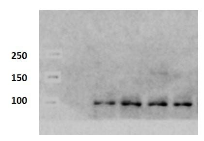
Detection of Mouse MMP‑9 by Western Blot.
Western blot shows lysates of mouse spleen tissue and mouse lung tissue. PVDF membrane was probed with 0.25 µg/mL of Goat Anti-Mouse MMP-9 Antigen Affinity-purified Polyclonal Antibody (Catalog # AF909) followed by HRP-conjugated Anti-Goat IgG Secondary Antibody (HAF019). A specific band was detected for MMP-9 at approximately 100 kDa (as indicated). This experiment was conducted under reducing conditions and using Immunoblot Buffer Group 1.
MMP‑9 in Mouse Splenocytes.
MMP-9 was detected in immersion fixed mouse splenocytes using Goat Anti-Mouse MMP-9 Antigen Affinity-purified Polyclonal Antibody (Catalog # AF909) at 5 µg/mL for 3 hours at room temperature. Cells were stained using the NorthernLights™ 557-conjugated Anti-Goat IgG Secondary Antibody (red; NL001) and counterstained with DAPI (blue). Specific staining was localized to cytoplasm. View our protocol for Fluorescent ICC Staining of Non-adherent Cells.
Detection of Mouse MMP‑9 by Simple WesternTM.
Simple Western lane view shows lysates of mouse lung tissue and mouse spleen tissue, loaded at 0.2 mg/mL. A specific band was detected for MMP-9 at approximately 155 kDa (as indicated) using 5 µg/mL of Goat Anti-Mouse MMP-9 Antigen Affinity-purified Polyclonal Antibody (Catalog # AF909) followed by 1:50 dilution of HRP-conjugated Anti-Goat IgG Secondary Antibody (HAF109). This experiment was conducted under reducing conditions and using the 12-230 kDa separation system.
MMP‑9 in Rat Brain.
MMP-9 was detected in perfusion fixed frozen sections of rat brain (cingulate cortex) using 1.7 µg/mL Goat Anti-Mouse MMP-9 Antigen Affinity-purified Polyclonal Antibody (Catalog # AF909) overnight at 4 °C. Tissue was stained with the Anti-Goat HRP-DAB Cell & Tissue Staining Kit (brown; CTS008) and counterstained with hematoxylin (blue). View our protocol for Chromogenic IHC Staining of Frozen Tissue Sections.
Detection of Mouse MMP-9 by Immunohistochemistry
Remodeled ECM enhances the invasive behaviors of ovarian cancer cells. (a) H&E stain of the tumor lesions from WT and ApoE−/− mice at two weeks post ID8 engraftment (left). Graph represents the mean size of the lesions calculated from ten random fields (right). (b) Masson’s Trichrome stain of tumor lesions (left). The mean percentage of positive regions in ten random fields was calculated (right). (c) The cytokines/chemokines profile in the supernatants of ascites from WT and ApoE−/− mice. Four groups of mouse cytokine dot-blot arrays are shown. Dot-blots with significant changes are shown in boxed areas (red). (d) The top four pathways enriched among the molecules with significant changes using KEGG pathway analysis. (e) MMP-10 and MMP-9 protein expression in tumor lesions, determined by blinded IHC analysis (left). Box plot of the IHC score of MMP-10 and MMP-9 (right). Box represents the 25th–75th percentile while whiskers indicate the 5th–95th percentile. The black box represents tumor lesions from WT mice and the grey box represents tumor lesions from ApoE−/− mice. Each experiment included data from 5 mice. Bar represents 50 μm. *P < 0.05; **P < 0.005 Image collected and cropped by CiteAb from the following publication (https://pubmed.ncbi.nlm.nih.gov/29458390), licensed under a CC-BY license. Not internally tested by R&D Systems.
Detection of Mouse MMP-9 by Immunohistochemistry
Remodeled ECM promotes malignancy of ovarian cancer via FAK-ERK-MMP activation. (a) Specimens from Fig. 5D were immunoassayed for p-FAKY397 (red), and nuclei were stained with Hoechst (blue). (b) IHC of active ERK (p-p44/42 MAPKThr202/Tyr204) in tumor lesions from WT and ApoE−/− mice with PBS or BAPN treatment two weeks after ID8 engraftment. (c) PBS or PD-325901 was administrated to ApoE−/− mice once ID8-Luciferase cells were intraperitoneally injected and treatment continued for one month. The representative images (top) and quantification data (bottom) of MMP-9 protein expression in tumor lesions two months after ID8 engraftment. (d) In vivo luciferase expression was determined two weeks or two months post treatment. Luminescence (right panel) is represented as the radiance (p/s/cm2/sr). Each experiment included data from 4 mice. Bar represents 50 μm. *P < 0.05; **P < 0.005 Image collected and cropped by CiteAb from the following publication (https://pubmed.ncbi.nlm.nih.gov/29458390), licensed under a CC-BY license. Not internally tested by R&D Systems.
Detection of Mouse MMP-9 by Western Blot
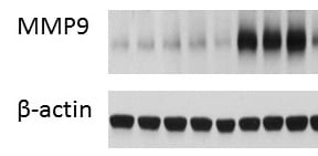
Fibrotic deposit and cardiac dysfunction correlate with decreasing CVPC presence in mdx heart. (A) Representative images of histological analysis stained using Masson trichrome technique showing myocytes (in red) and collagenous fibrotic tissue (in blue) in the left ventricle of WT and mdx hearts at 9, 24, and 52 wo. Line represents 100 µm. (B) The ratio of red and blue stained tissue was evaluated in WT hearts (open bars and black dots, n = 4–11 slices/3 animals per group) and mdx hearts (black bars and grey dots, n = 3–16 slices/3 animals per group) at the age of 24 wo and further at 52 wo. Statistical significance was calculated by Kruskal–Wallis test and Dunn‘s multiple comparison post-hoc test (*** p < 0.001, **** p < 0.0001). (C) Western blot analysis of collagen proteins and inflammatory proteins in the cardiac tissues. Left panel shows representative images of collagen 1A1 (Coll 1A1), collagen 3 (Coll 3), cyclooxygenase 2 (COX-2), and matrix metalloproteinase 9 (MMP-9) compared to the GAPDH control. The right panels show the normalized densitometry of each protein normalized by GAPDH content of WT (open bars, n = 2 animals) and mdx (black bars, n = 2 animals). Image collected and cropped by CiteAb from the following publication (https://pubmed.ncbi.nlm.nih.gov/34068508), licensed under a CC-BY license. Not internally tested by R&D Systems.
Detection of Mouse MMP-9 by Immunocytochemistry/Immunofluorescence
MMP2, MMP9, and RANKL expression in AAAs.A. Immunohistochemical staining for indicated proteins of serial sections of aortas one week after CaCl2 treatment. Elastic van Gieson staining is also shown. SM alpha -actin and F4/80 were stained to locate SMCs and macrophages, respectively. Shown are representative images of 4 or more samples in each group. Scale bars, 50 µm. B. Relative positive staining area of MMP2, MMP9, and RANKL in sections from control diet and EPA diet groups. n = 4–5. *P<0.05. Image collected and cropped by CiteAb from the following publication (https://dx.plos.org/10.1371/journal.pone.0096286), licensed under a CC-BY license. Not internally tested by R&D Systems.
Detection of Mouse MMP-9 by Immunohistochemistry
BAPN treatment delays ovarian cancer progression by reducing adhesions. (a) Experimental design: PBS or BAPN was intraperitoneally administrated to 20-weeks-old female ApoE−/− mice each day and continued for four weeks. A cohort of mice was sacrificed for further experiments. For the remaining mice, the drug treatment was stopped for two weeks before the establishment of ID8 allografts. (b) Hydroxyproline was measured in the plasma and diaphragm. (c) Masson’s Trichrome stain after BAPN treatment (left). The positive-staining percentage of 10 random fields was calculated (right). Bar represents 50 μm. (d) Cells adhesive to the omentum were analyzed four hours after ID8 intraperitoneal injection by fluorescence microscopy (left). The adhesive cells were determined from the total fluorescent intensity after digestion (right). Bar represents 200 μm. (e) In vivo luciferase measured at two weeks (top) and two months (bottom) post establishment in ApoE−/− mice with PBS or BAPN pre-treatment. Quantification of luminescence is represented as the radiance. (f) MMP-9 expression measured by IHC in tumor lesions of ApoE−/− mice with PBS or BAPN treatment. Each experiment includes data from 4 mice. Bar represents 50 μm. *P < 0.05; **P < 0.005 Image collected and cropped by CiteAb from the following publication (https://pubmed.ncbi.nlm.nih.gov/29458390), licensed under a CC-BY license. Not internally tested by R&D Systems.
Detection of MMP‑9 in Mouse Thymus.
MMP‑9 was detected in immersion fixed paraffin-embedded sections of mouse thymus using Goat Anti-Mouse MMP‑9 Antigen Affinity-purified Polyclonal Antibody (Catalog # AF909) at 1.7 µg/ml for 1 hour at room temperature followed by incubation with the Anti-Goat IgG VisUCyte™ HRP Polymer Antibody (Catalog # VC004). Before incubation with the primary antibody, tissue was subjected to heat-induced epitope retrieval using VisUCyte Antigen Retrieval Reagent-Basic (Catalog # VCTS021). Tissue was stained using DAB (brown) and counterstained with hematoxylin (blue). Specific staining was localized to the cytoplasm and macrophages. View our protocol for Chromogenic IHC Staining of Paraffin-embedded Tissue Sections.
Detection of Mouse MMP-9 by Flow Cytometry
Single cell RNA-sequencing analysis of healing bone fractures. A Preparation of non-haematopoietic stromal cells from PFD14 and control bone for scRNA-seq analysis. b UMAP plots showing colour-coded cell clusters from control and PFD14 bone. Cell types and numbers per cluster are displayed on the right. c–e Heatmap showing the top 5 marker genes for each cluster (c). Feature blots showing markers for non-haematopoietic bone cells: Acan—chondrocytes (CHO); Igfbp6—fibroblasts (FB); Pdgfrb—bone mesenchymal stromal cells (BMSCs); Col22a1—osteoblast lineage cells (OBs); Emcn—ECs; Rgs5—Smooth muscle cells (SMCs) (d). Comparison of marker gene expression in PFD14 and control samples (e). Haematopoietic cells (HCs-1,2,3,4). f, g UMAP plots showing colour-coded BMSC subclusters from control and PFD14 bone (f). Fabp5 and Mmp9 expressing cells are increased in PFD14 samples (g). h, i UMAP plot showing mpMSCs and SCs in the control and PFD14 mpMSC subcluster (h). Fracture-derived SCs show higher expression of Mmp9 and Fabp5 (i). Image collected and cropped by CiteAb from the following open publication (https://pubmed.ncbi.nlm.nih.gov/35091558), licensed under a CC-BY license. Not internally tested by R&D Systems.
Detection of Mouse MMP-9 by Flow Cytometry
Single cell RNA-sequencing analysis of healing bone fractures. A Preparation of non-haematopoietic stromal cells from PFD14 and control bone for scRNA-seq analysis. b UMAP plots showing colour-coded cell clusters from control and PFD14 bone. Cell types and numbers per cluster are displayed on the right. c–e Heatmap showing the top 5 marker genes for each cluster (c). Feature blots showing markers for non-haematopoietic bone cells: Acan—chondrocytes (CHO); Igfbp6—fibroblasts (FB); Pdgfrb—bone mesenchymal stromal cells (BMSCs); Col22a1—osteoblast lineage cells (OBs); Emcn—ECs; Rgs5—Smooth muscle cells (SMCs) (d). Comparison of marker gene expression in PFD14 and control samples (e). Haematopoietic cells (HCs-1,2,3,4). f, g UMAP plots showing colour-coded BMSC subclusters from control and PFD14 bone (f). Fabp5 and Mmp9 expressing cells are increased in PFD14 samples (g). h, i UMAP plot showing mpMSCs and SCs in the control and PFD14 mpMSC subcluster (h). Fracture-derived SCs show higher expression of Mmp9 and Fabp5 (i). Image collected and cropped by CiteAb from the following open publication (https://pubmed.ncbi.nlm.nih.gov/35091558), licensed under a CC-BY license. Not internally tested by R&D Systems.Applications for Mouse MMP-9 Antibody
Application
Recommended Usage
Immunocytochemistry
5-15 µg/mL
Sample: Immersion fixed mouse splenocytes
Sample: Immersion fixed mouse splenocytes
Immunohistochemistry
5-15 µg/mL
Sample: Perfusion fixed frozen sections of rat brain (cingulate cortex), and Immersion fixed paraffin-embedded sections of mouse thymus
Sample: Perfusion fixed frozen sections of rat brain (cingulate cortex), and Immersion fixed paraffin-embedded sections of mouse thymus
Immunoprecipitation
25 µg/mL
Sample: Conditioned cell culture medium spiked with Recombinant Mouse MMP‑9 (Catalog # 909-MM), see our available Western blot detection antibodies
Sample: Conditioned cell culture medium spiked with Recombinant Mouse MMP‑9 (Catalog # 909-MM), see our available Western blot detection antibodies
Simple Western
5 µg/mL
Sample: Mouse lung tissue and mouse spleen tissue
Sample: Mouse lung tissue and mouse spleen tissue
Western Blot
0.25 µg/mL
Sample: Mouse spleen tissue and mouse lung tissue
Sample: Mouse spleen tissue and mouse lung tissue
Reviewed Applications
Read 4 reviews rated 4.5 using AF909 in the following applications:
Formulation, Preparation, and Storage
Purification
Antigen Affinity-purified
Reconstitution
Reconstitute at 0.2 mg/mL in sterile PBS. For liquid material, refer to CoA for concentration.
Loading...
Formulation
Lyophilized from a 0.2 μm filtered solution in PBS with Trehalose. *Small pack size (SP) is supplied either lyophilized or as a 0.2 µm filtered solution in PBS.
Shipping
Lyophilized product is shipped at ambient temperature. Liquid small pack size (-SP) is shipped with polar packs. Upon receipt, store immediately at the temperature recommended below.
Stability & Storage
Use a manual defrost freezer and avoid repeated freeze-thaw cycles.
- 12 months from date of receipt, -20 to -70 °C as supplied.
- 1 month, 2 to 8 °C under sterile conditions after reconstitution.
- 6 months, -20 to -70 °C under sterile conditions after reconstitution.
Calculators
Background: MMP-9
(Catalog # http://www.rndsystems.com/product_results.aspx?k=911-MP">911-MP), the mouse enzyme contains extra sequences in the linker region and in the hemopexin-like domain, respectively.
Long Name
Matrix Metalloproteinase 9
Alternate Names
CLG4B, Gelatinase B, GELB, MANDP2, MMP9
Gene Symbol
MMP9
UniProt
Additional MMP-9 Products
Product Documents for Mouse MMP-9 Antibody
Certificate of Analysis
To download a Certificate of Analysis, please enter a lot or batch number in the search box below.
Note: Certificate of Analysis not available for kit components.
Product Specific Notices for Mouse MMP-9 Antibody
For research use only
Citations for Mouse MMP-9 Antibody
 Powered by Bioz
Customer Reviews for Mouse MMP-9 Antibody (4)
4.5 out of 5
4 Customer Ratings
Have you used Mouse MMP-9 Antibody?
Submit a review and receive an Amazon gift card!
$25/€18/£15/$25CAN/¥2500 Yen for a review with an image
$10/€7/£6/$10CAN/¥1110 Yen for a review without an image
Submit a review
Customer Images
Showing
1
-
4 of
4 reviews
Showing All
Filter By:
-
Application: Western BlotSample Tested: Lung tissueSpecies: MouseVerified Customer | Posted 10/27/2017we tried other mmp9 antibody for mouse, this one works best of all!

-
Application: Western BlotSample Tested: Kidney tissueSpecies: MouseVerified Customer | Posted 09/01/2017
-
Application: Western BlotSample Tested: See PMID 23766088Species: MouseVerified Customer | Posted 01/12/2015
-
Application: Immunohistochemistry-FrozenSample Tested: See PMID 23060524Species: MouseVerified Customer | Posted 01/12/2015
There are no reviews that match your criteria.
Protocols
Find general support by application which include: protocols, troubleshooting, illustrated assays, videos and webinars.
- Antigen Retrieval Protocol (PIER)
- Antigen Retrieval for Frozen Sections Protocol
- Appropriate Fixation of IHC/ICC Samples
- Cellular Response to Hypoxia Protocols
- Chromogenic IHC Staining of Formalin-Fixed Paraffin-Embedded (FFPE) Tissue Protocol
- Chromogenic Immunohistochemistry Staining of Frozen Tissue
- ClariTSA™ Fluorophore Kits
- Detection & Visualization of Antibody Binding
- Fluorescent IHC Staining of Frozen Tissue Protocol
- Graphic Protocol for Heat-induced Epitope Retrieval
- Graphic Protocol for the Preparation and Fluorescent IHC Staining of Frozen Tissue Sections
- Graphic Protocol for the Preparation and Fluorescent IHC Staining of Paraffin-embedded Tissue Sections
- Graphic Protocol for the Preparation of Gelatin-coated Slides for Histological Tissue Sections
- ICC Cell Smear Protocol for Suspension Cells
- ICC Immunocytochemistry Protocol Videos
- ICC for Adherent Cells
- IHC Sample Preparation (Frozen sections vs Paraffin)
- Immunocytochemistry (ICC) Protocol
- Immunocytochemistry Troubleshooting
- Immunofluorescence of Organoids Embedded in Cultrex Basement Membrane Extract
- Immunofluorescent IHC Staining of Formalin-Fixed Paraffin-Embedded (FFPE) Tissue Protocol
- Immunohistochemistry (IHC) and Immunocytochemistry (ICC) Protocols
- Immunohistochemistry Frozen Troubleshooting
- Immunohistochemistry Paraffin Troubleshooting
- Immunoprecipitation Protocol
- Preparing Samples for IHC/ICC Experiments
- Preventing Non-Specific Staining (Non-Specific Binding)
- Primary Antibody Selection & Optimization
- Protocol for Heat-Induced Epitope Retrieval (HIER)
- Protocol for Making a 4% Formaldehyde Solution in PBS
- Protocol for VisUCyte™ HRP Polymer Detection Reagent
- Protocol for the Fluorescent ICC Staining of Cell Smears - Graphic
- Protocol for the Fluorescent ICC Staining of Cultured Cells on Coverslips - Graphic
- Protocol for the Preparation & Fixation of Cells on Coverslips
- Protocol for the Preparation and Chromogenic IHC Staining of Frozen Tissue Sections
- Protocol for the Preparation and Chromogenic IHC Staining of Frozen Tissue Sections - Graphic
- Protocol for the Preparation and Chromogenic IHC Staining of Paraffin-embedded Tissue Sections
- Protocol for the Preparation and Chromogenic IHC Staining of Paraffin-embedded Tissue Sections - Graphic
- Protocol for the Preparation and Fluorescent ICC Staining of Cells on Coverslips
- Protocol for the Preparation and Fluorescent ICC Staining of Non-adherent Cells
- Protocol for the Preparation and Fluorescent ICC Staining of Stem Cells on Coverslips
- Protocol for the Preparation and Fluorescent IHC Staining of Frozen Tissue Sections
- Protocol for the Preparation and Fluorescent IHC Staining of Paraffin-embedded Tissue Sections
- Protocol for the Preparation of Gelatin-coated Slides for Histological Tissue Sections
- Protocol for the Preparation of a Cell Smear for Non-adherent Cell ICC - Graphic
- R&D Systems Quality Control Western Blot Protocol
- TUNEL and Active Caspase-3 Detection by IHC/ICC Protocol
- The Importance of IHC/ICC Controls
- Troubleshooting Guide: Immunohistochemistry
- Troubleshooting Guide: Western Blot Figures
- Western Blot Conditions
- Western Blot Protocol
- Western Blot Protocol for Cell Lysates
- Western Blot Troubleshooting
- Western Blot Troubleshooting Guide
- View all Protocols, Troubleshooting, Illustrated assays and Webinars
FAQs for Mouse MMP-9 Antibody
Showing
1
-
1 of
1 FAQ
Showing All
-
Q: Does Mouse MMP-9 antibody, catalog # AF909, detect the pro or active form of MMP-9?
A: This antibody can detect both the pro and active form of mouse MMP-9.


