Noggin was originally cloned based on its dorsalizing activity in Xenopus embryos. Mammalian Noggins were subsequently identified and cloned from human, mouse and rat cDNA libraries. Mouse Noggin cDNA encodes a 232 amino acid (aa) residue precursor protein with 19 aa residue putative signal peptide that is cleaved to generate the 213 aa residue mature protein which is secreted as a homodimeric glycoprotein. Noggin is a highly conserved molecule. Mature mouse Noggin shares 99% and 83% aa sequence identity with human and Xenopus Noggin, respectively. Noggin has a complex pattern of expression during embryogenesis. In the adult, Noggin is expressed in the central nervous system and in several adult peripheral tissues such as lung, skeletal muscle and skin. Noggin has been shown to be a high-affinity BMP (bone morphogenetic protein) binding protein that antagonizes BMP bioactivities.

Key Product Details
Species Reactivity
Validated:
Mouse
Cited:
Human, Mouse, Rat, Avian - Chicken, Rabbit
Applications
Validated:
Immunohistochemistry, Western Blot, Immunocytochemistry
Cited:
Immunohistochemistry, Immunohistochemistry-Paraffin, Immunohistochemistry-Frozen, Immunocytochemistry, Functional Assay
Label
Unconjugated
Antibody Source
Polyclonal Goat IgG
Loading...
Product Specifications
Immunogen
Mouse myeloma cell line NS0-derived recombinant mouse Noggin
Leu20-Cys232
Accession # P97466
Leu20-Cys232
Accession # P97466
Specificity
Detects mouse Noggin in direct ELISAs and Western blots. In direct ELISAs, approximately 30% cross-reactivity with recombinant human Noggin is observed.
Clonality
Polyclonal
Host
Goat
Isotype
IgG
Scientific Data Images for Mouse Noggin Antibody
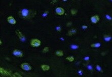
Noggin in Embryonic Mouse Cardiac Tissue.
Noggin was detected in immersion fixed frozen sections of embryonic mouse cardiac tissue (11 d.p.c.) using 15 µg/mL Goat Anti-Mouse Noggin Antigen Affinity-purified Polyclonal Antibody (Catalog # AF719) overnight at 4 °C. Tissue was stained with the Anti-Goat HRP-DAB Cell & Tissue Staining Kit (brown; Catalog # CTS008) and counterstained with hematoxylin (blue). View our protocol for Chromogenic IHC Staining of Frozen Tissue Sections.
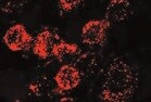
Noggin in PC‑3 Human Cell Line.
Noggin was detected in immersion fixed PC-3 human prostate cancer cell line using Goat Anti-Mouse Noggin Antigen Affinity-purified Polyclonal Antibody (Catalog # AF719) at 10 µg/mL for 3 hours at room temperature. Cells were stained using the NorthernLights™ 557-conjugated Anti-Goat IgG Secondary Antibody (red, upper panel; Catalog # NL001) and counterstained with DAPI (blue, lower panel). Specific staining was localized to cytoplasm. View our protocol for Fluorescent ICC Staining of Cells on Coverslips.
Detection of Human Noggin by Immunohistochemistry
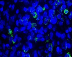
Myo/Nog cells contain ink in human tattooed skin.Tissue sections of tattooed skin were double labeled with the G8 mAb and antibodies to Noggin (Nog) (A-D), MyoD (F-I) or alpha -SMA (K-N). The colors of the secondary antibodies are indicated in the photographs. Nuclei were stained with Hoechst dye (blue). Images were produced with the epifluorescence (A-E, J and O) and confocal microscopes (F-I and K-N) with 60x lenses. Overlap of green and red fluorescence appears yellow in merged images (C, D, H, I, M and N). Fluorescent photomicrographs were merged with the corresponding DIC image to visualize the ink (black in D, I and N). Some double labeled cells appeared to contain ink (arrows in D, I and N). All ink laden G8+ cells were alpha -SMA+ (N). Smooth muscle cells of blood vessels also contained alpha -SMA (K). Minimal to no background fluorescence was visible after staining with the anti-goat (E), anti-IgM and anti-IgG (I), and anti-rabbit (O) secondary antibodies only. Bar = 28 μM in E and 5.6 μM in A-D and G-O. Image collected and cropped by CiteAb from the following open publication (https://pubmed.ncbi.nlm.nih.gov/32833999), licensed under a CC-BY license. Not internally tested by R&D Systems.
Detection of Human Noggin by Immunohistochemistry
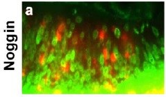
Myo/Nog cells contain ink in human tattooed skin.Tissue sections of tattooed skin were double labeled with the G8 mAb and antibodies to Noggin (Nog) (A-D), MyoD (F-I) or alpha -SMA (K-N). The colors of the secondary antibodies are indicated in the photographs. Nuclei were stained with Hoechst dye (blue). Images were produced with the epifluorescence (A-E, J and O) and confocal microscopes (F-I and K-N) with 60x lenses. Overlap of green and red fluorescence appears yellow in merged images (C, D, H, I, M and N). Fluorescent photomicrographs were merged with the corresponding DIC image to visualize the ink (black in D, I and N). Some double labeled cells appeared to contain ink (arrows in D, I and N). All ink laden G8+ cells were alpha -SMA+ (N). Smooth muscle cells of blood vessels also contained alpha -SMA (K). Minimal to no background fluorescence was visible after staining with the anti-goat (E), anti-IgM and anti-IgG (I), and anti-rabbit (O) secondary antibodies only. Bar = 28 μM in E and 5.6 μM in A-D and G-O. Image collected and cropped by CiteAb from the following open publication (https://pubmed.ncbi.nlm.nih.gov/32833999), licensed under a CC-BY license. Not internally tested by R&D Systems.
Detection of Human Noggin by Immunohistochemistry
Myo/Nog cells contain ink in human tattooed skin.Tissue sections of tattooed skin were double labeled with the G8 mAb and antibodies to Noggin (Nog) (A-D), MyoD (F-I) or alpha -SMA (K-N). The colors of the secondary antibodies are indicated in the photographs. Nuclei were stained with Hoechst dye (blue). Images were produced with the epifluorescence (A-E, J and O) and confocal microscopes (F-I and K-N) with 60x lenses. Overlap of green and red fluorescence appears yellow in merged images (C, D, H, I, M and N). Fluorescent photomicrographs were merged with the corresponding DIC image to visualize the ink (black in D, I and N). Some double labeled cells appeared to contain ink (arrows in D, I and N). All ink laden G8+ cells were alpha -SMA+ (N). Smooth muscle cells of blood vessels also contained alpha -SMA (K). Minimal to no background fluorescence was visible after staining with the anti-goat (E), anti-IgM and anti-IgG (I), and anti-rabbit (O) secondary antibodies only. Bar = 28 μM in E and 5.6 μM in A-D and G-O. Image collected and cropped by CiteAb from the following open publication (https://pubmed.ncbi.nlm.nih.gov/32833999), licensed under a CC-BY license. Not internally tested by R&D Systems.Applications for Mouse Noggin Antibody
Application
Recommended Usage
Immunocytochemistry
5-15 µg/mL
Sample: Immersion fixed PC‑3 human prostate cancer cell line
Sample: Immersion fixed PC‑3 human prostate cancer cell line
Immunohistochemistry
5-15 µg/mL
Sample: Immersion fixed frozen sections of embryonic mouse cardiac tissue (11 d.p.c.)
Sample: Immersion fixed frozen sections of embryonic mouse cardiac tissue (11 d.p.c.)
Western Blot
0.1 µg/mL
Sample: Recombinant Mouse Noggin (Catalog # 1967-NG)
Sample: Recombinant Mouse Noggin (Catalog # 1967-NG)
Reviewed Applications
Read 5 reviews rated 4.8 using AF719 in the following applications:
Formulation, Preparation, and Storage
Purification
Antigen Affinity-purified
Reconstitution
Reconstitute at 0.2 mg/mL in sterile PBS. For liquid material, refer to CoA for concentration.
Loading...
Formulation
Lyophilized from a 0.2 μm filtered solution in PBS with Trehalose. *Small pack size (SP) is supplied either lyophilized or as a 0.2 µm filtered solution in PBS.
Shipping
Lyophilized product is shipped at ambient temperature. Liquid small pack size (-SP) is shipped with polar packs. Upon receipt, store immediately at the temperature recommended below.
Stability & Storage
Use a manual defrost freezer and avoid repeated freeze-thaw cycles.
- 12 months from date of receipt, -20 to -70 °C as supplied.
- 1 month, 2 to 8 °C under sterile conditions after reconstitution.
- 6 months, -20 to -70 °C under sterile conditions after reconstitution.
Calculators
Background: Noggin
References
- Smith, W.C. and R.M. Harland (1992) Cell 70:829.
- Valenzuela, D.M. et al. (1995) J. Neurosci. 15:6077.
- Brunet, L.J. et al. (1998) Science 280:1455.
Alternate Names
NOG, SYM1, SYNS1, SYNS1A
Gene Symbol
NOG
UniProt
Additional Noggin Products
Product Documents for Mouse Noggin Antibody
Certificate of Analysis
To download a Certificate of Analysis, please enter a lot or batch number in the search box below.
Note: Certificate of Analysis not available for kit components.
Product Specific Notices for Mouse Noggin Antibody
For research use only
Related Research Areas
Citations for Mouse Noggin Antibody
 Powered by Bioz
Customer Reviews for Mouse Noggin Antibody (5)
4.8 out of 5
5 Customer Ratings
Have you used Mouse Noggin Antibody?
Submit a review and receive an Amazon gift card!
$25/€18/£15/$25CAN/¥2500 Yen for a review with an image
$10/€7/£6/$10CAN/¥1110 Yen for a review without an image
Submit a review
Customer Images
Showing
1
-
5 of
5 reviews
Showing All
Filter By:
-
Application: Immunocytochemistry/ImmunofluorescenceSample Tested: Epithelial cellsSpecies: MouseVerified Customer | Posted 10/15/2021

-
Application: Immunocytochemistry/ImmunofluorescenceSample Tested: MacrophagesSpecies: MouseVerified Customer | Posted 07/14/2021
-
Application: Immunocytochemistry/ImmunofluorescenceSample Tested: fibroblastsSpecies: MouseVerified Customer | Posted 07/01/2021
-
Application: Immunocytochemistry/ImmunofluorescenceSample Tested: IC-21 mouse macrophage cell lineSpecies: MouseVerified Customer | Posted 12/16/2020
-
Application: Western BlotSample Tested: See PMID 24042462Species: MouseVerified Customer | Posted 01/09/2015
There are no reviews that match your criteria.
Protocols
Find general support by application which include: protocols, troubleshooting, illustrated assays, videos and webinars.
- Antigen Retrieval Protocol (PIER)
- Antigen Retrieval for Frozen Sections Protocol
- Appropriate Fixation of IHC/ICC Samples
- Cellular Response to Hypoxia Protocols
- Chromogenic IHC Staining of Formalin-Fixed Paraffin-Embedded (FFPE) Tissue Protocol
- Chromogenic Immunohistochemistry Staining of Frozen Tissue
- ClariTSA™ Fluorophore Kits
- Detection & Visualization of Antibody Binding
- Fluorescent IHC Staining of Frozen Tissue Protocol
- Graphic Protocol for Heat-induced Epitope Retrieval
- Graphic Protocol for the Preparation and Fluorescent IHC Staining of Frozen Tissue Sections
- Graphic Protocol for the Preparation and Fluorescent IHC Staining of Paraffin-embedded Tissue Sections
- Graphic Protocol for the Preparation of Gelatin-coated Slides for Histological Tissue Sections
- ICC Cell Smear Protocol for Suspension Cells
- ICC Immunocytochemistry Protocol Videos
- ICC for Adherent Cells
- IHC Sample Preparation (Frozen sections vs Paraffin)
- Immunocytochemistry (ICC) Protocol
- Immunocytochemistry Troubleshooting
- Immunofluorescence of Organoids Embedded in Cultrex Basement Membrane Extract
- Immunofluorescent IHC Staining of Formalin-Fixed Paraffin-Embedded (FFPE) Tissue Protocol
- Immunohistochemistry (IHC) and Immunocytochemistry (ICC) Protocols
- Immunohistochemistry Frozen Troubleshooting
- Immunohistochemistry Paraffin Troubleshooting
- Preparing Samples for IHC/ICC Experiments
- Preventing Non-Specific Staining (Non-Specific Binding)
- Primary Antibody Selection & Optimization
- Protocol for Heat-Induced Epitope Retrieval (HIER)
- Protocol for Making a 4% Formaldehyde Solution in PBS
- Protocol for VisUCyte™ HRP Polymer Detection Reagent
- Protocol for the Fluorescent ICC Staining of Cell Smears - Graphic
- Protocol for the Fluorescent ICC Staining of Cultured Cells on Coverslips - Graphic
- Protocol for the Preparation & Fixation of Cells on Coverslips
- Protocol for the Preparation and Chromogenic IHC Staining of Frozen Tissue Sections
- Protocol for the Preparation and Chromogenic IHC Staining of Frozen Tissue Sections - Graphic
- Protocol for the Preparation and Chromogenic IHC Staining of Paraffin-embedded Tissue Sections
- Protocol for the Preparation and Chromogenic IHC Staining of Paraffin-embedded Tissue Sections - Graphic
- Protocol for the Preparation and Fluorescent ICC Staining of Cells on Coverslips
- Protocol for the Preparation and Fluorescent ICC Staining of Non-adherent Cells
- Protocol for the Preparation and Fluorescent ICC Staining of Stem Cells on Coverslips
- Protocol for the Preparation and Fluorescent IHC Staining of Frozen Tissue Sections
- Protocol for the Preparation and Fluorescent IHC Staining of Paraffin-embedded Tissue Sections
- Protocol for the Preparation of Gelatin-coated Slides for Histological Tissue Sections
- Protocol for the Preparation of a Cell Smear for Non-adherent Cell ICC - Graphic
- R&D Systems Quality Control Western Blot Protocol
- TUNEL and Active Caspase-3 Detection by IHC/ICC Protocol
- The Importance of IHC/ICC Controls
- Troubleshooting Guide: Immunohistochemistry
- Troubleshooting Guide: Western Blot Figures
- Western Blot Conditions
- Western Blot Protocol
- Western Blot Protocol for Cell Lysates
- Western Blot Troubleshooting
- Western Blot Troubleshooting Guide
- View all Protocols, Troubleshooting, Illustrated assays and Webinars

