Mouse B7 homolog 1(B7-H1), also called programmed death ligand 1 (PD-L1) and programmed cell death 1 ligand 1 (PDCD1L1), is a member of the B7 family of proteins that provide signals for regulating T-cell activation and tolerance (1-4). Other family members include B7-1, B7-2, B7-H2, B7-H3 and PD-L2. B7 proteins are immunoglobulin (Ig) superfamily members with extracellular Ig-V-like and Ig-C-like domains and a short cytoplasmic region. Among the family members, they share from 20-40% amino acid (aa) sequence identity. The cloned mouse B7-H1/PD-L1 cDNA encodes a 290 aa type I membrane precursor protein with a putative 18 aa signal peptide, a 220 aa extracellular region containing one V-like and one C-like Ig domain, a 22 aa transmembrane region, and a 30 aa cytoplasmic domain. Mouse and human B7-H1/PD-L1 share approximately 70% aa sequence identity. B7-H1/PD-L1 is one of two ligands for programmed death-1 (PD-1), a member of the CD28 family of immunoreceptors. The other identified ligand is PD-L2. Mouse B7-H1/PD-L1 and PD-L2 share approximately 34% aa sequence identity and have similar functions. B7-H1/PD-L1 is constitutively expressed in various lymphoid and non-lymphoid organs including placenta, heart, pancreas, lung, liver, and endothelium
(1‑4). The expression of B7-H1/PD-L1 is detected on B cells, T cells, monocytes, dendritic cells and thymic epithelial cells. IFN-gamma treatment induces B7-H1/PD-L1 expression in monocytes, dendritic cells, and endothelial cells. B7-H1/PD-L1 expression is also upregulated in a variety of tumor cell lines. On previously activated T cells, B7-H1/PD-L1 interaction with PD-1 inhibits TCR-mediated proliferation and cytokine production, suggesting an inhibitory role in regulating immune responses. In contrast, a costimulatory function for the PD-1 ligands on resting T cells has also beenreported (1-4).
Mouse PD-L1/B7-H1 Antibody (2096C)
R&D Systems | Catalog # MAB90781

Key Product Details
Validated by
Species Reactivity
Validated:
Cited:
Applications
Validated:
Cited:
Label
Antibody Source
Product Specifications
Immunogen
Met1-His239
Accession # Q9EP73
Specificity
Clonality
Host
Isotype
Scientific Data Images for Mouse PD-L1/B7-H1 Antibody (2096C)
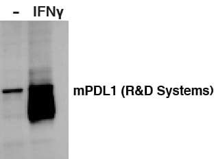
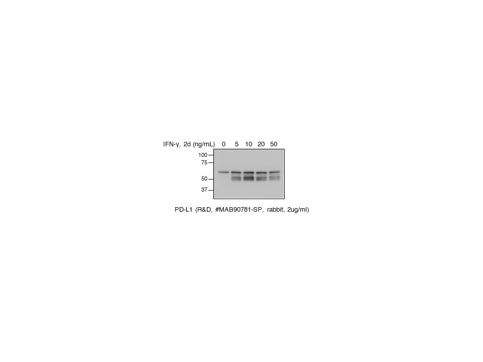
Detection of Mouse PD-L1/B7-H1 by Western Blot.
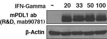
Western blot shows lysates of RAW 264.7 mouse monocyte/macrophage cell line and J774A.1 mouse reticulum cell sarcoma macrophage cell line untreated (-) or treated (+) with 10 µg/mL LPS for 4 hours. PVDF membrane was probed with 2 µg/mL of Rabbit Anti-Mouse PD-L1/B7-H1 Monoclonal Antibody (Catalog # MAB90781) followed by HRP-conjugated Anti-Rabbit IgG Secondary Antibody (Catalog # HAF008). A specific band was detected for PD-L1/B7-H1 at approximately 50-55 kDa (as indicated). This experiment was conducted under reducing conditions and using Immunoblot Buffer Group 1.
Detection of PD-L1/B7-H1 in RAW 264.7 Mouse Cell Line by Flow Cytometry.
RAW 264.7 mouse monocyte/macrophage cell line either treated with LPS overnight (filled histogram) or untreated (open histogram) was stained with Rabbit Anti-Mouse PD-L1/B7-H1 Monoclonal Antibody (Catalog # MAB90781), followed by Allophycocyanin-conjugated Anti-Rabbit IgG Secondary Antibody (Catalog # F0111). View our protocol for Staining Membrane-associated Proteins.
Detection of PD-L1/B7-H1 in HEK293 Human Cell Line Transfected with Mouse PD-L1/B7-H1 and eGFP by Flow Cytometry.
HEK293 human embryonic kidney cell line transfected with either (A) mouse PD-L1/B7-H1 or (B) irrelevant transfectants and eGFP was stained with Rabbit Anti-Mouse PD-L1/B7-H1 Monoclonal Antibody (Catalog # MAB90781) followed by Allophycocyanin-conjugated Anti-Rabbit IgG Secondary Antibody (Catalog # F0111). Quadrant markers were set based on control antibody staining (Catalog # MAB1050). View our protocol for Staining Membrane-associated Proteins.
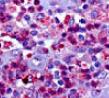
PD-L1/B7-H1 in Mouse Thymus.
PD-L1/B7-H1 was detected in perfusion fixed frozen sections of mouse thymus using Rabbit Anti-Mouse PD-L1/B7-H1 Monoclonal Antibody (Catalog # MAB90781) at 5 µg/mL for 1 hour at room temperature followed by incubation with the Anti-Rabbit IgG VisUCyte™ HRP Polymer Antibody (Catalog # VC003). Tissue was stained using DAB (brown) and counterstained with hematoxylin (blue). Specific staining was localized to thymocytes. View our protocol for IHC Staining with VisUCyte HRP Polymer Detection Reagents.Applications for Mouse PD-L1/B7-H1 Antibody (2096C)
Flow Cytometry
Sample: RAW 264.7 mouse monocyte/macrophage cell line treated with LPS and HEK293 human embryonic kidney cell line transfected with mouse B7-H1/PD-L1
Immunohistochemistry
Sample:
Perfusion fixed frozen sections of mouse thymus
Western Blot
Sample: RAW 264.7 mouse monocyte/macrophage cell line and J774A.1 mouse reticulum cell sarcoma macrophage cell line treated with LPS
Reviewed Applications
Read 10 reviews rated 4.4 using MAB90781 in the following applications:
Flow Cytometry Panel Builder
Bio-Techne Knows Flow Cytometry
Save time and reduce costly mistakes by quickly finding compatible reagents using the Panel Builder Tool.
Advanced Features
- Spectra Viewer - Custom analysis of spectra from multiple fluorochromes
- Spillover Popups - Visualize the spectra of individual fluorochromes
- Antigen Density Selector - Match fluorochrome brightness with antigen density

Formulation, Preparation, and Storage
Purification
Reconstitution
Reconstitute at 0.5 mg/mL in sterile PBS. For liquid material, refer to CoA for concentration.
Formulation
Shipping
Stability & Storage
- 12 months from date of receipt, -20 to -70 °C as supplied.
- 1 month, 2 to 8 °C under sterile conditions after reconstitution.
- 6 months, -20 to -70 °C under sterile conditions after reconstitution.
Calculators
Background: PD-L1/B7-H1
References
- Tamura, H. et al. (2001) Blood 97:1809.
- Freeman, G. et al. (2000) J. Exp. Med. 192:1027.
- Sharpe, A.H. and G. J. Freeman (2002) Nat. Rev. Immunol. 2:116.
- Coyle, A. and J. Gutierrez-Ramos (2001) Nat. Immunol. 2:203.
Long Name
Alternate Names
Entrez Gene IDs
Gene Symbol
UniProt
Additional PD-L1/B7-H1 Products
Product Documents for Mouse PD-L1/B7-H1 Antibody (2096C)
Certificate of Analysis
To download a Certificate of Analysis, please enter a lot or batch number in the search box below.
Note: Certificate of Analysis not available for kit components.
Product Specific Notices for Mouse PD-L1/B7-H1 Antibody (2096C)
For research use only
Citations for Mouse PD-L1/B7-H1 Antibody (2096C)
 Powered by Bioz
Customer Reviews for Mouse PD-L1/B7-H1 Antibody (2096C) (10)
Have you used Mouse PD-L1/B7-H1 Antibody (2096C)?
Submit a review and receive an Amazon gift card!
$25/€18/£15/$25CAN/¥2500 Yen for a review with an image
$10/€7/£6/$10CAN/¥1110 Yen for a review without an image
Submit a review
Customer Images


-
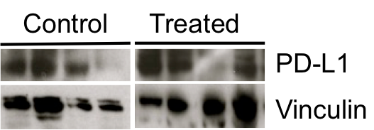
Application: Western BlotSample Tested: J774A.1 mouse reticulum cell sarcoma macrophage cell lineSpecies: MouseVerified Customer | Posted 09/26/2021

-
Application: Western BlotSample Tested: Mouse osteosarcoma cell lineSpecies: MouseVerified Customer | Posted 06/08/2020
-
Application: Immunohistochemistry-ParaffinSample Tested: Breast cancer tissueSpecies: MouseVerified Customer | Posted 04/03/2020
-
Application: Western BlotSample Tested: Mouse Breast cancer cell lineSpecies: MouseVerified Customer | Posted 12/09/2019
-
Application: Western BlotSample Tested: mouse breast cancer cell line Eo771Species: MouseVerified Customer | Posted 02/28/2019
-
Application: Western BlotSample Tested: 4T1 mouse breast cancer cell line and B16-F10 mouse melanoma cell lineSpecies: MouseVerified Customer | Posted 10/28/2018
-
Application: Western BlotSample Tested: EMT6 cells and CT26Species: MouseVerified Customer | Posted 10/17/2018
-
Application: Western BlotSample Tested: Mammary gland tissue and AdenocarcinomaSpecies: MouseVerified Customer | Posted 04/13/2018Tissue lysates from mammary gland tumors
-
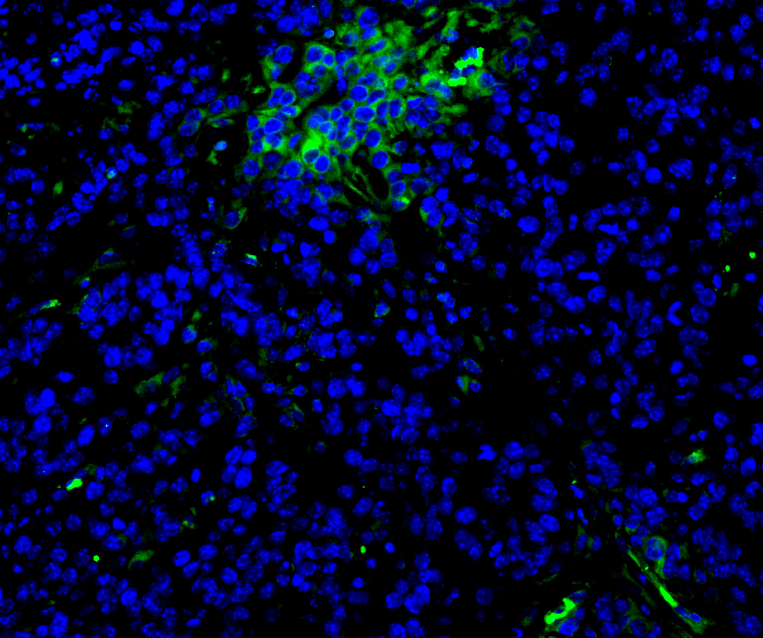
Application: Immunocytochemistry/ImmunofluorescenceSample Tested: Pancreatic cancer tissueSpecies: MouseVerified Customer | Posted 08/08/20171:50 concentration, Overnight incubation
-
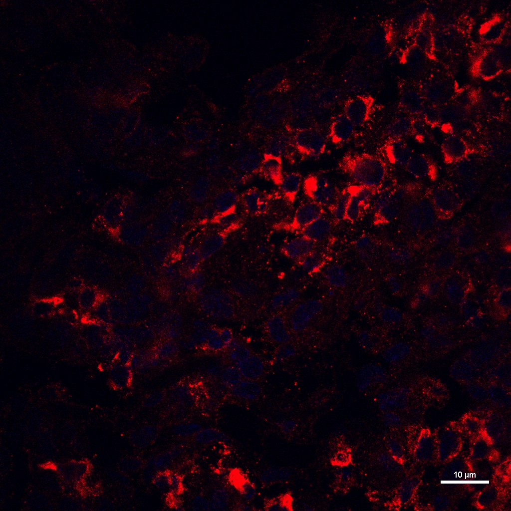
Application: ImmunocytochemistrySample Tested: Mouse Lymph NodeSpecies: MouseVerified Customer | Posted 05/16/2017
There are no reviews that match your criteria.
Protocols
Find general support by application which include: protocols, troubleshooting, illustrated assays, videos and webinars.
- 7-Amino Actinomycin D (7-AAD) Cell Viability Flow Cytometry Protocol
- Antigen Retrieval Protocol (PIER)
- Antigen Retrieval for Frozen Sections Protocol
- Appropriate Fixation of IHC/ICC Samples
- Cellular Response to Hypoxia Protocols
- Chromogenic IHC Staining of Formalin-Fixed Paraffin-Embedded (FFPE) Tissue Protocol
- Chromogenic Immunohistochemistry Staining of Frozen Tissue
- ClariTSA™ Fluorophore Kits
- Detection & Visualization of Antibody Binding
- Extracellular Membrane Flow Cytometry Protocol
- Flow Cytometry Protocol for Cell Surface Markers
- Flow Cytometry Protocol for Staining Membrane Associated Proteins
- Flow Cytometry Staining Protocols
- Flow Cytometry Troubleshooting Guide
- Fluorescent IHC Staining of Frozen Tissue Protocol
- Graphic Protocol for Heat-induced Epitope Retrieval
- Graphic Protocol for the Preparation and Fluorescent IHC Staining of Frozen Tissue Sections
- Graphic Protocol for the Preparation and Fluorescent IHC Staining of Paraffin-embedded Tissue Sections
- Graphic Protocol for the Preparation of Gelatin-coated Slides for Histological Tissue Sections
- IHC Sample Preparation (Frozen sections vs Paraffin)
- Immunofluorescent IHC Staining of Formalin-Fixed Paraffin-Embedded (FFPE) Tissue Protocol
- Immunohistochemistry (IHC) and Immunocytochemistry (ICC) Protocols
- Immunohistochemistry Frozen Troubleshooting
- Immunohistochemistry Paraffin Troubleshooting
- Intracellular Flow Cytometry Protocol Using Alcohol (Methanol)
- Intracellular Flow Cytometry Protocol Using Detergents
- Intracellular Nuclear Staining Flow Cytometry Protocol Using Detergents
- Intracellular Staining Flow Cytometry Protocol Using Alcohol Permeabilization
- Intracellular Staining Flow Cytometry Protocol Using Detergents to Permeabilize Cells
- Preparing Samples for IHC/ICC Experiments
- Preventing Non-Specific Staining (Non-Specific Binding)
- Primary Antibody Selection & Optimization
- Propidium Iodide Cell Viability Flow Cytometry Protocol
- Protocol for Heat-Induced Epitope Retrieval (HIER)
- Protocol for Liperfluo
- Protocol for Making a 4% Formaldehyde Solution in PBS
- Protocol for VisUCyte™ HRP Polymer Detection Reagent
- Protocol for the Characterization of Human Th22 Cells
- Protocol for the Characterization of Human Th9 Cells
- Protocol for the Preparation & Fixation of Cells on Coverslips
- Protocol for the Preparation and Chromogenic IHC Staining of Frozen Tissue Sections
- Protocol for the Preparation and Chromogenic IHC Staining of Frozen Tissue Sections - Graphic
- Protocol for the Preparation and Chromogenic IHC Staining of Paraffin-embedded Tissue Sections
- Protocol for the Preparation and Chromogenic IHC Staining of Paraffin-embedded Tissue Sections - Graphic
- Protocol for the Preparation and Fluorescent IHC Staining of Frozen Tissue Sections
- Protocol for the Preparation and Fluorescent IHC Staining of Paraffin-embedded Tissue Sections
- Protocol for the Preparation of Gelatin-coated Slides for Histological Tissue Sections
- Protocol: Annexin V and PI Staining by Flow Cytometry
- Protocol: Annexin V and PI Staining for Apoptosis by Flow Cytometry
- R&D Systems Quality Control Western Blot Protocol
- TUNEL and Active Caspase-3 Detection by IHC/ICC Protocol
- The Importance of IHC/ICC Controls
- Troubleshooting Guide: Fluorokine Flow Cytometry Kits
- Troubleshooting Guide: Immunohistochemistry
- Troubleshooting Guide: Western Blot Figures
- Western Blot Conditions
- Western Blot Protocol
- Western Blot Protocol for Cell Lysates
- Western Blot Troubleshooting
- Western Blot Troubleshooting Guide
- View all Protocols, Troubleshooting, Illustrated assays and Webinars