CD47 N-terminal IgV-like Extracellular Domain Antibody
R&D Systems | Catalog # AF1866

Key Product Details
Validated by
Biological Validation
Species Reactivity
Validated:
Mouse, Rat
Cited:
Human, Mouse
Applications
Validated:
Immunohistochemistry, Western Blot
Cited:
Immunohistochemistry, Western Blot, Flow Cytometry, Immunofluorescence, Immunocytochemistry, Immunoprecipitation
Label
Unconjugated
Antibody Source
Polyclonal Goat IgG
Loading...
Product Specifications
Immunogen
Mouse myeloma cell line NS0-derived recombinant mouse CD47 N-terminal IgV-like
Gln19-Pro158
Accession # NP_034711
Gln19-Pro158
Accession # NP_034711
Specificity
Detects mouse CD47 N-terminal IgV-like in direct ELISAs and Western blots. In direct ELISAs, less than 15% cross-reactivity with recombinant human CD47 is observed.
Clonality
Polyclonal
Host
Goat
Isotype
IgG
Scientific Data Images for CD47 N-terminal IgV-like Extracellular Domain Antibody
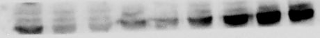
Detection of Mouse CD47 by Western Blot.
Western blot shows lysates of mouse ovary tissue and mouse thymus tissue. PVDF membrane was probed with 0.25 µg/mL of Goat Anti-Mouse/Rat CD47 N-terminal IgV-like Extracellular Domain Antigen Affinity-purified Polyclonal Antibody (Catalog # AF1866) followed by HRP-conjugated Anti-Goat IgG Secondary Antibody (HAF019). Specific bands were detected for CD47 at approximately 40-60 kDa (as indicated). This experiment was conducted under reducing conditions and using Immunoblot Buffer Group 1.
Detection of Rat CD47 by Western Blot.
Western blot shows lysates of rat placenta tissue. PVDF membrane was probed with 1 µg/mL of Goat Anti-Mouse/Rat CD47 N-terminal IgV-like Extracellular Domain Antigen Affinity-purified Polyclonal Antibody (Catalog # AF1866) followed by HRP-conjugated Anti-Goat IgG Secondary Antibody (HAF017). Specific bands were detected for CD47 at approximately 40-60 kDa (as indicated). This experiment was conducted under reducing conditions and using Immunoblot Buffer Group 1.
Detection of Mouse CD47 by Western Blot
CD47 regulates thrombospondin-1, TGF-beta 1, and collagen deposition after injury. a Whole cell lysates from freshly isolated intestinal epithelial cells from Cd47f/f and Cd47 delta IEC mice were subjected to SDS–PAGE and immunoblot for thrombospondin-1/TSP-1, TGF-beta 1, and phosphorylated SMAD2 and SMAD3. Results are representative of three independent experiments. b Representative Masson’s trichrome staining of wounds beds and chronic DSS-colitis colons from Cd47f/f and Cd47 delta IEC mice. Scale bars = 50 μm. Results representative of three independent experiments with 5–7 mice per group. Source data are provided as a Source Data file Image collected and cropped by CiteAb from the following publication (https://pubmed.ncbi.nlm.nih.gov/31676794), licensed under a CC-BY license. Not internally tested by R&D Systems.
Detection of CD47 in Mouse Spleen.
CD47 was detected in immersion fixed paraffin-embedded sections of Mouse Spleen using Goat Anti-Mouse/Rat CD47 N-terminal IgV-like Extracellular Domain Antigen Affinity-purified Polyclonal Antibody (Catalog # AF1866) at 1 µg/mL for 1 hour at room temperature followed by incubation with the HRP-conjugated Anti-Goat IgG Secondary Antibody (Catalog # HAF017). Before incubation with the primary antibody, tissue was subjected to heat-induced epitope retrieval using VisUCyte Antigen Retrieval Reagent-Basic (Catalog # VCTS021). Tissue was stained using DAB (brown) and counterstained with hematoxylin (blue). Specific staining was localized to Cell surface and cytoplasm. View our protocol for Chromogenic IHC Staining of Paraffin-embedded Tissue Sections.
Detection of Mouse CD47 by Western Blot
CD47 associates with beta 1 integrin and promotes focal adhesion formation. a In situ proximity ligation assay utilizing antibodies against CD47 and beta 1 integrin indicating close association between CD47 and beta 1 integrin in the colonic epithelium. Positive PLA signals are shown in green, beta-catenin in magenta and DAPI/nuclei in blue. Crypts are indicated by dashed lines. Arrowheads and asterisks (*) indicate positive PLA signals on IECs and immune cells in the lamina propria, respectively. Strong PLA signals are detected in IECs that are mainly concentrated at the base of the crypts in Cd47f/f colon in contrast to very few PLA signals are observed in crypts IECs in Cd47 delta IEC colons. Scale bars = 20 μm. The specificity of the PLA signals was evaluated in Supplementary figure 6 demonstrating no PLA signals in Cd47−/− colons. b Whole-cell lysates from freshly isolated intestinal epithelial cells from Cd47f/f and Cd47 delta IEC mice were subjected to SDS–PAGE and immunoblot for signaling molecules downstream of beta 1 integrin-dependent cell adhesion, revealing decreased beta 1 integrin protein expression and reduced phosphorylation of SrcY416, FAKY397/Y861, and p130CASY410 in cells from Cd47 delta IEC mice. Results are representative of three independent experiments. c Murine enteroid-derived primary epithelial cell monolayers were scratch-wounded and harvested at the indicated time points, then analyzed by SDS–PAGE and immunoblot. CD47-deficient epithelial cells show a reduction in phosphorylated SrcY416, FAKY397/Y861, and p130CASY410 upon wounding, whereas maintaining reduced beta 1 integrin protein baseline expression. d Epithelial cells immediately adjacent to wounds from Cd47f/f and Cd47 delta IEC mice were imaged by confocal microscopy, showing disrupted basal co-staining for phosphorylated FAKY861 and beta 1 integrin (apical surface indicated by dashed line). Arrows indicate reduced colocalization of phospho-FAKY861 and beta 1 integrin in the absence of CD47 expression. Scale bars = 10 μm. Results are re
Detection of Mouse CD47 by Immunocytochemistry/Immunofluorescence
Loss of CD47 in IEC does not induce immune-mediated mucosal damage. a–c Naive Cd47 delta IEC and tamoxifen-treated Cd47ER delta IEC mice housed in pathogen-free conditions were analyzed for intestinal epithelial CD47 expression. Cd47ER delta IEC mice were treated with tamoxifen to induce acute CD47 deletion in intestinal epithelial cells, and analyzed 2 weeks later. a Tissue sections from naive Cd47 delta IEC and tamoxifen-treated Cd47ER delta IEC mice were stained with anti-CD47 antibodies (green) with DAPI counterstain (blue). CD47 expression is absent in the epithelium but retained in the lamina propria and submucosa. Scale bars = 100 μm upper panels, 50 μm lower panels. b IECs were isolated from the terminal ileum of naive mice. Protein lysates were analyzed by SDS–PAGE and immunoblot for CD47. c IECs were isolated from Cd47ER delta IEC mice treated with vehicle (corn oil) or tamoxifen, and analyzed as in b. Results are representative of three independent experiments with 3–5 mice per group. d Paraffin-embedded colon tissue sections from Cd47 delta IEC mice were stained with Hematoxylin and Eosin counterstain for histological examination. Gross mucosal architecture is intact in the absence of epithelial CD47 expression. Scale bars = 50 μm upper panels, 100 μm lower panels. e Colon tissue digests from naive Cd47 delta IEC mice were stained and analyzed by flow cytometry, showing no significant differences in major lamina propria immune cell populations. Points represent individual samples each containing two mice (n = 4 mice per group). Data are means ± SEM and are representative of three independent experiments. Source data are provided as a Source Data file Image collected and cropped by CiteAb from the following publication (https://pubmed.ncbi.nlm.nih.gov/31676794), licensed under a CC-BY license. Not internally tested by R&D Systems.
Detection of Mouse CD47 by Immunocytochemistry/Immunofluorescence
IEC-specific deletion of CD47 results in impaired mucosal healing. Utilizing a miniature video endoscope and biopsy scissors, 5–7 wounds were created in the dorsal aspect of the descending colon mucosa of anesthetized mice. Cd47ER delta IEC mice were wounded 2 weeks after tamoxifen treatment. a Digital measurement of wound surface area at 24 and 72 h post wounding revealed a striking impairment in wound closure in Cd47 delta IEC and tamoxifen-treated Cd47ER delta IEC mice. Points represent mean value within all wounds from an individual mouse. Data are representative of two independent experiments with 5–6 mice per group. Data are means ± SEM. ***p < 0.001; one-way ANOVA. b Tissue sections taken from day 3 wounds were stained with the epithelial-specific marker E-Cadherin (green), plus DAPI counterstain (blue). Re-epithelialization of the wound is disorganized in the absence of CD47 (Cd47 delta IEC) compared with control Cd47f/f. c Tissue sections taken from day 3 wounds were stained with the epithelial-specific marker E-Cadherin (green), brush border protein Villin (magenta), and DAPI (blue). Insets of epithelial cells on top of the wound bed show polarized wound-associated epithelial cells (WAE) expressing Villin (arrows) in Cd47f/f wounds while cells are not polarized in Cd47 delta IEC wounds. Scale bars = 100 μm. d Ki67 staining of frozen sections of wounded colon mucosa 3 days post-wounding (red) revealed similar proliferation rates in crypt epithelial cells immediately adjacent to wounds in the absence of epithelial CD47. Sections were counterstained with E-Cadherin (green) and DAPI (blue). Scale bars = 50 μm. Points represent the average number of Ki67-positive cells for four crypts adjacent to wounds for each individual mouse. Data are means ± SEM and are representative of two independent experiments with 4–6 mice per group. Source data are provided as a Source Data file Image collected and cropped by CiteAb from the following publication (https://pubmed.ncbi.nlm.nih.gov/31676794), licen
Detection of Mouse CD47 by Western Blot
CD47 associates with beta 1 integrin and promotes focal adhesion formation. a In situ proximity ligation assay utilizing antibodies against CD47 and beta 1 integrin indicating close association between CD47 and beta 1 integrin in the colonic epithelium. Positive PLA signals are shown in green, beta-catenin in magenta and DAPI/nuclei in blue. Crypts are indicated by dashed lines. Arrowheads and asterisks (*) indicate positive PLA signals on IECs and immune cells in the lamina propria, respectively. Strong PLA signals are detected in IECs that are mainly concentrated at the base of the crypts in Cd47f/f colon in contrast to very few PLA signals are observed in crypts IECs in Cd47 delta IEC colons. Scale bars = 20 μm. The specificity of the PLA signals was evaluated in Supplementary figure 6 demonstrating no PLA signals in Cd47−/− colons. b Whole-cell lysates from freshly isolated intestinal epithelial cells from Cd47f/f and Cd47 delta IEC mice were subjected to SDS–PAGE and immunoblot for signaling molecules downstream of beta 1 integrin-dependent cell adhesion, revealing decreased beta 1 integrin protein expression and reduced phosphorylation of SrcY416, FAKY397/Y861, and p130CASY410 in cells from Cd47 delta IEC mice. Results are representative of three independent experiments. c Murine enteroid-derived primary epithelial cell monolayers were scratch-wounded and harvested at the indicated time points, then analyzed by SDS–PAGE and immunoblot. CD47-deficient epithelial cells show a reduction in phosphorylated SrcY416, FAKY397/Y861, and p130CASY410 upon wounding, whereas maintaining reduced beta 1 integrin protein baseline expression. d Epithelial cells immediately adjacent to wounds from Cd47f/f and Cd47 delta IEC mice were imaged by confocal microscopy, showing disrupted basal co-staining for phosphorylated FAKY861 and beta 1 integrin (apical surface indicated by dashed line). Arrows indicate reduced colocalization of phospho-FAKY861 and beta 1 integrin in the absence of CD47 expression. Scale bars = 10 μm. Results are re
Detection of Mouse CD47 by Immunocytochemistry/Immunofluorescence
Loss of CD47 in IEC results in impaired recovery from DSS-induced colitis. Age- and sex-matched Cd47 delta IEC and tamoxifen-treated Cd47ER delta IEC mice were treated with three consecutive cycles of 2.5% DSS in drinking water for either 4 days (Cd47 delta IEC) or 3 days (Cd47ER delta IEC), followed by 5 days of water recovery. Cd47ER delta IEC mice were treated with DSS 2 weeks after tamoxifen treatment. a–b Disease activity index scores are represented as an average of scores 0–4 for percent weight loss, stool consistency, and presence of blood in stools. Cyclical treatment of aCd47 delta IEC and b tamoxifen-treated Cd47ER delta IEC mice with 2.5% DSS in drinking water, followed by a plain water recovery period, induced greater DAI scores in the absence of epithelial CD47. Data are representative of two independent experiments with 5–6 mice per group and are expressed as means ± SEM. a *p = 0.02, b *p = 0.036 by two-way ANOVA. c Histological scoring of hematoxylin and eosin (H&E)-stained tissue sections of colonic mucosa: percentage of injury/ulceration represents a ratio of the length of injured/ulcerated areas (≥ 50% crypt loss) relative to the entire colon length, as assessed in Swiss roll mounts of the entire colon. Results indicate greater damage in the absence of epithelial CD47. Points represent individual mice. Data are representative of two independent experiments with 5–6 mice per group and are expressed as means ± SEM. Significance determined by two-tailed Student’s t test. *p = 0.016, ***p = 0.001. d, e Representative H&E staining of colon tissue sections after three cycles of DSS/water revealed extensive crypt destruction in distal colon of Cd47 delta IEC and Cd47ER delta IEC mice. Large areas of ulcerated mucosa with granulocytic infiltrates were present in the mid colon in Cd47 delta IEC and Cd47ER delta IEC mice, in comparison with littermate controls. Scale bars = 100 μm upper panels, 300 μm lower panels. Results are representative of at least two independent experiments with 5–6 mice per group. Source data are
Detection of Mouse CD47 by Immunocytochemistry/Immunofluorescence
IEC-specific deletion of CD47 results in impaired mucosal healing. Utilizing a miniature video endoscope and biopsy scissors, 5–7 wounds were created in the dorsal aspect of the descending colon mucosa of anesthetized mice. Cd47ER delta IEC mice were wounded 2 weeks after tamoxifen treatment. a Digital measurement of wound surface area at 24 and 72 h post wounding revealed a striking impairment in wound closure in Cd47 delta IEC and tamoxifen-treated Cd47ER delta IEC mice. Points represent mean value within all wounds from an individual mouse. Data are representative of two independent experiments with 5–6 mice per group. Data are means ± SEM. ***p < 0.001; one-way ANOVA. b Tissue sections taken from day 3 wounds were stained with the epithelial-specific marker E-Cadherin (green), plus DAPI counterstain (blue). Re-epithelialization of the wound is disorganized in the absence of CD47 (Cd47 delta IEC) compared with control Cd47f/f. c Tissue sections taken from day 3 wounds were stained with the epithelial-specific marker E-Cadherin (green), brush border protein Villin (magenta), and DAPI (blue). Insets of epithelial cells on top of the wound bed show polarized wound-associated epithelial cells (WAE) expressing Villin (arrows) in Cd47f/f wounds while cells are not polarized in Cd47 delta IEC wounds. Scale bars = 100 μm. d Ki67 staining of frozen sections of wounded colon mucosa 3 days post-wounding (red) revealed similar proliferation rates in crypt epithelial cells immediately adjacent to wounds in the absence of epithelial CD47. Sections were counterstained with E-Cadherin (green) and DAPI (blue). Scale bars = 50 μm. Points represent the average number of Ki67-positive cells for four crypts adjacent to wounds for each individual mouse. Data are means ± SEM and are representative of two independent experiments with 4–6 mice per group. Source data are provided as a Source Data file Image collected and cropped by CiteAb from the following publication (https://pubmed.ncbi.nlm.nih.gov/31676794), licen
Detection of Mouse CD47 by Immunocytochemistry/Immunofluorescence
CD47 associates with beta 1 integrin and promotes focal adhesion formation. a In situ proximity ligation assay utilizing antibodies against CD47 and beta 1 integrin indicating close association between CD47 and beta 1 integrin in the colonic epithelium. Positive PLA signals are shown in green, beta-catenin in magenta and DAPI/nuclei in blue. Crypts are indicated by dashed lines. Arrowheads and asterisks (*) indicate positive PLA signals on IECs and immune cells in the lamina propria, respectively. Strong PLA signals are detected in IECs that are mainly concentrated at the base of the crypts in Cd47f/f colon in contrast to very few PLA signals are observed in crypts IECs in Cd47 delta IEC colons. Scale bars = 20 μm. The specificity of the PLA signals was evaluated in Supplementary figure 6 demonstrating no PLA signals in Cd47−/− colons. b Whole-cell lysates from freshly isolated intestinal epithelial cells from Cd47f/f and Cd47 delta IEC mice were subjected to SDS–PAGE and immunoblot for signaling molecules downstream of beta 1 integrin-dependent cell adhesion, revealing decreased beta 1 integrin protein expression and reduced phosphorylation of SrcY416, FAKY397/Y861, and p130CASY410 in cells from Cd47 delta IEC mice. Results are representative of three independent experiments. c Murine enteroid-derived primary epithelial cell monolayers were scratch-wounded and harvested at the indicated time points, then analyzed by SDS–PAGE and immunoblot. CD47-deficient epithelial cells show a reduction in phosphorylated SrcY416, FAKY397/Y861, and p130CASY410 upon wounding, whereas maintaining reduced beta 1 integrin protein baseline expression. d Epithelial cells immediately adjacent to wounds from Cd47f/f and Cd47 delta IEC mice were imaged by confocal microscopy, showing disrupted basal co-staining for phosphorylated FAKY861 and beta 1 integrin (apical surface indicated by dashed line). Arrows indicate reduced colocalization of phospho-FAKY861 and beta 1 integrin in the absence of CD47 expression. Scale bars = 10 μm. Results are re
Detection of Mouse CD47 by Immunocytochemistry/Immunofluorescence
Loss of CD47 in IEC results in impaired recovery from DSS-induced colitis. Age- and sex-matched Cd47 delta IEC and tamoxifen-treated Cd47ER delta IEC mice were treated with three consecutive cycles of 2.5% DSS in drinking water for either 4 days (Cd47 delta IEC) or 3 days (Cd47ER delta IEC), followed by 5 days of water recovery. Cd47ER delta IEC mice were treated with DSS 2 weeks after tamoxifen treatment. a–b Disease activity index scores are represented as an average of scores 0–4 for percent weight loss, stool consistency, and presence of blood in stools. Cyclical treatment of aCd47 delta IEC and b tamoxifen-treated Cd47ER delta IEC mice with 2.5% DSS in drinking water, followed by a plain water recovery period, induced greater DAI scores in the absence of epithelial CD47. Data are representative of two independent experiments with 5–6 mice per group and are expressed as means ± SEM. a *p = 0.02, b *p = 0.036 by two-way ANOVA. c Histological scoring of hematoxylin and eosin (H&E)-stained tissue sections of colonic mucosa: percentage of injury/ulceration represents a ratio of the length of injured/ulcerated areas (≥ 50% crypt loss) relative to the entire colon length, as assessed in Swiss roll mounts of the entire colon. Results indicate greater damage in the absence of epithelial CD47. Points represent individual mice. Data are representative of two independent experiments with 5–6 mice per group and are expressed as means ± SEM. Significance determined by two-tailed Student’s t test. *p = 0.016, ***p = 0.001. d, e Representative H&E staining of colon tissue sections after three cycles of DSS/water revealed extensive crypt destruction in distal colon of Cd47 delta IEC and Cd47ER delta IEC mice. Large areas of ulcerated mucosa with granulocytic infiltrates were present in the mid colon in Cd47 delta IEC and Cd47ER delta IEC mice, in comparison with littermate controls. Scale bars = 100 μm upper panels, 300 μm lower panels. Results are representative of at least two independent experiments with 5–6 mice per group. Source data are
Detection of Mouse CD47 by Immunocytochemistry/Immunofluorescence
IEC-specific deletion of CD47 results in impaired mucosal healing. Utilizing a miniature video endoscope and biopsy scissors, 5–7 wounds were created in the dorsal aspect of the descending colon mucosa of anesthetized mice. Cd47ER delta IEC mice were wounded 2 weeks after tamoxifen treatment. a Digital measurement of wound surface area at 24 and 72 h post wounding revealed a striking impairment in wound closure in Cd47 delta IEC and tamoxifen-treated Cd47ER delta IEC mice. Points represent mean value within all wounds from an individual mouse. Data are representative of two independent experiments with 5–6 mice per group. Data are means ± SEM. ***p < 0.001; one-way ANOVA. b Tissue sections taken from day 3 wounds were stained with the epithelial-specific marker E-Cadherin (green), plus DAPI counterstain (blue). Re-epithelialization of the wound is disorganized in the absence of CD47 (Cd47 delta IEC) compared with control Cd47f/f. c Tissue sections taken from day 3 wounds were stained with the epithelial-specific marker E-Cadherin (green), brush border protein Villin (magenta), and DAPI (blue). Insets of epithelial cells on top of the wound bed show polarized wound-associated epithelial cells (WAE) expressing Villin (arrows) in Cd47f/f wounds while cells are not polarized in Cd47 delta IEC wounds. Scale bars = 100 μm. d Ki67 staining of frozen sections of wounded colon mucosa 3 days post-wounding (red) revealed similar proliferation rates in crypt epithelial cells immediately adjacent to wounds in the absence of epithelial CD47. Sections were counterstained with E-Cadherin (green) and DAPI (blue). Scale bars = 50 μm. Points represent the average number of Ki67-positive cells for four crypts adjacent to wounds for each individual mouse. Data are means ± SEM and are representative of two independent experiments with 4–6 mice per group. Source data are provided as a Source Data file Image collected and cropped by CiteAb from the following publication (https://pubmed.ncbi.nlm.nih.gov/31676794), licen
Detection of Mouse CD47 by Immunohistochemistry
IEC-specific deletion of CD47 results in impaired mucosal healing. Utilizing a miniature video endoscope and biopsy scissors, 5–7 wounds were created in the dorsal aspect of the descending colon mucosa of anesthetized mice. Cd47ER delta IEC mice were wounded 2 weeks after tamoxifen treatment. a Digital measurement of wound surface area at 24 and 72 h post wounding revealed a striking impairment in wound closure in Cd47 delta IEC and tamoxifen-treated Cd47ER delta IEC mice. Points represent mean value within all wounds from an individual mouse. Data are representative of two independent experiments with 5–6 mice per group. Data are means ± SEM. ***p < 0.001; one-way ANOVA. b Tissue sections taken from day 3 wounds were stained with the epithelial-specific marker E-Cadherin (green), plus DAPI counterstain (blue). Re-epithelialization of the wound is disorganized in the absence of CD47 (Cd47 delta IEC) compared with control Cd47f/f. c Tissue sections taken from day 3 wounds were stained with the epithelial-specific marker E-Cadherin (green), brush border protein Villin (magenta), and DAPI (blue). Insets of epithelial cells on top of the wound bed show polarized wound-associated epithelial cells (WAE) expressing Villin (arrows) in Cd47f/f wounds while cells are not polarized in Cd47 delta IEC wounds. Scale bars = 100 μm. d Ki67 staining of frozen sections of wounded colon mucosa 3 days post-wounding (red) revealed similar proliferation rates in crypt epithelial cells immediately adjacent to wounds in the absence of epithelial CD47. Sections were counterstained with E-Cadherin (green) and DAPI (blue). Scale bars = 50 μm. Points represent the average number of Ki67-positive cells for four crypts adjacent to wounds for each individual mouse. Data are means ± SEM and are representative of two independent experiments with 4–6 mice per group. Source data are provided as a Source Data file Image collected and cropped by CiteAb from the following publication (https://pubmed.ncbi.nlm.nih.gov/31676794), licenApplications for CD47 N-terminal IgV-like Extracellular Domain Antibody
Application
Recommended Usage
Immunohistochemistry
1-15 µg/mL
Sample: Immersion fixed paraffin-embedded sections of Mouse Spleen
Sample: Immersion fixed paraffin-embedded sections of Mouse Spleen
Western Blot
0.25-1 µg/mL
Sample: Mouse ovary tissue, mouse thymus tissue, and rat placenta tisssue
Sample: Mouse ovary tissue, mouse thymus tissue, and rat placenta tisssue
Reviewed Applications
Read 4 reviews rated 5 using AF1866 in the following applications:
Formulation, Preparation, and Storage
Purification
Antigen Affinity-purified
Reconstitution
Reconstitute at 0.2 mg/mL in sterile PBS. For liquid material, refer to CoA for concentration.
Loading...
Formulation
Lyophilized from a 0.2 μm filtered solution in PBS with Trehalose. See Certificate of Analysis for details.
*Small pack size (-SP) is supplied either lyophilized or as a 0.2 µm filtered solution in PBS.
*Small pack size (-SP) is supplied either lyophilized or as a 0.2 µm filtered solution in PBS.
Shipping
Lyophilized product is shipped at ambient temperature. Liquid small pack size (-SP) is shipped with polar packs. Upon receipt, store immediately at the temperature recommended below.
Stability & Storage
Use a manual defrost freezer and avoid repeated freeze-thaw cycles.
- 12 months from date of receipt, -20 to -70 °C as supplied.
- 1 month, 2 to 8 °C under sterile conditions after reconstitution.
- 6 months, -20 to -70 °C under sterile conditions after reconstitution.
Calculators
Background: CD47
References
- Sarfati, M. et al. (2009) Curr. Drug Targ. 9:842.
- Isenberg, J.S. et al. (2008) Arterioscler. Thromb. Vasc. Biol. 28:615.
- Lindberg, F.P. et al. (1993) J. Cell Biol. 123:485.
- Maile, L.A. et al. (2008) Mol. Endocrinol. 22:1226.
- Oldenborg, P.-A. et al. (2000) Science 288:2051.
- Liu, Y. et al. (2002) J. Biol. Chem. 277:10028.
- Stefanidakis, M. et al. (2008) Blood 112:1280.
- de Vries, H.E. et al. (2002) J. Immunol. 168:5832.
- Manna, P.P. et al. (2005) J. Biol. Chem. 280:29637.
- Avice, M.-N. et al. (2001) J. Immunol. 167:2459.
- Bouguermouh, S. et al. (2008) J. Immunol. 180:8073.
- Barazi, H.O. et al. (2002) J. Biol. Chem. 277:42859.
- Isenberg, J.S. et al. (2009) Nitric Oxide 21:52.
- Isenberg, J.S. et al. (2009) Matrix Biol. 28:110.
- Kukreja, A. et al. (2009) Blood 114:3413.
Alternate Names
CD47, IAP, MER6, OA3
Entrez Gene IDs
Gene Symbol
CD47
UniProt
Additional CD47 Products
Product Documents for CD47 N-terminal IgV-like Extracellular Domain Antibody
Certificate of Analysis
To download a Certificate of Analysis, please enter a lot or batch number in the search box below.
Note: Certificate of Analysis not available for kit components.
Product Specific Notices for CD47 N-terminal IgV-like Extracellular Domain Antibody
For research use only
Citations for CD47 N-terminal IgV-like Extracellular Domain Antibody
 Powered by Bioz
Customer Reviews for CD47 N-terminal IgV-like Extracellular Domain Antibody (4)
5 out of 5
4 Customer Ratings
Have you used CD47 N-terminal IgV-like Extracellular Domain Antibody?
Submit a review and receive an Amazon gift card!
$25/€18/£15/$25CAN/¥2500 Yen for a review with an image
$10/€7/£6/$10CAN/¥1110 Yen for a review without an image
Submit a review
Customer Images


Showing
1
-
4 of
4 reviews
Showing All
Filter By:
-
Application: Western BlotSample Tested: 4T1 cells whole cell lysate and EMT6 cellsSpecies: MouseVerified Customer | Posted 04/03/2020

-
Application: Western BlotSample Tested: 4T1 mouse breast cancer cell lineSpecies: MouseVerified Customer | Posted 10/17/2018
-
Application: Western BlotSample Tested: Blood mononuclear cells (PBMCs)Species: MouseVerified Customer | Posted 09/05/2018
-
Application: Western BlotSample Tested: Peripheral blood mononuclear cells (PBMCs)Species: MouseVerified Customer | Posted 07/19/2018CD47 expression is highly variable in PBMC of mice exposed to different inflammatory and anti-inflammatory stimuli
There are no reviews that match your criteria.
Protocols
Find general support by application which include: protocols, troubleshooting, illustrated assays, videos and webinars.
- Antigen Retrieval Protocol (PIER)
- Antigen Retrieval for Frozen Sections Protocol
- Appropriate Fixation of IHC/ICC Samples
- Cellular Response to Hypoxia Protocols
- Chromogenic IHC Staining of Formalin-Fixed Paraffin-Embedded (FFPE) Tissue Protocol
- Chromogenic Immunohistochemistry Staining of Frozen Tissue
- ClariTSA™ Fluorophore Kits
- Detection & Visualization of Antibody Binding
- Fluorescent IHC Staining of Frozen Tissue Protocol
- Graphic Protocol for Heat-induced Epitope Retrieval
- Graphic Protocol for the Preparation and Fluorescent IHC Staining of Frozen Tissue Sections
- Graphic Protocol for the Preparation and Fluorescent IHC Staining of Paraffin-embedded Tissue Sections
- Graphic Protocol for the Preparation of Gelatin-coated Slides for Histological Tissue Sections
- IHC Sample Preparation (Frozen sections vs Paraffin)
- Immunofluorescent IHC Staining of Formalin-Fixed Paraffin-Embedded (FFPE) Tissue Protocol
- Immunohistochemistry (IHC) and Immunocytochemistry (ICC) Protocols
- Immunohistochemistry Frozen Troubleshooting
- Immunohistochemistry Paraffin Troubleshooting
- Preparing Samples for IHC/ICC Experiments
- Preventing Non-Specific Staining (Non-Specific Binding)
- Primary Antibody Selection & Optimization
- Protocol for Heat-Induced Epitope Retrieval (HIER)
- Protocol for Making a 4% Formaldehyde Solution in PBS
- Protocol for VisUCyte™ HRP Polymer Detection Reagent
- Protocol for the Preparation & Fixation of Cells on Coverslips
- Protocol for the Preparation and Chromogenic IHC Staining of Frozen Tissue Sections
- Protocol for the Preparation and Chromogenic IHC Staining of Frozen Tissue Sections - Graphic
- Protocol for the Preparation and Chromogenic IHC Staining of Paraffin-embedded Tissue Sections
- Protocol for the Preparation and Chromogenic IHC Staining of Paraffin-embedded Tissue Sections - Graphic
- Protocol for the Preparation and Fluorescent IHC Staining of Frozen Tissue Sections
- Protocol for the Preparation and Fluorescent IHC Staining of Paraffin-embedded Tissue Sections
- Protocol for the Preparation of Gelatin-coated Slides for Histological Tissue Sections
- R&D Systems Quality Control Western Blot Protocol
- TUNEL and Active Caspase-3 Detection by IHC/ICC Protocol
- The Importance of IHC/ICC Controls
- Troubleshooting Guide: Immunohistochemistry
- Troubleshooting Guide: Western Blot Figures
- Western Blot Conditions
- Western Blot Protocol
- Western Blot Protocol for Cell Lysates
- Western Blot Troubleshooting
- Western Blot Troubleshooting Guide
- View all Protocols, Troubleshooting, Illustrated assays and Webinars
Loading...
Associated Pathways
