
Key Product Details
Species Reactivity
Validated:
Mouse, Rat
Cited:
Human, Mouse, Canine, Transgenic Mouse
Applications
Validated:
Western Blot, Immunocytochemistry, Simple Western
Cited:
Immunohistochemistry, Immunohistochemistry-Frozen, Western Blot, Neutralization, Flow Cytometry, Immunocytochemistry
Label
Unconjugated
Antibody Source
Polyclonal Goat IgG
Loading...
Product Specifications
Immunogen
Chinese hamster ovary cell line CHO-derived recombinant mouse Integrin alpha 8
Phe38-Phe1007
Accession # A28ARA8
Phe38-Phe1007
Accession # A28ARA8
Specificity
Detects mouse and rat Integrin alpha 8 in Western blots. In direct ELISAs and Western blots, approximately 30% cross-reactivity with recombinant human (rh) Integrin alpha 8 is observed, and less than 1% cross-reactivity with rhIntegrin alpha 5, recombinant mouse (rm) Integrin alpha 5, rhIntegrin alpha V and rmIntegrin alpha V is observed.
Clonality
Polyclonal
Host
Goat
Isotype
IgG
Scientific Data Images for Integrin alpha 8 Antibody
Detection of Mouse Integrin alpha 8 by Western Blot.
Western blot shows lysates of mouse lung tissue. PVDF membrane was probed with 0.25 µg/mL of Goat Anti-Mouse/Rat Integrin a8 Antigen Affinity-purified Polyclonal Antibody (Catalog # AF4076) followed by HRP-conjugated Anti-Goat IgG Secondary Antibody (HAF017). A specific band was detected for Integrin a8 at approximately 150 kDa (as indicated). This experiment was conducted under reducing conditions and using Immunoblot Buffer Group 1.
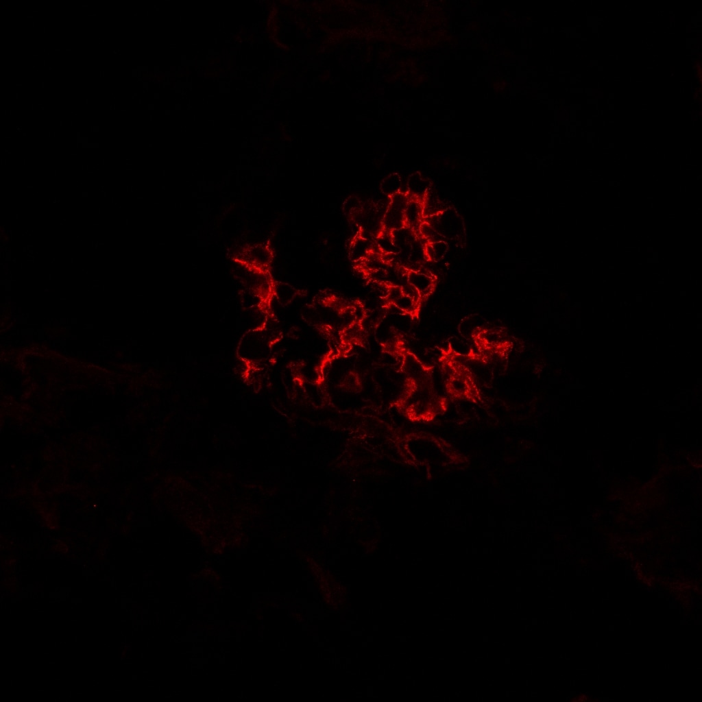
Integrin alpha 8 in 4T1 Mouse Breast Cancer Cell Line.
Integrin a8 was detected in immersion fixed 4T1 mouse breast cancer cell line using Goat Anti-Mouse/Rat Integrin a8 Antigen Affinity-purified Polyclonal Antibody (Catalog # AF4076) at 10 µg/mL for 3 hours at room temperature. Cells were stained using the NorthernLights™ 557-conjugated Anti-Goat IgG Secondary Antibody (red; NL001) and counterstained with DAPI(blue). Specific staining was localized to cytoplasm and cell surface. View our protocol for Fluorescent ICC Staining of Cells on Coverslips.
Detection of Mouse and Rat Integrin alpha 8 by Simple WesternTM.
Simple Western lane view shows lysates of mouse lung tissue and rat lung tissue, loaded at 0.2 mg/mL. A specific band was detected for Integrin a8 at approximately 150 kDa (as indicated) using 10 µg/mL of Goat Anti-Mouse/Rat Integrin a8 Antigen Affinity-purified Polyclonal Antibody (Catalog # AF4076) followed by 1:50 dilution of HRP-conjugated Anti-Goat IgG Secondary Antibody (HAF109). This experiment was conducted under reducing conditions and using the 12-230 kDa separation system.
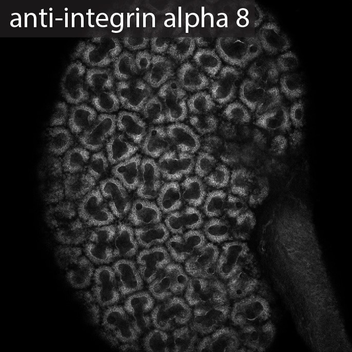
Detection of Canine Integrin alpha 8 by Immunocytochemistry/Immunofluorescence
Integrin alpha 8 co-localizes with laminin 211 in the GBM of AS but not WT dogs.A-C: Dual immunofluorescence immunostaining of kidney from a WT dog; 63x 1.4 n.a. oil. The GBM was localized with anti-collagen alpha 5 ( alpha 5(IV)) and the mesangium was localized with anti-integrin alpha 8 (INT alpha 8). D-I: Dual immunofluorescence immunostaining of kidney tissue from an AS dog at milestone 2. Laminin 211, produced by mesangial cells, was labeled with anti-laminin alpha 2 (LAMA2) and mesangial cells were localized with anti-integrin alpha 8 (INT alpha 8), demonstrating co-localization of laminin 211 with mesangial cell extension in capillary loops. Images D-F were taken with 40x1.3 n.a. oil; images G-I were taken with 63x1.4 n.a. oil with 2X zoom. Image collected and cropped by CiteAb from the following publication (https://dx.plos.org/10.1371/journal.pone.0168343), licensed under a CC-BY license. Not internally tested by R&D Systems.
Detection of Canine Integrin alpha 8 by Immunocytochemistry/Immunofluorescence
Integrin alpha 8 co-localizes with laminin 211 in the GBM of AS but not WT dogs.A-C: Dual immunofluorescence immunostaining of kidney from a WT dog; 63x 1.4 n.a. oil. The GBM was localized with anti-collagen alpha 5 ( alpha 5(IV)) and the mesangium was localized with anti-integrin alpha 8 (INT alpha 8). D-I: Dual immunofluorescence immunostaining of kidney tissue from an AS dog at milestone 2. Laminin 211, produced by mesangial cells, was labeled with anti-laminin alpha 2 (LAMA2) and mesangial cells were localized with anti-integrin alpha 8 (INT alpha 8), demonstrating co-localization of laminin 211 with mesangial cell extension in capillary loops. Images D-F were taken with 40x1.3 n.a. oil; images G-I were taken with 63x1.4 n.a. oil with 2X zoom. Image collected and cropped by CiteAb from the following publication (https://dx.plos.org/10.1371/journal.pone.0168343), licensed under a CC-BY license. Not internally tested by R&D Systems.Applications for Integrin alpha 8 Antibody
Application
Recommended Usage
Immunocytochemistry
5-15 µg/mL
Sample: Immersion fixed 4T1 mouse breast cancer cell line
Sample: Immersion fixed 4T1 mouse breast cancer cell line
Simple Western
10 µg/mL
Sample: Mouse lung tissue and rat lung tissue
Sample: Mouse lung tissue and rat lung tissue
Western Blot
0.25 µg/mL
Sample: Mouse lung tissue
Sample: Mouse lung tissue
Reviewed Applications
Read 3 reviews rated 5 using AF4076 in the following applications:
Formulation, Preparation, and Storage
Purification
Antigen Affinity-purified
Reconstitution
Reconstitute at 0.2 mg/mL in sterile PBS. For liquid material, refer to CoA for concentration.
Loading...
Formulation
Lyophilized from a 0.2 μm filtered solution in PBS with Trehalose. *Small pack size (SP) is supplied either lyophilized or as a 0.2 µm filtered solution in PBS.
Shipping
Lyophilized product is shipped at ambient temperature. Liquid small pack size (-SP) is shipped with polar packs. Upon receipt, store immediately at the temperature recommended below.
Stability & Storage
Use a manual defrost freezer and avoid repeated freeze-thaw cycles.
- 12 months from date of receipt, -20 to -70 °C as supplied.
- 1 month, 2 to 8 °C under sterile conditions after reconstitution.
- 6 months, -20 to -70 °C under sterile conditions after reconstitution.
Calculators
Background: Integrin alpha 8
Alternate Names
ITGA8
Gene Symbol
ITGA8
UniProt
Additional Integrin alpha 8 Products
Product Documents for Integrin alpha 8 Antibody
Certificate of Analysis
To download a Certificate of Analysis, please enter a lot or batch number in the search box below.
Note: Certificate of Analysis not available for kit components.
Product Specific Notices for Integrin alpha 8 Antibody
For research use only
Related Research Areas
Citations for Integrin alpha 8 Antibody
 Powered by Bioz
Customer Reviews for Integrin alpha 8 Antibody (3)
5 out of 5
3 Customer Ratings
Have you used Integrin alpha 8 Antibody?
Submit a review and receive an Amazon gift card!
$25/€18/£15/$25CAN/¥2500 Yen for a review with an image
$10/€7/£6/$10CAN/¥1110 Yen for a review without an image
Submit a review
Customer Images
Showing
1
-
3 of
3 reviews
Showing All
Filter By:
-
Application: Immunocytochemistry/ImmunofluorescenceSample Tested: Embryonic kidneySpecies: MouseVerified Customer | Posted 08/10/2017

-
Application: Immunocytochemistry/ImmunofluorescenceSample Tested: Kidney tissueSpecies: MouseVerified Customer | Posted 05/13/2016
-
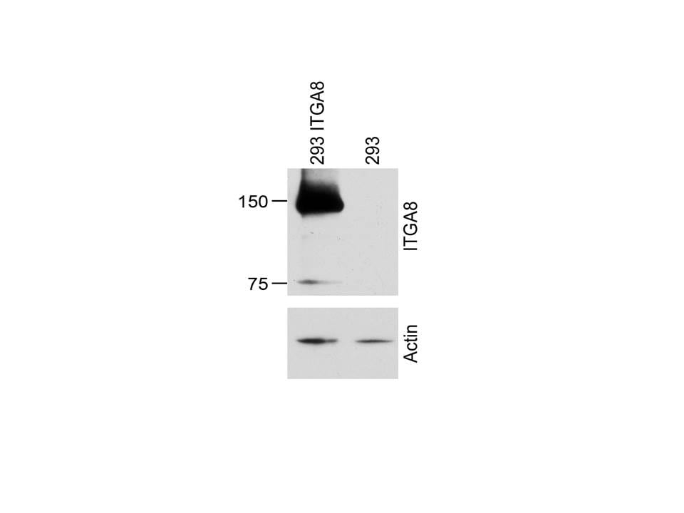
Application: Western BlotSample Tested: Stable HEK293 cells expressing full length ITGA8Species: input species here, HEK293 cells and MouseVerified Customer | Posted 02/22/2016Stables HEK293 expressing ITGA8 were lysed in RIPA and used for western blot analysis. Blot was incubated overnight with 5% milk blocking solution followed by an overnight incubation with 0.4ug/ml of goat anti-ITGA8 in 5% milk. Blot was incubated the next day with 1:3,000 anti-goat HRP secondary antibody and developed employing a film processor. 293 ITGA8: Stable cell line. 293: untransfected cells. Actin was used as loading control.
There are no reviews that match your criteria.
Protocols
Find general support by application which include: protocols, troubleshooting, illustrated assays, videos and webinars.
- Appropriate Fixation of IHC/ICC Samples
- Cellular Response to Hypoxia Protocols
- ClariTSA™ Fluorophore Kits
- Detection & Visualization of Antibody Binding
- ICC Cell Smear Protocol for Suspension Cells
- ICC Immunocytochemistry Protocol Videos
- ICC for Adherent Cells
- Immunocytochemistry (ICC) Protocol
- Immunocytochemistry Troubleshooting
- Immunofluorescence of Organoids Embedded in Cultrex Basement Membrane Extract
- Immunohistochemistry (IHC) and Immunocytochemistry (ICC) Protocols
- Preparing Samples for IHC/ICC Experiments
- Preventing Non-Specific Staining (Non-Specific Binding)
- Primary Antibody Selection & Optimization
- Protocol for VisUCyte™ HRP Polymer Detection Reagent
- Protocol for the Fluorescent ICC Staining of Cell Smears - Graphic
- Protocol for the Fluorescent ICC Staining of Cultured Cells on Coverslips - Graphic
- Protocol for the Preparation and Fluorescent ICC Staining of Cells on Coverslips
- Protocol for the Preparation and Fluorescent ICC Staining of Non-adherent Cells
- Protocol for the Preparation and Fluorescent ICC Staining of Stem Cells on Coverslips
- Protocol for the Preparation of a Cell Smear for Non-adherent Cell ICC - Graphic
- R&D Systems Quality Control Western Blot Protocol
- TUNEL and Active Caspase-3 Detection by IHC/ICC Protocol
- The Importance of IHC/ICC Controls
- Troubleshooting Guide: Western Blot Figures
- Western Blot Conditions
- Western Blot Protocol
- Western Blot Protocol for Cell Lysates
- Western Blot Troubleshooting
- Western Blot Troubleshooting Guide
- View all Protocols, Troubleshooting, Illustrated assays and Webinars
Loading...